Menu

An artificial intelligence predicts therapeutic response in patients with advanced rectal cancer treated with preoperative radiochemotherapy

Cancer colorectal humain et les cellules immunitaires. Crédits: Inserm/Galon, Jérôme

The digestive oncology teams from European Georges Pompidou Hospital, Cochin and Ambroise Paré AP-HP, and laboratory “Information Sciences and personalized medicine” of 1138 Unit Research Center Cordeliers INSERM and Paris-Descartes University, have developed an artificial intelligence system that predict therapeutic response to preoperative chemoradiotherapy in patients monitored for rectal cancer. The work, coordinated by Dr. Jean-Emmanuel Bibault of oncology radiotherapy European Hospital Georges Pompidou AP-HP, would propose a conservative treatment rather than full rectal ablation patients complete therapeutic response.

These results thus contribute to the development of more personalized care in oncology. They were the subject of a publication August 22, 2018 in the journalScientific Reports (Nature Review Group) . 

The standard treatment of locally advanced rectal cancer comprises a preoperative chemoradiotherapy followed by surgical resection (removal) full rectum. About a quarter of patients in complete response after chemoradiotherapy they could avoid radical surgery and benefit instead of conservative treatment (surveillance or endoscopic resection) resulting in less sequelae. However, the only way to know whether these patients respond perfectly to chemoradiation currently remains the operation with removal of the entire rectum.

This study, led by Dr. Jean-Emmanuel Bibault, the radiation oncology department of the European Hospital Georges Pompidou AP-HP, Professor Philippe Giraud, the radiotherapy service HEGP and Professor Anita Burgun, head of the laboratory “information Science and personalized medicine” Unit 1138 Cordeliers research Center INSERM and Université Paris Descartes, therefore aimed to develop an artificial intelligence device which predict in advance the patients with a complete response to preoperative radiochemotherapy, to prevent their operation.

The teams have relied on clinical patient data and images radiation scanners. They then used a method of artificial intelligence-type “Deep Learning” (or “deep neural network”) that has been set for identifying patients with complete response to treatment. The “Deep Learning” is part of machine learning methods that model with a high level of data abstraction.

The algorithm developed was then evaluated on patient data already supported at European Hospital Georges Pompidou, at Cochin Hospital and Ambroise Paré Hospital AP-HP. He was accurate in 80% of cases analyzed. 22 of the 95 patients included in the study had a complete response to preoperative radiochemotherapy.

This study shows that the artificial intelligence algorithms type “deep learning” could be used to predict the effectiveness of cancer treatments to further customize the care of patients.

These results may lead to further research to integrate this device into the treatment of locally advanced cancers of the rectum.

A Hormone Produced During Exercise Could Improve Muscle Capacity in the Elderly

© David Monje – Unsplash 

How can we limit age-related decrease in muscle capacity (sarcopenia), which is a major cause of loss of autonomy in the elderly? Researchers from Inserm, Université Toulouse III – Paul Sabatier and the Gérontopôle of Toulouse University Hospital might have found, within the muscles themselves, a formidable ally in fighting this disease: apelin. This hormone, whose production decreases with age, is secreted during physical activity and improves muscle capacity. This research published in Nature Medicine makes it possible to envisage apelin both as a diagnostic tool for sarcopenia and as a solution for its treatment.

In 2016, WHO finally recognized the decrease in muscle capacity –also known as sarcopenia or muscle loss with aging– as a disease. Indeed, maintaining functional capacity in the elderly is essential to preserve their independence and quality of life. Associated with limited mobility, sarcopenia appears to be a primary cause of the progressive loss of autonomy and the development of age-related pathologies (osteoporosis, cardiac and/or cognitive impairment) and as a consequence one of the main reasons for medical institutionalization.

Sarcopenia is the degenerative loss of skeletal muscle mass, quality and strength. Current treatment strategies produce varying levels of efficacy, often associated with side effects. There is also a lack of early diagnosis tools. Physical activity –while presenting the major disadvantage of often being impractical or fruitless in individuals with reduced motor capacity– is often considered to be the most effective approach. In previous studies it was shown that by stimulating activation of the stem cells at the origin of muscle cells, the muscle contraction generated by physical activity helped renew the muscle fibers (myofibers) and improve their metabolism.

The Inserm research team from the Institute of Cardiovascular and Metabolic Diseases, I2MC (U1048 Inserm/Université Toulouse III – Paul Sabatier) in collaboration with teams from the Gérontopôle of Toulouse University Hospital looked at the relationship between these mechanisms and the development of sarcopenia. They identified a hormone, apelin, produced by the contraction of the muscle during physical activity, and which appears to be capable of maintaining and even restoring muscle capacity.

Indeed, when apelin was administered to elderly mice, the researchers saw their muscle capacity improve and their myofibers return to normal. This improvement is thought to be due to the ability of apelin to stimulate both the cellular metabolism in the muscle and the regeneration of myofibers from stem cells.

Finally, the researchers observed that the production of apelin in response to physical activity diminishes with age. According to Philippe Valet, co-director of the study and professor at Université Toulouse III – Paul Sabatier “in the years to come, apelin could be used for therapeutic purposes in sarcopenia because the results of the study in mice show that treatment with this hormone significantly improves muscle faculties. With this research we can envisage apelin both as a tool for the early diagnosis of sarcopenia and as a promising treatment in the fight against age-related loss of function.”

Clinical trials will be conducted from 2019 by the Gérontopôle of Toulouse University Hospital as part of the IHU-Inspire project which focuses on prevention, aging in good health and regenerative medicine.

Chronic Malnutrition In Children: A New Gut Microbial Signature

Projet Afribiota à Bangui RCA en avril 2017. © Institut Pasteur de Bangui / JM Zokoué

Chronic malnutrition, usually associated with an inflammation of the small intestine, affects one in every four children under the age of five. It is the leading cause of child mortality in low-income countries and is also responsible for severe stunting. The Afribiota project, led by the Institut Pasteur in Paris, the Institut Pasteur in Madagascar and the Institut Pasteur in Bangui, in collaboration with the University of British Colombia, Inserm and the Collège de France, was set up to advance our understanding of the underlying mechanisms of chronic malnutrition and improve treatment. A first study recently demonstrated microbiota disorders in malnourished children, revealing the existence of a surprising microbial signature in the gut, characterized by the widespread presence of bacteria that are normally found in the nose and mouth. The findings were published in the journal PNAS on the 20th of August, 2018.

Chronic malnutrition affects one in every four children under the age of five worldwide. It claims more than 3 million lives every year and results in impaired cognitive and physical development, especially stunted growth, which can be difficult to overcome.

With traditional treatment, in other words providing affected children with micronutrients, a balanced diet and ample food while treating the underlying infections, we can only correct 30% of growth delays,” explains Pascale Vonaesch, holder of a PhD in Microbiology and a member of the Molecular Microbial Pathogenesis Unit at the Institut Pasteur. “There really do seem to be a number of phenomena that have not yet been elucidated.

Chronic malnutrition is not only linked to a lack of food; it is also associated with immune problems and chronic gut inflammation, the workings of which are not yet fully understood. The Afribiota project, carried out in collaboration with the Institut Pasteur in Paris, the Institut Pasteur in Madagascar and the Institut Pasteur in Bangui, was set up in 2016 precisely to find out more about the underlying mechanisms of these disorders and to develop more effective treatment.[1]

In this first study, the scientists focused on the children’s gut flora, one of their aims being to characterize the bacterial populations that colonize the small intestine in malnourished children.

We know that these children suffer from gut inflammation. The villi in the small intestine in particular become blunted, preventing the intestine from effectively carrying out its role in digestion and the absorption of nutrients,” says Philippe Sansonetti, a physician and microbiology researcher in the Molecular Microbial Pathogenesis Unit, which he leads at the Institut Pasteur. “But we didn’t know to what extent the usual bacterial populations were being altered.

To shed light on this question, the stools and duodenal juice of 400 children living in Antananarivo (Madagascar) and Bangui (Central African Republic), both with and without chronic malnutrition, were analyzed. Bacterial cultures and metagenomic analyses aimed at revealing all the microbial species present were also performed, yielding surprising results.

We expected to see an increase in enteropathogenic bacteria such as Campylobacter, Shigella or even Salmonella in the malnourished children,” comments Pascale Vonaesch, “but we certainly didn’t expect to see oropharyngeal bacteria!” “What also surprised us was the sheer number of these bacteria,” adds Philippe Sansonetti. “We had already observed this type of phenomenon for some inflammatory bowel diseases and colon cancer, but never such large-scale migrations. There are 10 to 100 times more bacteria than in the control patients.

Oropharyngeal bacteria, some of which are known for their inflammatory properties, seem to have literally crossed the barriers that usually keep them in the nasopharynx and the mouth, migrating towards and colonizing the stomach and the intestine. This unusual, large-scale migration was observed in malnourished children from both Madagascar and the Central African Republic, in other words independently of their origins, eating habits and environment. 

The origins and consequences of this microbial signature in the gut, characteristic of chronic malnutrition, are yet to be elucidated, even if initial theories are already emerging. “We know that children affected by malnutrition also often have poor oral hygiene and suffer from repeated colds. So there could be an overgrowth of oral and nasopharyngeal flora which may then be swallowed and, in the absence of effective controls, arrive in the digestive system,” suggests Philippe Sansonetti. “It is important to clarify this information so that we can issue effective prevention messages.

In the long term, this gut microbial signature, together with data from the epidemiological, biological and anthropological research carried out for the Afribiota project, should help identify the causes of chronic malnutrition, facilitate diagnosis and ultimately improve treatment of this global health threat.

The Afribiota project is supported by the Total Corporate Foundation, Odyssey Reinsurance Company, the Institut Pasteur, the Nutricia Research Foundation and the Petram Foundation.
Find out more: https://www.pasteur.fr/fr/institut-pasteur/institut-pasteur-monde/programmes-recherche-internationaux/malnutrition-infantile

When a Gut Bacterium Aggravates Metabolic Syndrome and a Probiotic Relieves It

©AdobeStock

While it is evident that obesity, type 2 diabetes and other metabolic complications are current public health issues, their prevalence is much less clear. A team of researchers from Inra, Danone, the Paris public hospital system (AP-HP), Inserm and Sorbonne Université have recently revealed, in an in vivo preclinical study, that the metabolic disorders linked to a high fat diet are aggravated by the proliferation of Bilophila wadsworthia, a pro-inflammatory gut bacterium which contributes to the deterioration of the intestinal barrier. These effects are attenuated by a probiotic bacterium known as Lactobacillus rhamnosus CNCM I-3690. The findings of this study pave the way for the development of nutritional approaches and probiotics which target the microbiota. They were published on July 18, 2018 in Nature Communications.

The name Bilophila wadsworthia probably does not ring any bells for the vast majority of us. It is true that, in a healthy individual, it represents less than 0.1 ‰ of the gut microbiota. However, it is significantly more abundant in individuals consuming a high fat diet. Yet, changes in the composition of the microbiota are commonly associated with metabolic dysfunction without the underlying mechanism of this relationship being properly understood.

In an in vivo preclinical study, researchers from Inra, Danone, the Paris public hospital system (AP-HP), Inserm, Sorbonne Université and their colleagues showed that a high fat diet creates conditions conducive to the proliferation of gut bacteria such as B. wadsworthia. This proliferation is accompanied by an aggravation of the various parameters which characterize metabolic syndrome (e.g. impaired glucose tolerance, reduced sensitivity to insulin and increased blood or hepatic lipids). It is also linked to gut inflammation, intestinal barrier dysfunction as well as bile salt metabolism disorders, favorable to the development of this bacterium.

The scientists then explored the therapeutic potential of a probiotic bacterium, Lactobacillus rhamnosus, revealing the utility of a specific strain known as CNCM I-3690. This strain limits the proliferation of B. wadsworthia, protects the intestinal barrier from its pro-inflammatory effects and improves the parameters of glucose regulation.

This research highlights the role of a gut bacterium, B. wadsworthia, in aggravating the metabolic effects of a high-fat diet. These findings, if they are confirmed in humans, pave the way for the preventive and therapeutic use of probiotic strains likely to ward off the specter of inflammatory and metabolic diseases such as type 2 diabetes and obesity, by restoring the functions ensured by a balanced gut microbiota and by contributing to improve dietary quality.

Obesity: Using the Microbiota to Fight the Harmful Effects of Fatty Liver

Detail of a stéatose, an accumulation of a fat, a triglyceride, in the hepatic cell. Copyright: Inserm/Hadchouel, Michelle

Day after day, the microbiota continues to yield its secrets. In a new study published in Nature Medicine, Rémy Burcelin, Inserm Research Director at the Institute of Cardiovascular and Metabolic Diseases (Inserm/UPS) along with researchers from Inserm Paris, Imperial College London, Girona Hospital and University of Rome Tor Vergata, show how certain gut bacteria cause the accumulation of fat in the liver and play a major role in fatty liver disease (hepatic steatosis). This research could ultimately lead to the availability of biomarkers to predict the disease and proposed therapeutic solutions based on nutritional, and pharmacological approaches and a new generation of probiotics.

Seventy to 80% of people with obesity and type 2 diabetes have “fatty liver”. Fat accumulated in this organ rapidly, when associated with inflammation, leads to the development of liver failure, a diminished ability to filter environmental and dietary toxins resulting in the risk of cancer.

No drug treatments exist at present. The only solutions involve diet and, in the most extreme cases, liver transplant.

Faced with this therapeutic dead-end, FLORINASH, a European consortium of researchers from France (Inserm), Italy and the UK decided to create and compile data from two large cohorts of 800 obese men and women, separating the groups according to the presence or absence of fatty liver. Various medical data were collected from the participants. Then, in a smaller subgroup of around 100 obese women, molecular analyses were carried out on liver biopsies and urine, plasma and fecal samples.

The objectives of this new study were to discover the biological pathway through which liver failure manifests, reveal biological markers to predict the risk of developing the disease in obese individuals, and find therapeutic solutions.

Existing findings from Inserm published in 2007 and 2011 had explained the process through which gut bacteria (microbiota) become harmful, leading to obesity and type 2 diabetes. The researchers were therefore wondering whether the microbiota also plays a role in the onset of the hepatic complications of type 2 diabetes and obesity.

Using the female patients’ biopsies and clinical data, the researchers applied a big data approach. An immense database containing the various molecular details of the microbiota composition, liver genes, plasma proteins and urine proteins was constituted. The researchers then developed algorithms able to identify a logical link between these data.

In other words, explains Rémy Burcelin, Inserm Research Director and study coordinator: “We wanted to see if we could identify, step by step, from the microbiota, the succession of mechanisms responsible for hepatic disease.”

Through this large-scale work in which the algorithms screened over three million bacterial genes, the researchers made two findings. The first is that the more the disease progresses,the greater the fall in microbial gene diversity, suggesting diminished microbiota composition even before the first symptoms appear. The second is that a specific compound of the microbiota, phenylacetic acid, potentiates fat accumulation in the liver while lipopolysaccharides trigger inflammation.

In order to prove the causal link between observation and effect, the researchers continued their work, this time in animals and human liver cells. This involved transferring into healthy mice the microbiota of human donors with fatty liver disease, leading to a drastic increase in their liver triglycerides. It was also shown that when phenylacetic acid was administered to mice, fat built up in their livers.

According to the team, it would be possible in the long-term, by manipulating intestinal bacteria, to prevent the hepatic complications of obesity. The idea is also to be able to lead to the development of a new generation of probiotics, food supplements and to a pharmacological strategy which interferes with the bacterial mechanisms responsible for the liver disease.

Chronic liver disease: discovery of the role of T cell mucosa-associated invariant (MAIT) in inflammation and fibrosis

Crédits: Inserm/Hadchouel, Michelle

In collaboration with the team “Inflammation and stress in liver disease” of the Research Center for Inflammation (UMR 1149-Inserm-Université Paris Diderot), teams of Hepatology Services, anesthesia and resuscitation ‘pathology Beaujon hospital AP-HP, Paris Diderot University and the Cochin Institute (1016 UMR Inserm-University Paris Descartes), demonstrated that a specific population of T cells, called “MAIT “played a major role in inflammation and fibrosis associated with chronic liver disease.

These cells could represent an interesting antifibrogénique strategy to develop new therapeutic approaches to chronic liver disease. This study was the 1 st June 2018 to be published in the journal Nature Communications.

Cirrhosis is the latest evolutionary stage of fibrosis associated with chronic liver diseases regardless of their cause (mainly alcohol abuse, chronic viral hepatitis and metabolic steatopathy in France). It is estimated that in France 200 000 to 500 000 individuals with cirrhosis and more than 170,000 deaths per year are linked to the disease in Europe. Eventually, cirrhosis leads to liver failure whose only cure is liver transplantation. Indeed, there is to date no molecule which antifibrotic effect has been validated in clinical practice.

Chronic liver disease is characterized by persistent inflammation that contributes to their progression to more severe stages. They can progress to liver fibrosis and cirrhosis, and then require a liver transplant. A treatment for a regulation of this inflammatory response could be an interesting anti-fibrogenic approach.

The team of Dr. Sophie Lotersztajn (center for research on inflammation Inserm-Université Paris Diderot), in collaboration with anaesthesiologists services (Dr Emmanuel Weiss), pathology (Prof. Valerie Paradise) and hepatology (Pr Pierre-Emmanuel Rautou) Beaujon hospital AP-HP and a team from the Institut Cochin – Paris Descartes University (Dr Agnes LEHUEN) have focused on the role of invariant T cells associated with mucosa (MAIT) which function just beginning to be decrypted.

This work shows that during cirrhosis, MAIT cells in the liver and blood are activated and they accumulate in the liver in contact with the fibrogenic cells in the fibrous septa. In a mouse model enriched MAIT cells, fibrosis is exacerbated.

Conversely, deficient mice are resistant MAIT fibrotic process. Finally, in vitro studies demonstrate that MAIT cells interact with macrophages by increasing inflammatory and hepatic myofibrolastes stimulating their profibrogéniques properties.

This study highlights the role of MAIT cells in inflammation and fibrosis associated with chronic liver diseases and suggests that targeting these cells could be an innovative therapeutic approach antifibrogénique.

A medical first: CLOVES Syndrome and overgrowth syndromes: remarkable improvement in the health of 19 paediatric and adult patients using a new therapeutic strategy

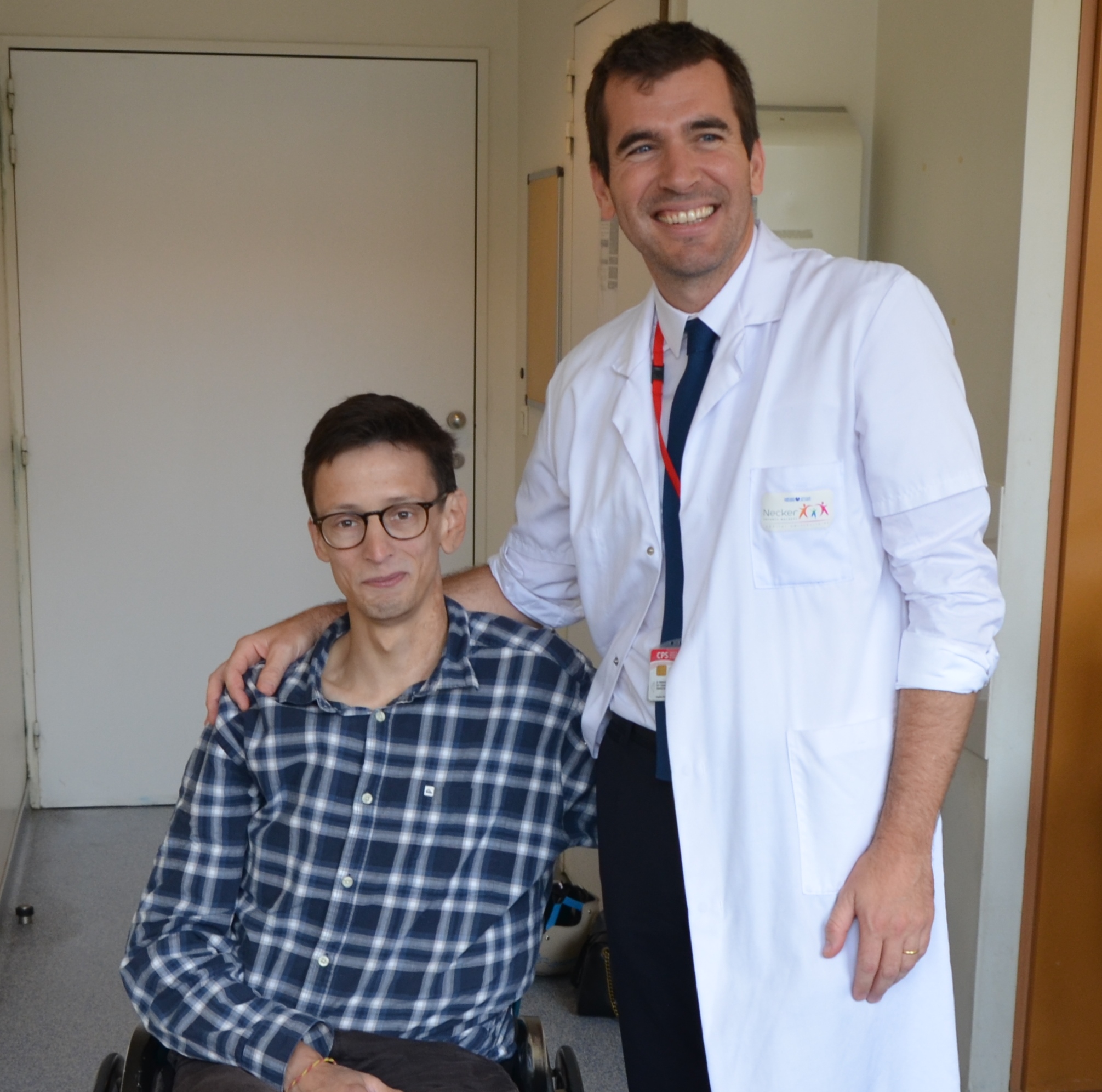
Credits: C Kowalski/Université Paris-Descartes

Dr Guillaume Canaud at the Necker-Enfants Malades Hospital AP-HP, the Paris Descartes University, Inserm (INEM Institute Necker Enfants Malades Centre for Molecular Medicine) and his team recently demonstrated the efficacy of a novel medication, a specific inhibitor called BYL719, in a cohort of 19 patients treated at the Necker-Enfants Malades Hospital AP-HP and suffering from CLOVES Syndrome (Congenital Lipomatous Overgrowth, Vascular Malformation, Epidermal Naevi) or similar disorders. This medication is currently undergoing therapeutic oncology trials (phase I/II). No significant side effects have been observed 18 months after commencement of treatment. This study, published in the journal Nature, is an example of precision medicine and demonstrates the major benefits of this therapeutic strategy for these patients, who have seen their health and quality of life improve significantly.

Patients suffering from CLOVES Syndrome (Congenital Lipomatous Overgrowth, Vascular Malformation, Epidermal Naevi) or similar disorders have major deformities and vascular masses due to mutations of a gene called PIK3CA. This gene regulates cell proliferation and growth. When overly activated, it causes the excessive growth of those parts of the body affected by the mutation. Accordingly, patients present with symptoms that vary greatly depending on the number of tissues affected, which may range from isolated macrodactyly (enlarged fingers or toes to very severe forms affecting the entire body such as CLOVES syndrome.

The most serious forms involve the overgrowth of fatty tissue, vascular malformations, scoliosis, skeletal anomalies such as major bone enlargement and the deformity of organs such as the brain or kidneys. Until now, no curative treatment has been available for those patients for whom the syndrome is potentially life-threatening in the short-to-medium-term and for whom the only therapeutic options have been symptomatic treatments. In the most severe cases patients undergo embolization procedures or mutilating surgery. Finally, it is important to note that these syndromes are frequently associated with chronic pain and have major repercussions on a patient’s quality of life and social life.

The PIK3CA gene frequently mutates in several cancers (particularly breast and colon cancers) and is a therapeutic target for the pharmaceutical industry. PIK3CA mutations in cancers are the same as those found in patients suffering from CLOVES syndrome and similar disorders.

At the end of 2015, a 29-year-old patient with a CLOVES syndrome was referred to Dr. Guillaume Canaud, a specialist of the PIK3CA pathway. This patient had a very advanced and life-threatening form of CLOVES syndrome and for whom no surgery or radiological embolization could be proposed. Dr. Guillaume Canaud then approached the Novartis laboratory working in the field of oncology on a specific inhibitor of the PIK3CA gene called BYL719. This medication is currently undergoing therapeutic oncology trials (phase I/II).

In January 2016, having obtained the authorisation of the ANSM (French National Agency for Medicines and Health Products Safety) to use this experimental treatment, Dr. Guillaume Canaud administered the treatment on this first patient. Very quickly, a positive effect was observed on all symptoms. In particular, a significant decrease was observed in the size of vascular masses and overgrowths from which the patient suffered and a major improvement was seen in his quality of life. Eighteen months later, this first patient had experienced only one side effect, hyperglycaemia, which was well controlled by diet.

At the same time, in order to better understand this disease and the mechanism of action of the medication, Dr Canaud and his research team within the INEM-Inserm Unit U1151) created the first mouse (murine) model that recapitulates all the lesions observed in patients with CLOVES syndrome. The mice were treated with the medication BYL719 and here too a major and rapid improvement in their health was observed.

On the basis of these results, in June 2016, Dr. Canaud quickly gathered a working group composed of ten medical and surgical specialisations to care for patients suffering from CLOVES Syndrome or similar syndromes at the Necker – Enfants Malades Hospital – AP-HP. The idea behind the group was to provide better care for patients.

During the summer of 2016, a 9-year old girl suffering from a serious form of CLOVES Syndrome with a life-threatening vascular mass for which no surgical intervention or embolization was possible, received this experimental treatment. Once again, BYL719 had a spectacular effect on all symptoms, deformities and the vascular mass. It is important to note that no side effects were observed in this first child, the first in the world to receive this treatment, and that her growth was not affected during the 12 months of treatment.

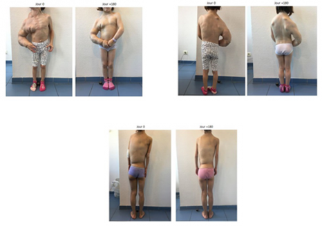
In February 2017, on the basis of these results, Dr. Canaud and his group were authorized by the ANSM to administer the BYL719 to17 additional patients (14 children and 3 adults with ages ranging from 4 to 50 years) followed at Necker hospital, who had life-threatening complications and/or were scheduled for debulking surgery. Very quickly after treatment initiation, they observed a spectacular improvement in their general health of all patients. All patients had a rapid reduction in the size and volume of vascular tumours, but also the enlarged veins and abnormal limbs, they also noticed an improvement in the skin appearance a decrease in fatigue. Moreover, an improvement in scoliosis was observed in all patients. Patients were accordingly able to return to physical activity, stop morphine-based treatment, go back to school, etc.

After six months of treatment, these 17 patients are still alive and have not undergone any surgical interventions. Minor side effects, such as mouth ulcers, were observed in three patients.


These 19 patients continue to receive BYL719 daily.

© Dr Canaud, AP-HP

This study, the protocol for which was approved by the French National Agency for Medicines and Health Products Safety (ANSM), demonstrates the efficacy of this therapeutic approach.

In the opinion of Dr Canaud, this treatment will radically change the future for patients suffering from overgrowth syndromes associated with PIK3CA mutation. The medication has enabled us to obtain results that exceeded our expectations with the regression of malformations that had existed for a number of years, and also an improvement in the quality of life for patients and their families. BYL719 therefore offers great hope as a treatment for even very severe forms of the disease. Finally, our study demonstrates the benefit of developing targeted treatments for genetic diseases to develop what is termed precision medicine, and  also the need for a very high degree of interaction between clinicians and researchers to advance knowledge and develop new medications.

 This work has led to a patent registration application being submitted by Inserm Transfert for AP-HP, Paris Descartes University and Inserm.

Influence of the Gut Microbiota on Tryptophan Metabolism and our Health

Crédits: Fotolia

Numerous compounds are involved in the complex interactions that exist between our body and its microbiota. One of these is the essential amino acid, tryptophan. On June 13, 2018, in the journal Cell Host and Microbe, a team from Inra, AP-HP, Sorbonne Université and Inserm gathered the most recent advances concerning the central role of tryptophan in the dialog with our gut microbiota. Data which opens opportunities for research and future therapeutic applications, particularly in the treatment of inflammatory bowel disease.

Back in 2016, a team from Inra, AP-HP, Inserm and Sorbonne Université studied the role of the CARD9 gene in susceptibility to inflammatory bowel disease (IBD)[1]. Without this gene, the mice studied were more sensitive to gut inflammation and their gut microbiota did not make effective use of tryptophan, an essential amino acid supplied only through diet whose metabolites are particularly implicated in the immune pathways. In addition, the researchers also showed that transferring the microbiota of mice without CARD9 to genetically normal mice also transmitted this susceptibility to gut inflammation. How is the microbiota capable of modulating immune response? How are genetics, immunity and gut bacteria linked? And, more specifically, what is the role of tryptophan in the dialog between the host and its microbiota?

To answer these questions, the same team two years later has gathered the most recent scientific advances on the role of tryptophan and its metabolites in the dialog with the gut microbiota. In their synthesis they address two aspects in particular: 1) the effects of the tryptophan derivatives produced directly by the microbiota bacteria, and 2) the indirect control of the host tryptophan metabolism by the gut microbiota. The researchers describe and analyze the three major pathways of tryptophan metabolism in the gut:

  • The aromatic hydrocarbons (AhR) pathway:

Tryptophan is transformed by certain microbiota bacteria into indole derivatives which are capable of activating AhR, a receptor present on immune cells and epithelial cells in the gut. By activating this receptor, the immune cells notably produce interleukin-22, which has an anti-inflammatory action and a protective role of the mucosa.

  • The serotonin pathway 

This neurotransmitter is produced from tryptophan and affects all parts of the body.  It is involved in various biological processes and in many pathologies. Yet more than 80% of the serotonin of our body is produced in the gut by specialist cells and under the influence of the microbiota.

  • The indoleamine 2,3-dioxygenase (IDO) pathway:

From tryptophan, the IDO pathway leads to the production, notably of kynurenine but also of many other metabolites which are involved in immune, metabolic and even neurological processes.

In decoding the complex balance between these various metabolic pathways, this research allows a better understanding of the development of intestinal and extra-intestinal diseases. They reveal new connections between the microbiota and our health and open opportunities for new therapies.

[1] Read the press release “Genetics and the Gut Microbiota together contribute to IBD”: https://presse.inra.fr/en/Press-releases/Genetics-and-the-gut-microbiota-together-contribute-to-IBD

Our Food Choices Predicted by our Brain Anatomy

©Katherine Chase – Unsplash 

Do you tend to go for cake or vegetables? If you sometimes find it difficult to eat healthily, a study conducted by a team of researchers from Inserm, CNRS and Sorbonne Université, including Liane Schmidt and Hilke Plassmann, at the Brain & Spine Institute (ICM) has established a link between the anatomy of certain regions of our brain and our ability to control our food choices. These results were published in The Journal of Neuroscience on June 4, 2018.

For many people, eating healthily is no easy feat. Individuals differ greatly in their ability to maintain a balanced diet and make healthy food choices.

From the cognitive perspective, making a choice involves two principal mechanisms – the first of which consists of attributing a value to each option. In the case of food, its taste and nutritional quality can, for example, determine its value. The second mechanism for our brain consists of analyzing the value given to each option in order to choose the most suitable – i.e. the food item to which we attribute the highest value.

How are these decisions expressed in the brain? To answer these questions, Liane Schmidt, Inserm researcher, Hilke Plassmann, and their colleagues Anita Tusche from the California Institute of Technology (USA), Cendri Hutcherson from the University of Toronto (Canada) and Todd Hare from the University of Zurich (Switzerland), pooled brain imaging data taken from four studies on food choices.

In three of these studies, the participants performed the same task which involved evaluating their appetite for a specific food according to three criteria: its taste, health benefits and their own natural preference. As such, the participants could base their choice either on taste or on nutritional value.

In the fourth study, the participants were asked to use a method of their choice (save money, eat organic, or lose weight) to reduce their desire for tasty products devoid of nutritional value. This last study “involved a more flexible control strategy not specifically focused on attributes of taste or health but on the various means of distancing oneself from a food or resisting desire” specifies Liane Schmidt, lead author of the study and Inserm researcher.

The researchers studied the variations in the quantity of gray matter in the brains of the participants using imaging data from the first three studies.

As such, they revealed a correlation between food choices and the quantity of gray matter in two regions of the frontal lobe: the dorsolateral prefrontal cortex (dlPFC), which governs the regulation of decisions, and the ventromedial prefrontal cortex (vmPFC), which is in charge of value attribution. They observed that people with more gray matter in these regions had a greater appetite for the foods they considered healthy.

The research team then sought to predict the food choices of the participants in the fourth study based on the quantity of gray matter detectable in the two regions identified previously. “The idea here was to see whether the correlations established in a context in which the control strategies are very clear – focus on taste or health – can be generalized to a situation in which the control strategies are more vague. ” continues Hilke Plassmann.

The researchers confirm these results, thereby establishing for the first time that neuroanatomical differences in the dlPFC and vmPFC regions play a role in individual food choices. These results open up avenues which should ultimately lead to the treatment of eating disorders associated with disrupted food control, such as bulimia nervosa or anorexia nervosa.

Immunoscore: a test to improve the care and treatment of colon cancer

Crédits: Inserm/Sénégas-Balas, Françoise

With Immunoscore, a test devised by a team of researchers from Inserm and Université Paris Descartes and doctors from the Paris AP-HP hospitals, disease progression in patients with colon cancer can now be defined more accurately. According to an international study conducted in more than 2,500 patients, Immunoscore has proved effective in predicting which patients are at high risk of tumor recurrence and, as such, would benefit from intensified treatment following surgery. These results have been published in The Lancet.

The seriousness of cancer, and particularly that of the colon, is essentially estimated according to the extent to which it has spread within the affected organ and the presence of metastasis. This estimation of the aggressiveness of the cancer and its risk of recurrence following treatment must however be improved.

For decades, it has been thought that the immune reaction developed by the patient against his or her cancer has a beneficial influence. Researchers from Inserm and doctors from the Paris AP-HP hospitals have demonstrated in recent years that infiltration of the cancerous tumor by immune cells is a good indication of the way in which colorectal cancer might develop, thereby representing a potential prognostic tool. The immune cell populations which provide the most information on disease progression were identified and the method of evaluating these populations optimized.

This method has led to the creation of an immunological test, applicable in clinical practice, called “Immunoscore”. It works by quantifying the density of two types of immune cells in the tumor and its invasive margin: total T-cells (CD3+) and killer T-cells (cytotoxic CD8+).

The objective of this international study published in The Lancet was to evaluate the prognostic value of Immunoscore in patients with colon cancer on a very large scale. For this, an international consortium of 14 immunology and pathology centers in 13 countries was formed.  A total of 2,681 patients from these centers were included in this analysis. The prognostic performance of Immunoscore, in which patients are classified into 3 groups (high, intermediate and low), was evaluated on the basis of recurrence risk (evaluated during the 5 years following the surgery) and survival. The statistical analyses were all performed by a group of external biostatisticians from the Mayo Clinic in the USA.

The results show that patients with a high Immunoscore present the lowest recurrence risk and prolonged survival. 

In the test group comprising 700 patients, only 8% of those with a high Immunoscore presented a recurrence after 5 years. However, the recurrence rate increased significantly in patients with intermediate and low Immunoscores, reaching 19% and 32%, respectively.  These findings were confirmed in the two other patient groups analyzed, representing 1,981 patients. Furthermore, Immunoscore had a stronger bearing on patient survival than the tumor criteria which are currently used to guide therapy. 

These findings show that Immunoscore provides an accurate and reliable estimation of recurrence risk in patients with colon cancer. The researchers consider that these results support the use of Immunoscore as a new component in the classification of cancer, in which recurrence risk is used to improve individual patient treatment strategies, particularly the modulation of chemotherapy.

In view of the highly positive results of this test in colon cancer, researchers are currently evaluating Immunoscore in other types of cancer and are studying its ability to predict patient response to the immunotherapies which are currently revolutionizing the treatment of cancer.

Nosocomial infections can also be caused by the Bacillus cereus bacterium

Bacillus cereus – Inserm/Leclerc, Henri, 1990 – Source : Inserm images


While Bacillus cereus is well known as a source of food infections, researchers from INRA and ANSES, working with doctors at nine French hospitals[1] including those in the Paris Public Hospital System (AP-HP), have demonstrated for the first time that this bacterium is also responsible for inter- and intra-hospital nosocomial contamination. This study, conducted in 39 patients between 2008 and 2012, also found strains of B. cereus in the hospital environment capable of causing infections that can sometimes be fatal. These results, published in PLOS ONE, suggest that more attention should be paid to these hospital infections, in order to improve patient care.

Bacillus cereus is found everywhere: in soil, in food, on almost all surfaces, on human skin, etc. In spore form, the bacterium is resistant to cooking and pasteurisation. B. cereus is the second most frequent agent responsible for foodborne infections in France and the third in Europe; characteristic symptoms are diarrhoea and vomiting. In rare but more severe cases, B. cereus may also be responsible for non-dietary clinical infections, particularly among vulnerable individuals (such as newborns and the elderly). However, the actual incidence of such clinical infections by B. cereus is unknown and there is little information available on the characteristics of the bacterial strains involved.

Over a five-year period, using clinical and epidemiological data collected from nine participating hospitals in France – including two in the Paris Public Hospital System (AP-HP) – and with the support of laboratories supervised by INSERM, researchers from INRA and ANSES conducted a study on B. cereus in the hospital to obtain an in-depth characterisation (phenotypic and genotypic) of the bacterial strains. This study was conducted in 39 mainly immunocompromised patients who had been infected by B. cereus, eight of whom died.

This work revealed nosocomial contamination by B. cereus and enabled an in-depth analysis of the genetic profile of the strains of B. cereus identified in the hospital. The same B. cereus strain was found in several patients between whom no link could be established, as well as in the hospital environment.

Eight groups of patients carrying the same strain were thus identified, with one strain infecting up to four patients. Moreover, the same B. cereus strain was identified two years apart in separate patients of the same hospital. The pathogen is therefore a source of infection for hospitalised patients, probably due to the ability of B. cereus to sporulate and/or to form biofilms.

Molecular characterisation of the strains also showed that a strain with the same genetic profile could be identified in several patients within the same hospital, as well as in different hospitals.

These results highlight the need for vigilance with regard to B. cereus in the hospital – especially in immunocompromised individuals – and for great thoroughness in cleaning and disinfection procedures. The work also offers insights on the development of diagnostic tests based on virulence factors that are able to determine whether or not the B. cereus strains present a danger to human health.

The in vitro effectiveness of the first-line antibiotic therapies recommended for treating B. cereus infections was also demonstrated. Thus, in order to improve patient care, early diagnosis of a serious B. cereus infection could enable antibiotic treatments to be adjusted, without waiting for the results of additional analyses.

[1] Micalis Institute and MaIAGE – Applied Mathematics and Computer Science, from Genomes to the Environment (INRA, AgroParisTech), ANSES, Institute for Digestive Health Research (University of Toulouse III – Paul Sabatier, INSERM, INRA, ENVT), Toulouse University Hospital (CHU), Centre for Immunology and Infectious Diseases (INSERM, UPMC), Pitié-Salpêtrière Hospital, AP-HP, Antoine-Béclère Hospital, AP-HP, Nice CHU, Strasbourg CHU, Chambéry CHU, Grenoble CHU.
fermer