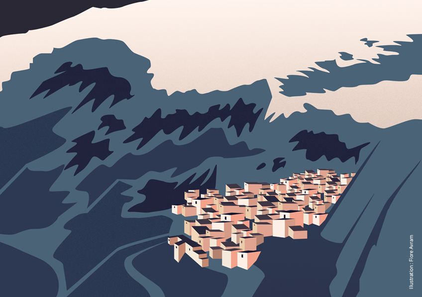
Menu

Syndrome Temple and Silver Russell understanding of epigenetic mechanisms regulating fetal growth

© Inserm/Alpha Pict/Caro, Daniel

A team Sorbonne / AP-HP / Inserm, led by Professor Irene Netchine, physiology professor at Sorbonne University and pediatrician at the Hospital Armand Trousseau AP-HP, studied the molecular mechanisms of the clinical similarity between syndrome Temple and Russell Silver syndrome. Their study, published in Science Advances , highlights the importance of gene network concept “imprinted” in the diagnosis and treatment of patients with these rare syndromes.

In the human genome, genetic information is carried by two copies, each inherited from one parent. For most genes, both copies (maternal and paternal) are expressed equivalently. However, some genes are known as ”  subject to parental imprinting  ” when only one of the two copies is expressed; the other copy extinguishing. The expression of these genes corresponds to an epigenetic mechanism, that is to say biochemical modifications of the DNA molecule, which are different on each of the two copies. These imprinted genes are usually involved in growth, metabolism and development .

The genes expressed by the mother copy have a tendency to restrict fetal growth to preserve its own reserves, while the genes expressed by the paternal copy foster meanwhile fetal growth to ensure healthy offspring and pass on its genes to the next generation.

Syndrome Temple and the syndrome Silver Russell are associated with impaired fetal growth is reduced , and are caused by genetic or epigenetic abnormalities in two regions containing imprinted genes, specifically on chromosomes 11 and 14 , respectively.

These patients also have metabolism and food intake disorders, as well as a premature puberty or advanced.

In his study, the team of Prof. Netchine analyzes the molecular mechanisms of the clinical similarity between the two diseases. In Russell Silver syndrome, a decrease in the expression of a gene ( IGF2 ) usually expressed from the paternal chromosome 11, is the origin of stunting. As to Temple syndrome, changes in genes on chromosome 14 (particularly some RNAs normally expressed from the maternal copy of chromosome 14) also impact gene expression IGF2 , while epigenetic marks are unmodified on the chromosome 11.

These discoveries help advance understanding of epigenetic regulation of gene expression, and consider a new approach to the physiology of fetal growth.

MON 810 and NK603 GM Maize: No Effects Detected on Rat Health or Metabolism

Photo by Charles Deluvio ???? on Unsplash


A diet based on MON 810 or NK603 transgenic maize does not affect the health or metabolism of rats, under the conditions of the GMO 90+1 project1. This unprecedented study performed by a research consortium led by Inra brought together a number of partners2, including Inserm. The research was performed as part of the Risk’OGM program funded by the French Ministry of Ecological and Inclusive Transition. For six months, rats were fed a diet containing either GM maize (MON 810 or NK603) or non-GM maize, in varying concentrations. The researchers, using high-throughput biology techniques, did not identify any significant biological markers related to the transgenic maize diet. Neither did anatomic pathology examination reveal any alteration of the liver, kidneys or reproductive system of the rats fed diets containing these GMOs. This research, published on December 10, 2018 in Toxicological Sciences, does not reveal any harmful effects related to the consumption of these two types of GM maize in the rat even after lengthy periods of exposure.

The researchers used two well-known types of GM maize: MON 810, which produces the protein Bt rendering it resistant to certain insects, and NK603 in which the modification of a gene renders it resistant to glyphosate. For 6 months, the rats were fed a diet containing either transgenic maize or non-GM control maize.  This time period, which is double that of the test required by European regulations, is equivalent to one third of the average lifespan of rats.

The objective of the researchers was to test for early biomarkers of biological function alterations in rats fed GM maize over periods of 3 and 6 months. For that, they used two ultra-sensitive high-throughput techniques: transcriptomics (gene expression) and metabolomics (study of the compounds derived from the body’s functioning). These techniques were used to identify and measure metabolites (amino acids, sugars and other small molecules) and to characterize the expression of messenger RNA and cellular microRNA. These methods are capable of detecting a broad spectrum of metabolic variations. The researchers identified markers able to differentiate the MON810 and NK603 diets. However, following the six-month period of the experiment, no significant differences were identified between the GM and non-GM diets, from the biological point of view.

In addition, in the rats fed the GM diets, anatomic pathology techniques (macro- and microscopic study of the tissues to detect potential abnormalities) revealed no alteration of the organs, particularly the liver, kidneys and reproductive system.

As such, the researchers did not detect any harmful effects of the MON810 and NK603 maize diets on the health and metabolism of the rodents, even after a lengthy exposure period.


1 The GMO 90+ project In 2010, the French Ministry of Ecology launched the Risk’OGM program, in the context of the 2008 law on genetically modified organisms for the establishment of a new legal and regulatory framework based notably on the principle of a triple evaluation of the impact of GMOs – from the health, environmental and socioeconomic standpoints. To set this dynamic in motion and fulfil public authority requirements in terms of expertise, guidance and completed research on GMOs, two calls for research proposals were held, in 2010 and 2013, respectively. The GMO 90+ project was selected during the 2013 call for proposals, with the following scope: to test for biomarkers predictive of biological effects in the study of the subchronic toxicity (3 and 6 months) of GMOs in the rat. Driven by a consortium pooling the various scientific competences, the purpose of this research was to determine whether the feeding of rats with genetically-modified maize led to metabolic changes which could be linked to early effect biomarkers (measurable biological characteristic). The objective was to provide key data which could be used in risk evaluation processes. https://recherche-riskogm.fr/fr/page/gmo90plus  
2 List of project partners: 1-Toxalim (Research Centre in Food Toxicology), Université de Toulouse, INRA, ENVT, INP-Purpan, UPS, Toulouse, France. 2-INSERM UMR-S1124, Toxicologie Pharmacologie et Signalisation cellulaire, Université Paris Descartes, USPC, Paris, France 3- Centre de Recherche sur l’Inflammation (CRI), INSERM UMRS 1149, Paris, France. 4- Laberca, ONIRIS, UMR INRA 1329, Nantes, France 5- Université de Rennes, Inserm, EHESP, Irset (Institut de recherche en santé, environnement et travail) – UMR_S 1085, Rennes, France. 6- Methodomics, France. 7- Institut de Mathématiques de Toulouse, UMR5219 – Université de Toulouse, CNRS – UPS IMT, Toulouse, France. 8- Anses, Maisons-Alfort, France. 9- Profilomic, Saclay/Gif sur Yvette, France 10- UMR1332 Biologie du Fruit et Pathologie, INRA, Université de Bordeaux, Villenave d’Ornon, France. 11- UR 1264, MycSA, INRA, Villenave d’Ornon, France. 12- Laboratoire Reproduction et Développement des Plantes, University Lyon, ENS de Lyon, UCB Lyon 1, CNRS, INRA, Lyon, France 13- CRO CitoxLAB, Evreux.

Type 2 Diabetes: A Therapeutic Avenue is Emerging

Physical contact between HSL and ChREBP in human adipose cells. Each red dot represents an interaction between the two proteins. The cell nucleus is stained in blue. (Credit: I2MC).

Restoring the action of insulin is one of the keys to fighting type 2 diabetes. Researchers from Inserm led by Dominique Langin at the Institute of Cardiovascular and Metabolic Diseases (Inserm/Université de Toulouse) are developing a therapeutic strategy that uses the properties of an enzyme (hormone-sensitive lipase) which, when stimulating fatty-acid synthesis in the fat cells, has a beneficial effect on insulin action. This research has been published in Nature Metabolism.

Diabetes is a disease in which blood sugar levels are high over a prolonged period (hyperglycemia). In the case of type 2 diabetes, this phenomenon which is caused by a disruption of the glucose metabolism develops progressively and insidiously. In France, the prevalence of diabetes is estimated at over 5 % of the 2015 population, with type 2 accounting for 90 % of cases. These figures do not include those who are unaware of their condition, particularly among the overweight or obese.

Hormone-sensitive lipase (HSL) is an enzyme which converts fats into fatty acids and releases them into the bloodstream. In obese patients, these fatty acids trigger the gradual insulin resistance at the origin of type 2 diabetes. Previous research by the Inserm team of Dominique Langin had shown that a decrease in HSL expression in the adipocytes led to a better response to insulin, a sign of good health for these cells.

Surprisingly, the researchers observed that the beneficial effect of a reduction in HSL was not actually due to the reduced release of fatty acid. It was explained by the increased synthesis of oleic acid, the principal fatty acid component of olive oil.

This initial observation gave a glimpse of an interesting avenue for treating obese patients who are at greater risk of developing type 2 diabetes.

To envisage a therapeutic strategy, it therefore had to be elucidated how reducing HSL exerted this beneficial effect on the action of insulin. The group of Prof. Langin discovered the existence of a physical interaction between HSL and a transcription factor responsible for the synthesis of fatty acids, ChREBP. HSL, when binding to ChREBP, blocks its activity. As such, a decrease in HSL leads to the release of this factor in the nucleus, promoting its activity, oleic acid synthesis and sensitivity to insulin.

Preliminary results indicate that a known inhibitor of HSL blocks the interaction with ChREBP. These data therefore pave the way for the development of molecules which target this interaction. In collaboration with global biopharmaceutical company AstraZeneca, the researchers in Toulouse are currently testing different approaches to block the interaction between HSL and ChREBP. Ultimately this project could lead to the development of new drugs to treat the increasing global epidemic of type 2 diabetes.

Expédition 5300: a scientific expedition to the world’s highest settlement

©Flore Avram/Inserm

A group of over fifteen scientists, led by Samuel Vergès, an Inserm researcher from the “Hypoxia and Cardiovascular and Respiratory Physiopathologies” group (Inserm/Université Grenoble Alpes), is taking part in a scientific expedition to investigate the effect of low oxygen levels on health. In early 2019 they will spend 6 weeks in the world’s highest settlement, situated in Peru at an altitude of 5,300 m, studying how its inhabitants have adapted physiologically to extreme living conditions and analyzing the specific altitude-related health problems they face. Eventually, the researchers hope to use the data in order to improve the understanding and treatment of diseases related to low oxygen levels.

Understanding how the human body is able to adapt to low oxygen levels, finding out why some individuals are unable to develop these mechanisms, and identifying ways to improve tolerance of low oxygen are key questions for both individuals traveling to or living at high altitude, and for the treatment of many diseases in less extreme environments.

Situated in Peru, on the border with Bolivia and at an altitude of 5,300 m, the town of La Rinconada’s economy is largely based on gold mining. The mine’s employees and their families live in extreme conditions, not only due to the altitude (which is higher than Mont Blanc), but also because it is impossible to connect the settlement to utilities such as electricity and running water.

High altitude represents an extreme environment that tests the human body’s capacity for adaptation, and its limitations. The reduced level of oxygen available at altitude (hypoxia) makes this environment a true open-air laboratory. This is why La Rinconada has attracted the attention of the scientists in Grenoble.

Scientific goals of the expedition

In February 2019, scientists from Inserm unit 1042 with specialist expertise in altitude and hypoxia research will work alongside other international groups to set up a temporary human physiology and biology laboratory in the heart of La Rinconada. With a view to improving treatment of the physiopathological effects of low oxygen levels, and related diseases, they hope to advance knowledge across a number of different areas.

Improving care

When an individual who normally resides at sea level travels to somewhere at altitude, three major conditions with damaging effects on health may occur:

  • acute mountain sickness, a syndrome that combines headache, nausea, fatigue, and disturbed sleep, and is experienced by half of all individuals traveling above an altitude of 4,000 m;
  • high-altitude pulmonary edema;
  • high-altitude cerebral edema.

The last two conditions are much more serious and can be fatal. At present they can only be treated by oxygen therapy supplied in cylinders or reoxygenation in a hyperbaric chamber. In order to combat these conditions, over time the inhabitants of La Rinconada appear to have developed genetic adaptations to living at altitude. However, they also appear to be more frequently affected by another kind of disease: chronic mountain sickness, which affects people who permanently reside at high altitudes.

The researchers’ main objective is to understand the mechanisms of adaptation to high-altitude hypoxia in order to treat both patients suffering from low oxygen levels in La Rinconada and treat diseases that are more common at sea level. These include diseases such as chronic obstructive pulmonary disease, sleep apnea, and sickle cell disease, which are characterized by patient exposure to inadequate oxygenation due to respiratory or hematological abnormalities. Understanding how the human body is able to tolerate hypoxia to a greater or lesser extent could enable the development of personalized care and improved therapeutic management.

Advancing technology

Unit 1042 is developing a number of diagnostic and therapeutic tools especially for low-oxygen conditions, whether at altitude or, for example, in respiratory disease. These include an altitude mask designed to improve oxygen diffusion via the lungs, which was developed by Dr. Vergès’s team during a previous expedition to the Himalayas. This mask is about to become commercially available for individuals living at sea level and traveling to places at altitude.

Reproducing conditions for life in space

Once the terrestrial environment is left behind, it is very difficult to reproduce the exact conditions of oxygen availability found in life on Earth. This means that future space travelers and dwellers will need to be able to live permanently in oxygenation conditions equivalent to those at high altitude, and thus to develop the mechanisms of adaptation to hypoxia that have been mastered over the course of generations by populations living at high altitude on Earth. Understanding these adaptation mechanisms, their benefits, and limitations will therefore make it possible to better imagine human adaptation to life outside Earth’s atmosphere.

Nearly 20 European and Peruvian scientific and medical experts will take part in the expedition.

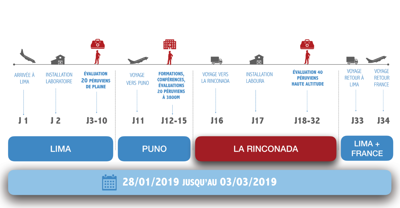
Phases of the expedition

The research team used an initial 10-day reconnaissance trip to Peru in early October to put in place a number of logistical elements (particularly demanding for this kind of expedition), to establish the necessary relationships with key people on the ground, and to work with Ivan Hancco, a Peruvian doctor in La Rinconada, to study the preliminary data collected over recent months from the town’s inhabitants. These data indicated very high levels of chronic mountain sickness compared to other populations living at altitude. Researchers also observed blood abnormalities reflecting extreme physiological responses to hypoxia.

In February 2019, the researchers will examine 40 of La Rinconada’s inhabitants from head to toe. The volunteer participants will be divided into two groups, one in good health and thus with high tolerance to altitude hypoxia, and the other showing symptoms of intolerance (such as headache, dizziness, palpitations, cyanosis, and varicose veins) and in all likelihood specific underlying diseases that require further investigation. Two further groups of 20 Peruvians living in Lima and, at lower altitude, in Puno (3,800 m) will also undergo the same kind of assessment, enabling comparison of group characteristics.

Over a 30-day period, the researchers will study the genetic profile of participants, their sleep quality, and capacity for physical exertion, and gather data on their blood, cardiovascular, and brain characteristics.

In concrete terms, this will involve setting up a scientific and medical laboratory for a period of 6 weeks, requiring several hundred kilos of specialist equipment to be transported by plane and ATV to the 3 study sites in Peru (Lima, Puno, and La Rinconada). The research group will see 6 to 8 volunteers a day, with each subject spending two days with the team in order to undergo all the assessments. Participants will also be studied during their sleep and will have to wear sensors for a 24-hour period. Some of the blood samples taken will undergo on-site laboratory testing (such as blood viscosity, for example), while others will be frozen and sent to France for specific analyses (including genetic and epigenetic testing).

The results obtained should enable the team to identify the mechanisms developed by the human body for living at lower oxygen levels: mechanisms developed over the course of generations by Peruvians living at altitude, including genetic adaptations, and mechanisms that might be manipulated to help individuals suffering from a lack of oxygen to maintain an adequate health status. The group’s preliminary results will be published from September 2019 onward.

Proven expertise

This scientific expedition follows other studies previously undertaken by the same research group:

  • in 2011, a fully working laboratory was set up in the Mont Blanc mountain range at an altitude of 4,300 m in order to study brain changes caused by exposure to high altitude in subjects living at sea level;
  • in 2015, the team led an international scientific expedition to the Himalayas, which brought together participants from 7 different countries to study cardiorespiratory and metabolic adaptations in 50 volunteer subjects trekking to an altitude above 5,000 m.

These two scientific studies resulted in the publication of numerous international scientific articles, as well as the development of a number of technologies and patents. These include two articles published in Neurology in 2013 and Frontiers in Physiology in 2016, which presented the first MRI evidence of the brain changes associated with a 1-week stay at an altitude over 4,000 m.

A patent for the mask mentioned above has been filed and developed in the laboratory and has already been tested in the previous two expeditions. Use of the mask increases saturation (the level of oxygen in the blood) by 8 to 9% at high altitude, which equates to providing the body with the oxygenation conditions of an altitude 1,000 to 1,500 m lower than the one at which the user is currently at.

A wide array of communication methods

Mindful of the unique nature of this scientific and humanitarian mission, the researchers will share their daily news on site thanks to support from communication specialists, some of whom will be based in Peru during the expedition. A website, Facebook page, and an Instagram account have been created for this purpose. It will also be possible to follow the expedition on Inserm’s social media accounts (Twitter and Facebook). A documentary video will be filmed on site by a professional videographer.

Development of an alternative to bone grafting for edentate patients

©Photo by rawpixel on Unsplash

Decreased jaw bone volume in edentate patients is the main difficulty which arises when fitting dental implants. Bone grafting is currently the most frequent solution for this problem. However, this method has several disadvantages, such as deterioration of the bone graft over time. Between 2010 and 2015, Pierre Layrolle, Inserm researcher, working with Norwegian and German teams as part of the REBORNE European project, tested an innovative technique for bone augmentation in 11 edentate patients, by combining a biomaterial with stem cells. The results, published in the journal Stem Cell Research & Therapy, demonstrate sufficient growth of viable bone in the treated area to allow implants to be fitted, together with the durable preservation of this bone after fitting dental prostheses.

Owing to the lack of mechanical stimulation resulting from mastication among edentate patients, bone wastage affecting the alveolar bone (surrounding and supporting the tooth) can be observed, which represents a third of contraindications for fitting dental implants. This procedure, in fact, requires sufficient bone volume to avoid impinging on the facial nerve, which would result in facial paralysis. Now, without sufficient bone volume in the jawbone to support the soft tissue in the gums, even dentures are proving relatively unsuitable.

While bone augmentation involving biomaterials is successfully used to fill the dental alveoli, bone grafting is currently the most commonly used technique for correcting longstanding multiple tooth loss, with a bone sample taken from the patient’s mandible or skull. In addition to the postoperative risks associated with creating two surgical sites, along with the limited quantity of bone material able to be sourced from a single patient, and postoperative pain, this technique appears relatively unsustainable over time. Transplanted bone tends to resorb very quickly. Owing to the lack of vascularization and continuity with the original underlying bone, it is degraded by immune cells which perceive it as a foreign body.

Pierre Layrolle, Inserm researcher heading up the “Inflammation and cellular communications in bone disorders” team (Unit 1238, Inserm/University of Nantes) worked with Norwegian and German teams as part of the Reborne project from 2010 to 2015, on a long-term solution for alveolar bone augmentation. This European clinical trial followed up 11 edentate (over several years) patients, treated with an innovative mandibular bone augmentation technique based on the “cell therapy” principle.

To replace the bone lamellae (containing the patient’s cells and growth factors) conventionally used in bone augmentation, the research team used a biomaterial, calcium phosphate, to which stem cells taken from patient hip bone marrow samples, cultured for amplification, had been affixed. Although calcium phosphate is unable to regenerate bone, it allows adhesion of stem cells – which have this function. This biomaterial is, moreover, highly resistant to the degrading action of immune cells. The mixture thus obtained was applied to the edentate part of the mandible.

For 5 months, researchers monitored positive bone formation around the graft, then generated a 3D model of the augmented part in order to select the type of implant suited to each patient. The sample taken from the 11 patients when fitting the implants showed that the tissue formed not only generated sufficient quantities of bone enabling dental implants to be fitted, but was also vascularized, unlike conventional bone grafts. The dental implants and prostheses were functional two years after fitting, in the 11 patients.

Further to these positive results, the research team launched the MAXIBONE European project in 2018, once again coordinated by Pierre Layrolle. This clinical trial aims to compare the results of maxillary bone augmentation using cell therapy with conventional bone grafting, on a wider scale on 150 patients, by evaluating the quantity of bone generated by the two types of transplants and their specific cost. This project will also examine the possibility of replacing the patient’s stem cells (autologous cells) with donor stem cells in the context of cell therapy. This could offset the technical difficulties related to the cost and complexity of sampling and storing autologous cells, but also mitigate the variations observed in terms of each individual’s ability to effectively generate bone.

The FP7-Reborne project, a phase 2 clinical trial, received European funding from 2010 to 2015.

The H2020-Maxibone project, a phase 3 clinical trial, has received two European grants and is scheduled to start in early 2019.

Pierre Layrolle is due to attend the Nantes international science fiction festival Les Utopiales from October 31 to November 5, 2018, to demonstrate 3D bioprinting. He will lead the round table “In foreign flesh: accepting a foreign body”, on Wednesday, October 31, at 14:00.

An Unbalanced Maternal Diet Affects the Digestive System of Offspring

©Inserm/Naveilhan, Philippe/U913/IMAD

Inadequate protein intake in a gestating female is linked to lasting digestive abnormalities in her offspring. When studying the link between perinatal malnutrition and digestive system in rats, a team of researchers from Inserm in conjunction with Inra, Université de Nantes and University Hospital Nantes discovered functional digestive abnormalities in young rats and an inappropriate response to stress. Although conducted in animals, this research once again demonstrates the incidence of perinatal stress on health in adulthood and draws our attention to the impacts of restrictive diet-related or involuntary deficiencies during pregnancy. This research has been published in The FASEB Journal.

Gestation is an extremely vulnerable time when it comes to normal fetal development and the maintenance of good health throughout life. Several studies have already revealed effects of maternal malnutrition on the cardiovascular and cognitive development of offspring, with repercussions in adulthood. This time, the focus was on the digestive system of young rats, just before they reached adulthood. This involved halving the protein intake of the mothers throughout the gestation and lactation periods and then reintroducing a normal diet once the offspring were weaned.

The researchers began by studying digestive system functioning in the young rats, particularly intestinal motility and permeability. Motility corresponds to the frequency and speed of stool passage. Permeability represents the capacity of nutrients and other molecules to pass through the intestinal wall into the bloodstream. What they observed was an increase in these two parameters in the animals whose mothers had had an inadequate protein intake. They also detected high levels of stress hormone.

To understand these phenomena, the animals were subjected to a model of psychological stress. In the control rats, the stress situation (mimicked by isolating them on a platform with no way out, placed in the middle of a basin of water) triggered accelerated motility and increased permeability. However, this response was altered in the young rats whose mothers had consumed insufficient protein. Their basic digestive activity was greater but did not increase during the stressful event. “Their stress response appeared inappropriate, as if perinatal stress had desensitized the response to acute stress during future life”, suggests Hélène Boudin, Inserm researcher and co-director of this research.

The researchers then examined whether the digestive nervous system had been modified. They observed that the stress hormone induced an excess of neurons stimulating intestinal motility and permeability. These neurons, in addition to their large number, present the disadvantage of being unable to eliminate waste and toxins naturally. And this is “indicative of a poor capacity to respond to stress” clarifies Boudin.

These various observations enabled the researchers to hypothesize that: perinatal nutritional deficiency leads to an increase in the stress hormone in offspring, itself inducing long-term remodeling of the digestive nervous system. This would be responsible for digestive disorders that can weaken the intestine and impact on wellbeing and quality of life.

This research also reinforces the hypothesis of the prenatal origin of certain digestive diseases and disorders. In addition, the mechanisms brought into play by the perinatal stress studied could be shared with those present in other diseases of malnutrition (overeating and undernourishment) and possibly also in neurodevelopmental diseases including some psychiatric diseases” clarifies Boudin. For her, this research once again shows the incidence of perinatal stress on health in adulthood and draws our attention to the impacts of restrictive diet-related or involuntary deficiencies during pregnancy.

Identifying a genetic factor causing lung fibrosis complicating rheumatoid arthritis

©Inserm/Cantagrel, Alain, 1993

Teams of rheumatology, respiratory medicine, genetics and the university hospital department FIRE Hospital Bichat Claude Bernard AP-HP, in collaboration with INSERM, Université Paris Diderot, the Broad Institute of MIT and Harvard, Cambridge, Massachusetts, USA, have discovered that a rare allele of rs35705950 variant gene  MUC5B  multiply by six the risk of occurrence of pulmonary fibrosis in patients with rheumatoid arthritis (RA). This broad study of genetic association demonstrates the existence of a common genetic basis between pulmonary fibrosis associated with rheumatoid arthritis and idiopathic pulmonary fibrosis (IPF). These results, obtained with the participation of national and international network of pulmonologists and rheumatologists, are published in the journal  The New England Journal of Medicine  October 20, 2018.  They are a first step in understanding the diffuse interstitial lung disease (PID) of rheumatoid arthritis, a serious complication whose therapeutic management is not currently codified.

The teams tested the influence of the major genetic risk factor for idiopathic pulmonary fibrosis (IPF): rs35705950 variant gene MUC5B . 

This genetic association study case-control, coordinated by Professor Philippe Dieudé in collaboration with Prof. Bruno Crestani, compared 620 patients with rheumatoid arthritis with diffuse interstitial lung disease (PID), 614 patients with rheumatoid arthritis and without PID 5448 control individuals, from 7 different countries (France, Greece, Netherlands, Japan, China, Mexico and USA).

The results of this international collaboration show a varying contribution MUC5Brs35705950 in the determination of the PID in rheumatoid arthritis. 

The presence of  the risk allele multiply by 3 the risk of occurrence of interstitial lung disease  diffuse (PID)  and  by 6 that of usual interstitial pneumonia (PIC) (the most severe form of PID). Finally, an exploratory phase exploring 12 more REIT susceptibility markers suggests the existence of a common genetic architecture between IPF and pulmonary fibrosis in rheumatoid arthritis.

This work is the first to show that there are  common pathways between the two diseases – pulmonary fibrosis in rheumatoid arthritis and idiopathic pulmonary fibrosis – and provides a strong argument for promoting therapeutic intervention studies in pulmonary fibrosis associated with rheumatoid arthritis, using anti-fibrotic drugs already validated in the idiopathic pulmonary fibrosis.


To know more

The diffuse interstitial lung disease (PID)  is a common extra-articular manifestation and severe  rheumatoid arthritis (RA) , which affects nearly 30% of RA patients, and gradually progresses to irreversible pulmonary fibrosis in about 40 to 50 % of cases. Thus, a PID occurrence is responsible for about 7 to 10% of deaths in patients with rheumatoid arthritis and median survival varies between 2 and 5 years after the onset of Respiratory symptoms. The risk factors and pathophysiological mechanisms underlying the onset of pulmonary fibrosis in RA remain largely unknown. 

Idiopathic pulmonary fibrosis (IPF)  is a lung disease characterized by progressive fibrosis, in which there is no extra-respiratory disorder for which the genetic origins are known. 

Pulmonary fibrosis in rheumatoid arthritis has similarities with idiopathic pulmonary fibrosis, including a  high prevalence of interstitial pneumonia (PIC) , shared environmental risk factors (such as smoking) and a very poor prognosis.

An Improved Diet Could Protect Against Depression

©Brooke Lark on Unsplash

Researchers from Inserm and Université de Montpellier have produced a meta-analysis of the links between a simple-to-use score measuring adherence to dietary guidelines and the development of depressive disorders. The researchers were able to show that adopting a Mediterranean diet (high in fruit, vegetables, fish and whole grains) was linked to a 33% reduction in the risk of depression. These results were published in Molecular Psychiatry.

Globally, more than 300 million people suffer from depression, representing a frequency of 7% for women and 4% for men. It is the costliest brain disorder in Europe. Using data from 36,556 adults, researchers from Inserm and Université de Montpellier were able to show that adopting a Mediterranean diet (high in fruit and vegetables, fish and whole grains) was linked to a 33% reduction in the risk of depression.

Their study also shows that a pro-inflammatory diet (high in saturated fatty acids, sugar, and processed foods) was associated with a higher risk of depression. The chronic inflammation potentially induced by this type of diet could be directly implicated in the onset of depression. According to Tasnime Akbaraly, Inserm researcher responsible for the study: “These findings support the hypothesis that avoiding pro-inflammatory foods (in favor of an anti-inflammatory diet) helps prevent depressive symptoms and depression.

Other studies have also shown the importance of diet in the functioning and composition of the gut microbiota, directly impacting the link between the gut and the brain – with this relationship playing a key role in depressive disorders.

For Tasnime Akbaraly: “The results of our study show the importance of our dietary habits in the development of depressive disorders and encourage the widespread provision of dietary advice at medical consultations”.

Additional clinical trials are necessary to evaluate the efficacy of diets such as the Mediterranean diet in reducing the risk, severity and recurrence of depressive episodes.

20% of reactions to radiologic contrast media are real allergies

©Adobestock

A team of Pole-Imaging Research Explorations-European Hospital Georges Pompidou AP-HP, Paris Descartes University and INSERM led by Professor Olivier Clément, and a team from Caen University Hospital and the University of Caen Normandy, led by Dr Dominique Laroche, conducted the first national prospective multicenter study on allergic reactions to contrast media in radiology. 31 centers in France bringing together radiologists investigators, allergists, anesthetists and biologists have investigated 245 cases of hypersensitivity to contrast media.

Promoted by the AP-HP, the study, funded by the Hospital Regional Program Clinical Research, 2003, shows that allergy is responsible for over 20% of hypersensitivity reactions to contrast media and recommends that patients diagnosed allergic, having a high risk of recurrence, are subject to monitoring based on skin tests performed in an allergist specializes in drug allergy.

This work was published in the journal EClinicalMedicine the Lancet in its issue of July 2018.

In radiology, patients may experience immediate hypersensitivity reactions to iodinated contrast media (for scanners) and gadolinés (for MRI) is injected them in the examination. The reactions such as hives, angioedema, bronchospasm, hypotension and anaphylactic shock. Severe reactions, rare, occur within minutes after injection and require from the imaging team a quick diagnosis and management.

For iodinated contrast agents, reactions have long been falsely labeled “iodine allergy” and mistaken reactions to seafood or skin disinfectants.

But the real allergy to contrast medium is diagnosed by elevated plasma markers tryptase and histamine in the first hour of reaction and intradermal skin tests to make between six weeks and six months after it. The few retrospective studies post on the performance of this type of skin test showed that between 13 and 65% of the responses were truly allergic in origin, according to the populations tested. However, these studies suffered from a lack of clinical data, in particular the name of the injected product, or incomplete or late tests performed, or they mixed the immediate reactions and delayed reactions.

A team of Pole-imaging research explorations-European Hospital Georges Pompidou AP-HP, Paris Descartes University and Inserm, led by Professor Olivier Clément, and a team from Caen University Hospital and the University of Caen Normandy, led by Dr Dominique Laroche studied prospectively immediate hypersensitivity reactions to iodinated products and gadolinés. This multicenter study was conducted in 31 French centers equipped to perform skin tests six weeks to six months after a reaction.

After receiving contrast media for radiology review, 245 patients with immediate reaction took a blood sample in the first hour after it to measure the levels of histamine and tryptase in their plasma. They are seen to offer six weeks after a visit to the allergist to test all existing contrast agents (10 gadolinés iodinated or 5).

Skin testing revealed three types of reactions: allergic (if positive test contrast diluted); potentially allergic (if positive test only to pure product) and nonallergic. They identified 41 patients allergic to iodine products and 10 patients allergic to gadolinés products.

The results showed that over the reaction was severe, the more allergic mechanism revealed by the skin test was frequent : 9.5% in the skin reactions; 22.9% in the moderate reactions; 52.9% in reactions involving life-threatening, and 100% when there was cardiac arrest. Similarly, the levels of histamine and tryptase plasma increased with the severity of the reaction. The presence of cardiovascular signs were also very strongly linked to allergic mechanism.

The group of potentially allergic patients showed clinical symptoms and histamine assays and tryptase intermediate between the group of patients allergic and non-allergic people. This suggests that some of them are truly allergic to the contrast material.

The teams also studied cross-reactions with other different contrast the one responsible for the reaction products: 62.7% of patients had allergic cross-reaction to one or more pure products tested.

This study shows that 21% of radiology hypersensitivity reactions are actually caused by an allergy to contrast media.

Allergic patients have a greater risk of recurrence if their is reinjected contrast agent giving a positive skin test.

Patients exhibited severe symptoms (anaphylactic or cardiovascular symptoms) should benefit from a dose of histamine and tryptase the waning of resuscitation and allergy testing in the six months to determine the allergic or not of their reaction, and especially to know which products will be shown against or authorized for future injections.

A molecule could relieve patients suffering from chronic diarrhea with familial amyloid neuropathy

The teams of hepato-gastroenterology department of adult Bicetre Hospital, AP-HP, in collaboration with the department of neurology adult, Inserm and the Université Paris-Sud, show that a derivative of somatostatin may be effective to treat chronic diarrhea suffered by patients with family amyloid neuropathy (NAF). This side effect concerns a quarter of patients after 5 years of follow-impairing their quality of life.

The results of this study, which highlight once again the expertise of the AP-HP in the treatment of rare diseases, are published in the journal Plos One .

Professor Franck Carbonnel and Dr. Michael Collins, Inserm researcher of Hepato-Gastroenterology department of adult Bicetre Hospital, AP-HP conducted a retrospective study of four patients with familial amyloid neuropathy (NAF). This is a rare progressive genetic disorder that causes multiple organ dysfunction and loss of autonomy.

The teams analyzed particularly gastrointestinal side effects such as diarrhea, nausea, vomiting, abdominal pain and incontinence. A quarter of patients after 5 years of follow suffer from chronic diarrhea. Diarrhea is a symptom very debilitating for patients.

The researchers show that similar to somatostatin, octreotide and lanreotide, appear effective for treating chronic diarrhea. Nine of the fourteen patients who received this treatment had a remission of chronic diarrhea after 6 months follow-up. These molecules are already available for other medical indications, including the treatment of neuroendocrine tumors, hemorrhagic complications of portal hypertension and some endocrine disorders. They are also used off-label in the treatment of severe dumping syndrome.

Their use in the refractory diarrhea had already been studied in the context of HIV, chemotherapy-induced diarrhea or diarrhea secondary to digestive suffering from scleroderma.

Induced diarrhea amylose is a multifactorial diarrhea, but involves disorders of the secretion of digestive hormones (including somatostatin) and of intestinal motility disorders.

Often humps transit aggravate constipation in these patients, associated with dysmotility. Sandostatin acts on intestinal secretion and intestinal motility in complex ways. Treatment with octreotide should be monitored and patients with amyloidosis warned of the risk of hypoglycemia.

This work was conducted in collaboration with the teams of Adult Neurology Service – reference center for family amyloid neuropathy and other neuropathies few devices – Professor Adams that showed the effectiveness of the drug candidate Patisiran, Interferent RNA, promising to treat and improve familial amyloid neuropathy transthyretin in July 2018.

To date, in France there are between 40,000 and 50,000 people with neuromuscular disease (NM).

The conditions under national chain of neuro muscular diseases FILNEMUS include muscle diseases (myopathy), diseases of the neuromuscular junction, rare diseases of the peripheral nerve and spinal muscular atrophy infantile.

How the Zika Virus Persists in Semen and Alters the Spermatozoa

Crédits: AdobeStock

While we already knew that the Zika virus is sexually transmitted and that it remains in the semen for several months after infection, what we did not know was why it persists and why there is a decrease in the number of spermatozoa in infected men. A team of Inserm researchers led by Nathalie Dejucq-Rainsford (Unit 1085 “Research Institute for Environmental and Occupational Health”, Irset), has shown that the virus infects several types of testicular cells –including the germ cells responsible for spermatozoa– which could alter the process of sex cell production. In addition, the low levels of immune defense of the testicle against Zika could be detrimental to eliminating the virus in the organ and as such contribute to its persistence in semen. This research was published in Journal of Clinical Investigation.

The mosquito-borne Zika virus leads to birth defects and can also be sexually transmitted from men to women. The virus can persist in semen for several months and be transmitted more than 40 days after symptoms disappear, which suggests infection of the male reproductive system. Animal models suggest that sexual transmission could facilitate infection of the fetus in pregnant women. Decreases in the number of spermatozoa and increases in abnormal spermatozoa have also been observed in the semen of infected men. However, the organ responsible for the persistence of the virus in semen remained to be elucidated, in addition to the origin of the modifications of the semen.

Using a novel human testicular culture model, the team of Dr. Dejucq-Rainsford at Inserm (Inserm Unit 1085 “Research Institute for Environmental and Occupational Health”, Irset) has shown that the Zika virus infects a number of testicular cell categories, including the germ cells responsible for spermatozoa. They did this by exposing to the virus testicular fragments from uninfected men and maintaining them in culture for 9 days to track the development of the infection. These findings were confirmed in Zika patients with the demonstration of infected testicular germ cells and spermatozoa in semen.

Following further analysis, the researchers suggest that the testicle could constitute a reservoir for the virus, which would explain why it is found in semen long after having disappeared from the rest of the body. Indeed, this organ has low levels of defense against the infection because it does not produce the proteins essential for an antiviral response (interferons) and that it produces only very small quantities of the molecules tasked with signaling the presence of pathogens to the immune system (pro-inflammatory cytokines).

In addition, and unlike other organs, the infected testicular cells do not die – they persist in the testicle. These various elements could enable the virus to be produced in the testicle for long periods without it being eliminated by the immune system.

Finally, infection of the germ cells themselves as well as of other testicular cells involved in the spermatozoa production process (Sertoli cells, Leydig cells) and in their expulsion from the testicle (peritubular contractile cells) could affect semen production. A hypothesis supported by the observation of modifications of semen recorded in infected men up to two months after contact with the Zika virus.

This study provides essential information on the manner in which the Zika virus can persist in semen and modify its parameters, as well as a valuable tool for testing the efficacy of antiviral agents on testicular infection ex vivo.

fermer