Menu

New therapeutic prospect for preeclampsia

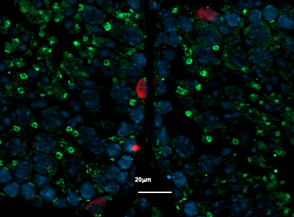
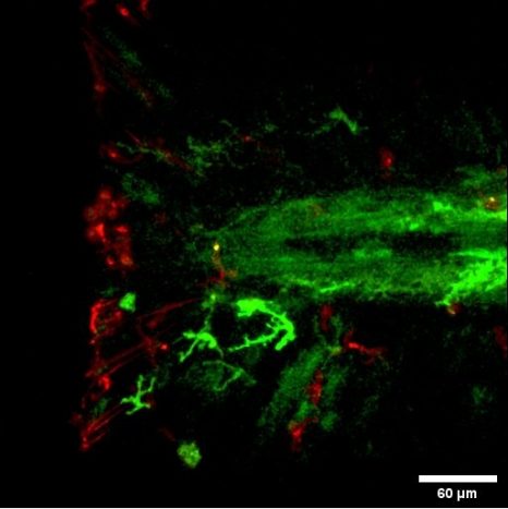
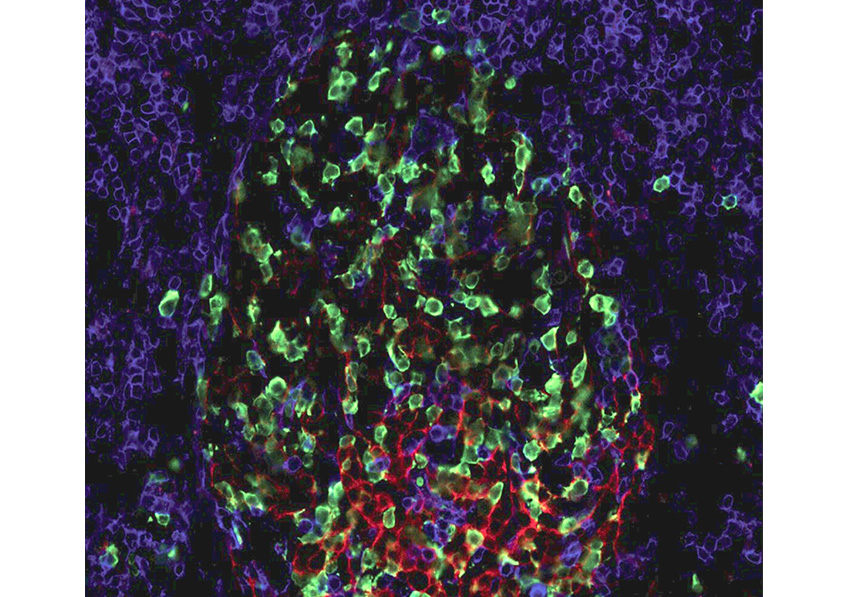
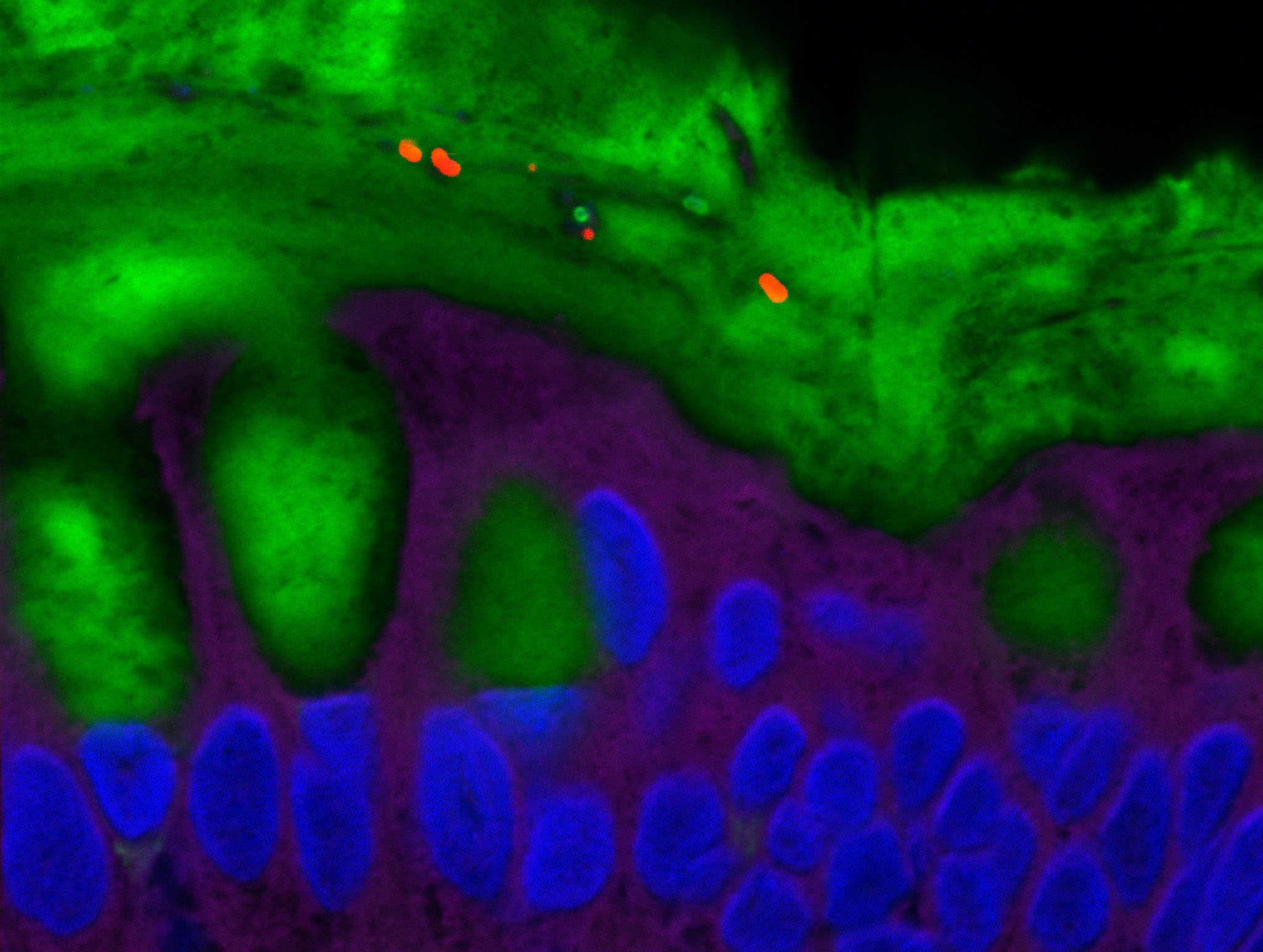
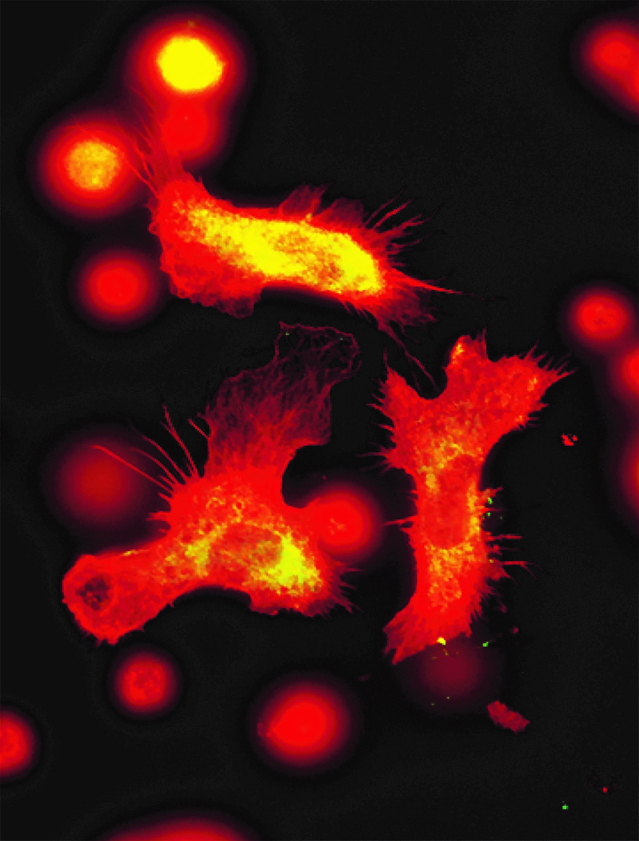
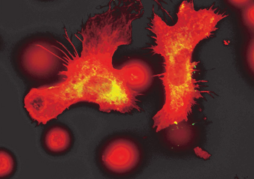
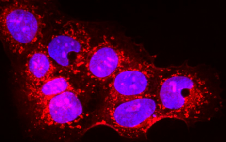
Placental cells are stained blue (nuclei) and the reactive oxygen species produced by mitochondria are stained red by fluorescence. The levels of these reactive species are altered during preeclampsia. © Laurent Chatre, Institut Pasteur/CNRS

Preeclampsia is a condition that affects the placenta during pregnancy and is dangerous for both the fetus and the mother. Scientists from the Institut Pasteur, Inserm and the CNRS have proposed a new therapy, tested in two rodent models, that corrects the defects identified in placental cells, and restores placental and fetal weight. The treatment successfully lowers blood pressure in the mother and resolves the characteristic preeclampsia symptoms of excess protein in urine and cardiovascular abnormalities. The research was published on July xx in the journal Redox Biology.

Preeclampsia is a placental dysfunction that affects approximately 2 to 8% of pregnant women worldwide. It can have fatal complications, with more than 50,000 maternal deaths each year and indirectly more than a million fetal or perinatal deaths worldwide. The primary symptoms of preeclampsia are arterial hypertension, proteinuria (increased levels of protein in the urine), abnormal coagulation in the placenta, cardiovascular abnormalities in the mother and fetal growth restriction. Preeclampsia can also have long-term effects on the cardiovascular system, brain, liver and kidneys of the mother several years after pregnancy. The current first-line treatment for preeclampsia is limited and involves the preventive use of aspirin for at-risk patients. This treatment reduces the procoagulant state in the placenta and partly relieves pressure on the vascular network.

Preeclampsia is characterized by a defective placenta caused by trophoblast dysfunction. Trophoblasts are specific cells in the placenta that help organize and manage the vascular network, allowing the provision of oxygen, nutrients and other elements that are essential for fetal growth. At the molecular level, preeclampsia is characterized by an uncontrolled increase in oxidative stress, with excessive production of various reactive species including reactive oxygen and nitrogen species. There is a genetic component: the first gene to be identified as being implicated in the genetic forms of preeclampsia was the STOX1 transcription factor, which controls the expression of thousands of genes, especially those involved in the production of nitric oxide (NO). In a transgenic mouse model, high accumulation of STOX1 in the placenta induced a preeclampsia-like syndrome. In preeclampsia, nitric oxide, a powerful vasodilator that dilates blood vessels to promote blood flow to the placenta, is mobilized to produce potentially toxic molecules (nitrosative stress) and its levels become insufficient in the placental vascular network, affecting trophoblast function and the vascular network and destabilizing other reactive species. This creates a vicious circle and causes uncontrollable oxidative/nitrosative stress with multiple complications, also affecting maternal blood vessel cells, with potentially fatal consequences.

NO is produced by a family of enzymes known as nitric oxide synthases (NOSs). Finding a way of restoring NO production in the placenta via NOSs could represent an effective new therapy to treat preeclampsia. A years-long collaboration between the team led by Dr. Daniel Vaiman (Institut Cochin, Inserm/CNRS/Université Paris Cité) and the team led by Dr. Miria Ricchetti (Department of Developmental & Stem Cell Biology, Institut Pasteur/CNRS) with Dr. Laurent Chatre*, and more recently an American team from Mississippi, gave rise to a potential solution. The scientists’ research was based on trophoblasts overexpressing STOX1 and on two rodent models of preeclampsia, one mimicking early-onset forms via placental overexpression of STOX1 and the other mimicking late-onset forms by partial occlusion of the lower abdominal aorta. The research revealed a cascade of events that ultimately led the scientists to propose a new therapy. Treating trophoblasts with BH4 (or tetrahydrobiopterin, a cofactor that stabilizes the NOS enzyme producing NO) corrected the defects identified in these cells, restoring production of NO rather than potentially toxic molecules. More importantly, administering BH4 to the two preclinical rodent models restored placental and fetal weight. Finally, in the early-onset STOX1 preclinical model with significant arterial hypertension and proteinuria, the BH4 treatment corrected blood pressure, excess protein in urine, and cardiovascular abnormalities in the mother. The results even suggest that the treatment may be effective in addressing the long-term effects of preeclampsia on mothers (vascular abnormalities in the brain, kidneys, heart and liver).

This research is the first step towards the development of a therapy for preeclampsia. The scientists also performed genetic (transcriptomic) analyses of placentas treated with BH4 and showed that it corrects the expression of several genes disrupted by excess STOX1 in a different way from the deregulation induced by aspirin in the placenta. In conclusion, the scientists propose that a treatment combining BH4 and aspirin could be the ultimate therapeutic solution for many cases of preeclampsia. This hypothesis needs to be validated in clinical trials.

*Dr. Laurent Chatre was previously a CNRS scientist at the Institut Pasteur. Since September 2018 he has been a CNRS scientist in the Imaging and Therapeutic Strategies for Cancers and Cerebral Tissues (ISTCT) laboratory (CNRS/University of Caen Normandy).

Infertility: New Avenues to Understand the Harmful Effects of Chemotherapy

Immunostaining of a mouse testicle section

Immunostaining of a mouse testicle section, with (in red) the undifferentiated germ cells and (in green) the GFP protein reflecting TGR5 receptor expression in this study model. ©David Volle/Inserm

Infertility is a public health problem affecting millions of couples in France. Among the possible causes, chemotherapy has been singled out as having particularly harmful effects on the fertility of both women and men. In order to better prevent and restore fertility in cancer survivors, understanding the mechanisms behind these negative effects is a priority. In a new study, researchers from Inserm, CNRS and Université Clermont Auvergne investigated a receptor found on male germ cells that produce gametes, their aim being to find out more about its role in chemotherapy-related infertility. Their findings, published in Advanced Science, pave the way for a better understanding of male infertility and the development of treatments to reduce the risk of sterility from chemotherapy.

Around 3.3 million people in France are directly affected by infertility. Concerning both men and women, it has continued to increase in recent years, making it a major public health problem [1].

While there are many causes of infertility, it is currently well established that cancer treatments, including chemotherapy, can have particularly harmful effects on male and female fertility. Although cancer therapies have improved in recent years, tackling this issue is becoming a matter of urgency, as an increasing number of cancer survivors will be affected by infertility problems.

For almost 15 years, Inserm researcher David Volle and his team at the Genetics, Reproduction and Development Laboratory (Inserm/CNRS/Université Clermont Auvergne) have sought to improve their understanding of the biological mechanisms underlying infertility. Part of their research focuses on the impact of chemotherapy on male fertility, with the longer-term objective of identifying avenues to counter the adverse effects of this treatment.

In their new study, the researchers looked at TGR5 receptors, which are present on cell membranes, in order to understand their role in the harmful effects of chemotherapy.

TGR5 receptors are widely studied in the context of metabolic diseases, such as diabetes and obesity. They are activated by bile acids – molecules produced in the liver that regulate certain physiological functions, including blood glucose and energy expenditure.

 However, previous research by the team had shown that these receptors are also present in germ cells, the cells that produce gametes. In mouse models mimicking liver disease, with elevated bile acid levels, the scientists had found that the TGR5 receptors on germ cells were activated – which was associated with increased sterility in animals.

Germ cell death

To further understand the impact of TGR5 on fertility in the context of chemotherapy, the scientists in their latest study exposed mice to a chemotherapy agent called busulfan. They then showed that the chemotherapy induces the death of some of the germ cells in healthy mice, thereby affecting their fertility. “The fact that it is the germ cells, at that point undifferentiated, which are affected is particularly problematic because we are talking about the reserve of cells that produce gametes. This can reduce their renewal and contribute to post-chemotherapy infertility,” says Volle.

However, in mice that have been genetically modified to have an absence of TGR5 receptors, the effects of chemotherapy on germ cells are attenuated. This results in an accelerated return of fertility in these busulfan-treated mice compared with the control mice.

Our study has therefore improved our understanding of the molecular mechanisms involved in the harmful effects of chemotherapies on germ cells and fertility. These findings show that TGR5 receptors play an important role in the harmful effects of chemotherapy on infertility,” adds Volle.

In the longer term, the objective is to develop methods to modulate TGR5 receptor activation in a targeted manner within germ cells, in order to protect them and restore fertility after chemotherapy.

The idea is also to assess whether these data can be extrapolated to other disease contexts in which TGR5 receptor activity could be modulated, such as obesity and diabetes, conditions known to impair fertility.

In addition, in parallel to this research, the team observed that even when fertility was maintained in mice exposed to chemotherapy, the quality of the gametes was affected. The scientists will therefore now endeavor to understand both the quantitative and qualitative impacts on germ cells in order to limit not just fertility disorders but also the longer-term consequences on the offspring of animals.


[1] A report requested by the French Minister of Health and the Secretary of State for Childhood and Family in February 2022 outlines a national strategy to combat infertility: https://solidarites- sante.gouv.fr/IMG/pdf/rapport_sur_les_causes_d_infertilite.pdf

Understanding Zebrafish Fin Regeneration Opens up Avenues in Regenerative Medicine

zebra fish _ poisson zèbre

The zebrafish (Danio rerio) is a tropical species commonly used as a model organism in research laboratories. © Adobe Stock


In the animal kingdom, several species share the extraordinary ability to regenerate their limbs or appendages following amputation. One of them is the zebrafish, which is particularly studied in research laboratories due to its ability to regenerate its caudal fin. This phenomenon is made possible by the formation of a blastema, a transient structure composed of undifferentiated cells, which initiates and controls the regeneration of the tissue. Improving our understanding of the cells that make up the blastema and deciphering their interactions opens up new avenues for improving our understanding of the regeneration processes, with the aim of developing clinical applications in the field of regenerative medicine. In a study published in Nature Communications, scientists from Inserm and Université de Montpellier have taken one step closer to this objective, by identifying within the blastema the cell population that coordinates the regeneration process in zebrafish.

The zebrafish, also known as Danio rerio, is a tropical species which has been commonly used as a model organism in research laboratories since the late 1990s. It offers many advantages to scientists, such as the transparency of the embryo and its external development, which is easier to observe than that of mammals. In addition, humans and zebrafish share 70% of the same genes. This genetic conservation with the other vertebrates makes Danio rerio a model of choice for deciphering several major biological processes and their conservation over the course of evolution.

Surprisingly, the zebrafish is also able to regenerate its caudal fin when amputated, thanks to the transient formation of a cell mass known as a “blastema”.

At the larval stage, this structure ensures regeneration of the sectioned appendage in only three days. This is enough to attract the interest of the scientific community, given that understanding the mechanisms associated with this process could pave the way for multiple regenerative medicine applications.

However, only a few cells of the blastema had been described until recently, with the underlying biological mechanisms poorly documented. In their previous work, Inserm Research Director Farida Djouad and her team had highlighted the unique role of macrophages – immune system cells – during the formation of zebrafish blastemas. The team had thus showed that macrophages coordinate the inflammatory processes necessary for the proliferation of the blastema cells and the regeneration of the caudal fin.


Identify the cell coordinating the regeneration

In their new study, these researchers went further in exploring the blastema and revealed the major involvement of a new cell population – cells derived from the neural crest[1]. These cells are found in all vertebrates, including in humans, and play a key role in embryonic development.

The scientists deployed several methodological approaches to observe and monitor the fate of the blastema cells. By combining the applications of real-time confocal imaging and single cell RNA-sequencing technology[2] on zebrafish larvae, the Montpellier-based team was able to demonstrate that cells derived from the neural crest coordinate the fin regeneration process, dialoguing with macrophages and other cells in the blastema in order to control and regulate their response. This dialog is conducted via a key factor called NRG1 (Neuregulin 1).

Interactions between the macrophages (in red) and neural crest cells (in green) during regeneration of the zebrafish larva caudal fin. © Farida Djouad

All of these data make it possible to go further in understanding the regeneration processes and their activation in zebrafish. Based on these findings, the next objective will be to understand why mammals, which also possess macrophages and cells derived from the neural crest, fail to regenerate their appendages.

We are continuing this work on other vertebrate models, including mice, in order to better understand when mammalian embryonic development loses this regeneration capacity, and why, whilst focusing on the role of cells derived from the neural crest,” says Djouad.

“The aim of this research on several animal models capable of regenerating is to identify ‘THE’ coordinating cell, which is common to all regeneration processes. A better understanding of its role, and especially of the factors it secretes, could open up new avenues to promote the regeneration of certain tissues in the treatment of degenerative diseases such as osteoarthritis.”


[1] The neural crest of vertebrates is a transient embryonic structure, involved in development, and capable of producing many tissues of the face and skull, particularly the cartilaginous and osteomembranous skeleton, the meninges, the vascular walls of the external and internal carotid system, the dermis… Source: French Academy of Medicine

[2] Single-cell sequencing is based on a set of molecular biology methods to analyze genetic information (DNA, RNA, epigenome…) on a single cell scale.

New therapeutic perspectives for patients with lymphatic abnormalities linked to a mutation in the PIK3CA gene

Anatomie 3d du système lymphatique © Fotalia

3d Anatomy of lymphatic system © Fotalia


The team of the adult nephrology-renal transplantation department of the Necker-Enfants Malades AP-HP hospital, Inserm and the University of Paris has carried out work, coordinated by Prof. Guillaume Canaud, which opens up new perspectives. therapies for patients with lymphatic abnormalities linked to a mutation in the PIK3CA gene.

The results of this study were published on October 6, 2021 in the journal Science Translational Medicine .

Lymphatic malformations, formerly called lymphangiomas, are malformations of the lymphatic system which can be localized (cutaneous, subcutaneous or mucous) or more rarely spread to the whole body. They are most often congenital and visible before the age of 2 years. They are frequently localized in the axillary and cervical regions. These malformations can be accompanied by painful “inflammatory flare-ups”, compression of organs, especially of the trachea, necessitating the installation of a tracheotomy, diffuse effusions in the pleura or serious infection. They can sometimes be life threatening. These malformations very often have a significant aesthetic impact and a strong impact on the integration of patients into society.

In the vast majority of cases, lymphatic malformations are due to a mutation in the PIK3CA gene acquired during embryonic development (in utero). Current treatments are based on percutaneous sclerosis guided by radiology and / or often decaying surgeries. They can be combined with supportive care such as corticosteroids to treat flare-ups, analgesics, antibiotics, nocturnal respiratory support, and nutritional and psychological support. In some cases, an immunosuppressive therapy, rapamycin sirolimus, is used with varying effectiveness. There is no approved treatment for this indication at this time.

The research work which has just been published in the journal Science Translational Medicine opens up new therapeutic perspectives for these patients with lymphatic abnormalities linked to a mutation in the PIK3CA gene.


The team created the first mouse model carrying a mutation in the PIK3CA gene specifically in lymphatic vessels which recapitulates the different types of lymphatic malformations presented by patients. This experimental model can develop very localized or, on the contrary, very diffuse malformations, depending on the needs.

The team then identified alpelisib (BYL719), a specific inhibitor of PIK3CA, and demonstrated its role as a therapeutic molecule of interest in this preclinical model. With very promising results in animals, the researchers then treated six patients, three children and three adults, with severe lymphatic malformations secondary to a PIK3CA mutation, who had resisted conventional treatments. 

In six months, treatment with alplesib was accompanied by an improvement in patients’ symptoms (pain, inflammatory flare-ups, oozing, discomfort on swallowing, etc.) and a 48% reduction in the volume of malformations measured by MRI. As previously reported in another indication, the treatment was well tolerated.

This work, thanks to the new experimental model created, makes it possible to better understand the pathophysiology of lymphatic malformations but above all opens up new very promising therapeutic perspectives.

This research work was supported by:

-European Research Council (CoG 2020 grant number 101000948 and PoC-2016 grant number 737546)

-National Research Agency – Future Investments Program (ANR-18-RHUS-005)

-National Research Agency – Collaborative Research Program (19-CE14-0030-01).

-CLOVES SYNDROME COMMUNITY (West Kennebunk, USA)

-Emmanuel BOUSSARD Foundation (London, UK)

-DAY SOLVAY Foundation (Paris, France) TOURRE Foundation (Paris, France)

-BETTENCOURT SCHUELLER Foundation (Paris, France)

-Foundation Simone and Cino DEL DUCA (Paris, France)

-Foundation Line RENAUD-Loulou GASTE (Paris, France)

– Schlumberger Foundation for Education and Research (Paris, France)

-Inserm

-Assistance Publique Hôpitaux de Paris

-University of Paris

-And many other generous donors

Persistence of rituximab-resistant memory B cells contributes to relapses in adult patients with immunologic thrombocytopenia

lymphocytes B

Image of a persistent germinal center (mouse spleen, 4 months after immunization), comprising B lymphocytes or memory B cells (green), strongly interacting with follicular dendritic cells (CD35, red), and follicular T helper cells (CD4, blue). ©Inserm/Reynaud, Claude-Agnès

Teams of Prof. Matthieu Mahévas from the reference center for autoimmune cytopenias in adults and the Mondor Biomedical Research Institute (Henri-Mondor AP-HP / Inserm / Université Paris-Est Créteil), Prof. Jean- Claude Weill and Dr Claude-Agnès Reynaud at the Institut Necker-Enfants Malades (Inserm / CNRS / University of Paris) studied the presence of self-reactive memory B lymphocytes before and after treatment with rituximab in adult patients with thrombocytopenia immunologic disease (ITP), a rare autoimmune disease.

The results of this study, which is the subject of a publication in the journal Science Translational Medicine on April 14, 2021 , show in particular that a fraction of memory B lymphocytes self-reactive towards platelet antigens resist treatment with rituximab, persist in the spleen for several months and participates in relapses. The discovery of these cells could open up new therapeutic avenues.    

Patients with B cell mediated autoimmune diseases, such as immunologic thrombocytopenia (ITP), may benefit from treatment with the anti-CD20 antibody, targeting B cells, rituximab. However, a significant proportion of patients relapse after this treatment.

The teams of Prof. Matthieu Mahévas from the internal medicine department of Henri-Mondor AP-HP hospital (Prof. Godeau and Prof. Michel), from the “Transfusion and red blood cell diseases” research unit of the Mondor Research Institute Biomedical (UPEC / Inserm), Prof. Jean-Claude Weill and Dr. Claude-Agnès Reynaud from the Necker-Enfants Malades Institute (Inserm / CNRS / University of Paris), in collaboration with numerous clinicians from the National Center for Auto Cytopenias -immunes of adults (CERECAI), sought to understand why by studying the presence of memory B lymphocytes reactive towards platelets in the spleen of patients splenectomized for a relapse of immunological thrombocytopenia after treatment with rituximab.

Several innovative experimental approaches have been carried out by Dr Crickx and Chappert in order to determine the phenotype, the transcriptional program and the specificity of these B cells capable of secreting anti-platelet antibodies in the spleen of patients during relapses. This work has made it possible to demonstrate that cells newly generated after B lymphocyte reconstitution and memory cells that have resisted treatment participate in relapses.

It thus appears that these pathogenic cells, resistant to rituximab, have lost the expression of CD20 on their surface but preserved the expression of CD19, specifically expressed by B lymphocytes, which could therefore constitute a new potential therapeutic target in this disease.

The persistence of immune memory is generally studied for the benefit it provides in terms of anti-infectious protection. This work demonstrates that memory cells can also persist during periods of remission of an autoimmune disease and contribute to subsequent relapses, suggesting new avenues to be explored to promote prolonged remissions in autoimmune diseases.

This work benefited from ANR funding (Auto-Immuni-B – ANR-18-CE15-0001).

A Major Breakthrough in Understanding the Predisposition of Newborns to Group B Streptococcal Meningitis


Every year throughout the world, Group B Streptococcal (GBS) meningitis affects thousands of newborns. Often fatal, the disease can also lead to severe after-effects in survivors. However, in adults, GBS is an uncommon cause of meningitis. Researchers from Inserm, Collège de France, CNRS, Institut Pasteur, Université de Paris and Paris hospitals AP-HP have now provided elements to explain the predisposition of newborns to GBS meningitis. They have identified and demonstrated that receptors for a bacterial protein enabling penetration of the blood-brain barrier[1] are overexpressed in newborns and absent in adults. The results of their research have been published in Journal of Clinical Investigation.

Group B Streptococcus (GBS) is present in the vaginal microbiota of 20-30% of women. To avoid infecting the baby during labor – which could lead to septicemia and, in the severest cases, meningitis – many developed countries, including France, perform vaginal screening a few weeks before birth. Women found to carry GBS are then given antibiotics during labor.

While this strategy has led to a strong reduction in the incidence of GBS infections during the first week of life, it has had no effect on those occurring between 1 week and 3 months of life.

What is more, many countries offer no such prenatal screening and large numbers of newborns die of GBS meningitis. It is therefore a major public health problem.


Predisposition of newborns

In order to better understand the disease and improve the care of mothers and children, Inserm researcher Julie Guignot and her team at Institut Cochin (Inserm/CNRS/Université de Paris)[2] sought to understand what predisposes newborns to this type of meningitis that affects children and adults only in exceptional cases.

In previous research, the scientists had shown that a variant of GBS was responsible for over 80% of meningitis cases in newborns. This variant expresses specific proteins on its surface, which play an essential role in crossing the blood-brain barrier that separates the blood from the brain.

Using complementary approaches, the researchers demonstrated that one of the proteins exclusively expressed by this variant specifically recognized two receptors present in the cerebral blood vessels that constitute the main element of the blood-brain barrier. Thanks to human samples, they have shown that these receptors are overexpressed in newborns. However, these brain receptors are not present in adults, which explains why GBS is only very rarely responsible for meningitis beyond the first year of life, given that the bacteria cannot reach the brain.

Using animal models of meningitis, the researchers confirmed their findings, showing that the expression of these receptors during the postnatal period contributed to the susceptibility of newborns to meningitis caused by the GBS variant.

For the researchers, these findings open up interesting therapeutic research avenues, particularly in regard to meningitis treatments. “The idea would be to develop treatments to target these receptors in the blood-brain barrier. In the longer term, we would like to study the individual factors of susceptibility leading to the development of these infections. This would make it possible to perform personalized monitoring of at-risk infants born to mothers colonized by this variant,” explains Guignot.


[1] Physiological barrier between the blood and the brain that protects the latter from toxic substances and pathogenic microorganisms

[2]The Structural Molecular Biology and Infectious Processes laboratory (CNRS/Institut Pasteur), the Center for Interdisciplinary Research in Biology, (CNRS/Collège de France/Inserm), the Institute for Advanced Biosciences (CNRS/Inserm/UGA), among others, also participated in this research

Maturation et persistance de la réponse lymphocytaire B mémoire anti-SARS-CoV-2


Food Emulsifiers Increase Pathogenicity of Certain Bacteria and Risk of Intestinal Inflammation

Certain bacteria of the intestinal microbiota (shown in red) are able to penetrate the normally sterile mucus layer (shown in green). © Benoit Chassaing

Diet is believed to play a role in triggering intestinal inflammation that can lead to the development of certain conditions, such as Crohn’s disease. Researchers from Inserm, CNRS and Université de Paris have shown that the emulsifiers present in many processed foods could have a harmful impact on specific bacteria in the gut microbiota, leading to chronic inflammation. Their findings have been published in Cell Reports.

The prevalence of chronic inflammatory bowel disease is increasing in all countries of the world and is thought to affect nearly 20 million people. Characterized by inflammation of the wall of part of the digestive tract, these conditions include Crohn’s disease and ulcerative colitis.

Several factors, both genetic and environmental, have been implicated in explaining the intestinal inflammation associated with these diseases. For several years, Inserm researcher Benoît Chassaing and his team at Institut Cochin (Inserm/CNRS/Université de Paris) have studied the role of diet, particularly the impact of certain additives such as emulsifiers.

Widely used by the food industry in many processed products, the purpose of emulsifiers[1] is to improve texture and extend shelf life. For example, lecithin and polysorbates ensure the smooth texture of mass-produced ice cream and prevent it from melting too quickly once served.

In previous studies based on animal models, the researchers had already shown that the consumption of dietary emulsifiers negatively alters the microbiota in such a way as to promote inflammation.

Moreover, in mouse models where the microbiota had been comprised of a low diversity of bacteria, they observed that the animals were protected against the negative effects of certain emulsifiers.

This led to their hypothesis that the emulsifiers would impact only specific bacteria, which are harmless under “normal” conditions but have the potential to cause disease. It is only in the presence of emulsifiers that these bacteria would be able to promote the development of chronic intestinal inflammation and its associated diseases.

E. coli as a model

As part of their study published in Cell Reports, the researchers used two mouse models: one without a microbiota and the other with a simple microbiota containing only eight species of bacteria. They colonized them with a strain of Escherichia coli (“AIEC bacteria”) associated with Crohn’s disease.

The researchers were interested in the effects of two emulsifiers administered following the colonization of the mice by the AIEC bacteria. Although the consumption of the emulsifiers had no harmful effects on the animals in the absence of these bacteria, they observed the development of chronic intestinal inflammation and metabolic deregulation when they were present. The presence of both the AIEC bacteria and the emulsifier was necessary and sufficient to induce chronic intestinal inflammation.

Further analysis revealed that when these bacteria were in contact with the emulsifiers, they over-expressed groups of genes that increased their virulence and propensity to induce inflammation. “We were able to identify a mechanism by which dietary emulsifiers can promote chronic intestinal inflammation in people who harbor certain bacteria, such as AIEC bacteria, in their digestive tract,” says Benoît Chassaing , who coordinated the study.

The next step is to list all the bacteria that have the same effects in contact with these food additives.

In the longer term, studies to identify and stratify patients according to the composition of their microbiota and risk of inflammation could be set up with the aims of taking a preventive approach and implementing personalized nutritional recommendations. People with specific microbiotas, sensitive to emulsifiers, could benefit from such recommendations.

“And while it is illusory to think that we can banish emulsifiers from our diet, the models and methodologies we have developed here will also allow us to test the action of several types of emulsifiers on the microbiota in order to identify those without harmful effects, and thus encourage their use,” concludes Chassaing.


[1] An emulsifier is a compound that has an affinity for both water and oil and allows the different phases of a compound to remain mixed together.

The Placenta Could Retain a Memory of Tobacco Exposure Prior to Pregnancy

©fotografierende on Unsplash

It is well-known that giving up smoking before pregnancy considerably reduces the health risks for both mother and child. Research by a team from Inserm, CNRS and Université Grenoble Alpes at the Institute for Advanced Biosciences, published in BMC Medicine, took a closer look at the subject showing for the first time that tobacco consumption, even when stopped before pregnancy, can have an impact on the placenta. By studying the placental DNA of 568 women, the team has shown that smoking not just during but also before pregnancy leads to epigenetic modifications (DNA methylation) which could have consequences on its course.

Although it has been shown that tobacco consumption during pregnancy has many negative health impacts for both mother and child, the mechanisms involved are still poorly understood. Previous studies have linked tobacco consumption during pregnancy to alterations in DNA methylation – a form of epigenetic modification (see boxed text) involved in gene expression – in umbilical cord blood and the cells of the placenta. Indeed, while the placenta plays a crucial role in the development of the fetus, it is vulnerable to many chemical compounds.

However, what had not been studied up until now was the impact on placental DNA methylation of tobacco exposure prior to pregnancy.

A team from Inserm, CNRS and Université Grenoble Alpes at the Institute for Advanced Biosciences measured and compared the impact of tobacco consumption on placental DNA methylation in pregnant women in the three months preceding pregnancy and/or during pregnancy.

The researchers studied DNA from samples of placenta collected at the time of delivery from 568 women of the EDEN1 cohort, who were divided into three categories: non-smokers (who had not smoked in the three months prior to pregnancy or during it), former smokers (who had given up smoking in the three months prior to pregnancy) and smokers (who had smoked both throughout pregnancy and in the three months prior to it).

In the smokers, the scientists observed that 178 regions of the placental genome showed alterations in DNA methylation. In the former smokers, they identified that DNA methylation was still altered in 26 of these 178 regions. Only in the women having smoked during pregnancy was methylation altered in the remaining 152 regions.

The regions that were altered most often corresponded to so-called enhancer zones, which remotely control the activation or repression of genes. In addition, some of them were located on genes known to play an important role in fetal development.

While many regions appear to have a normal methylation profile in women after they have stopped smoking, the presence of some DNA methylation changes in the placenta of those having stopped before pregnancy suggests the existence of an epigenetic memory of tobacco exposure,” says Inserm researcher Johanna Lepeule, who led this work. She suggests that changes in placental DNA methylation at the level of the genes related to fetal development and enhancer regions may partly explain the effects of smoking observed on the fetus and the subsequent health of the child.

The next steps in this research are aimed at determining whether these alterations impact the mechanisms involved in fetal development and whether they may have consequences for the health of the child.


[1] The pregnant women were recruited between 2003 and 2006 in the university hospitals of Nancy and Poitiers.


Learn more about epigenetic modifications and DNA methylation

Epigenetic modifications are materialized by biochemical marks present on DNA. Reversible, they do not lead to changes in DNA sequence but do induce changes in gene expression.  They are induced by the wider environment: the cell receives signals informing it about its environment, and specializes itself accordingly, or adjusts its activity. The best characterized epigenetic markers are DNA methylations, involved in the control of gene expression.

Blood Stem Cell Immune Memory: A New Research Avenue in COVID-19

Immune cells seen by fluorescence microscopy. Blood immune cells store information from past infections and then produce more immune cells like the macrophages captured in this image.© Sieweke lab/CIML.

Blood stem cells have a surprising ability. In addition to ensuring the continuous renewal of blood cells, they keep track of past infections so that faster and more effective immune responses can be triggered in the future. This is according to a new study co-led by Inserm researcher Sandrine Sarrazin and CNRS researcher Michael Sieweke at the Center of Immunology Marseille-Luminy (CNRS/Inserm/Aix-Marseille Université, France) and the Center for Regenerative Therapies Dresden (Germany). This discovery could have a significant impact on future vaccination strategies, particularly those being explored for COVID-19, and also further research into new treatments that modulate the immune system. These findings have been published in Cell Stem Cell.

It has long been known that the adaptive immune system has a memory. Following exposure to an infectious pathogen, lymphocytes in the blood become specific to it, with some of them remaining in the body long-term. The principles of vaccination are based on the knowledge of these immune mechanisms.

More recent studies suggest that the innate immune system, which enables immediate defense of the body in response to an infection, also has a form of memory. For example, researchers have shown that the innate immune system continues to be more efficient in the event of reinfection despite the very short lifespan of the immune cells, such as monocytes or granulocytes. They went on to suspect that this innate immune system memory is in fact inscribed in the blood stem cells, which have a very long lifespan and are at the origin of various mature immune cells.

To verify this hypothesis, scientists at the Center of Immunology Marseille-Luminy (CNRS/Inserm/Aix-Marseille Université) and the Center for Regenerative Therapies Dresden (Germany) carried out research whose findings have been

published in Cell Stem Cell. The researchers began by exposing mice to a molecule found on the surface of the E. coli bacterium (lipopolysaccharide or LPS), a pathogen which is commonly used in laboratories to mimic infections.

They then transferred blood stem cells taken from these animals to non-infected mice whose immune systems had previously been destroyed. The aim was to fully reconstitute their immune systems based on these stem cells.

The researchers then infected mice from this group with a live bacterium of the species P. aeruginosa, observing a mortality rate of just 25%. However, in the control mice whose stem cells had never been exposed to a pathogen, this rate was 75%. 

“This research strongly demonstrates that the blood stem cells have a memory function that we did not know existed. Initial exposure to a pathogen makes them better equipped to face subsequent infections”, explains Sandrine Sarrazin.

This mechanism is not specific to pathogens because, in another experiment, an initial exposure of the blood stem cells to a viral antigen protected the mice from secondary exposure to P. aeruginosa. The scientists made the surprising discovery that the protection afforded by this immune system memory extends beyond the infectious agent used for the first infection.

The researchers then looked at how this memory is coded. When studying the genome of the blood stem cells of the infected mice, they observed lasting modifications in its spatial organization. Changes that are likely to modify the expression of some genes implicated in the innate immune response. “At the time of first contact with the pathogen, genes required for the immune response are in fact put forward long-term so as to rapidly activate the immune system in the event of a second infection”, explains Bérengère de Laval, lead author of the study. Finally, the team looked for molecules implicated in this change of genome structure and discovered that the protein C/EBP beta played a major role.

Research relevant in fighting COVID-19?

These results are particularly relevant during this period of SARS-Cov-2 coronavirus pandemic.

Recent findings suggest that the BCG vaccine – it too known for inducing innate immune memory – also acts at blood stem cell level and offers a certain degree of protection from respiratory infections. Studies are ongoing in order to test its utility against COVID-19.

The team’s discoveries could elucidate the molecular mechanisms at play in this protection and open up new avenues for vaccines – particularly against COVID-19.

“Our discoveries represent a major contribution to understanding immune system memory and blood stem cell functions. They also point towards new strategies for stimulating or limiting immune response in various disease states and could make it possible to refine current vaccination strategies for better protection from various pathogens, including SARS-CoV-2″, hopes Michael Sieweke.

fermer