Menu

Bisphenol A: Alternative products that may also be harmful

Bisphenol F and bisphenol S, which are used as substitutes for bisphenol A in certain applications, have the same negative effect on human foetal testes as bisphenol A. This has recently been shown by René Habert and his colleagues at the Joint Research Unit 967 “Stem Cells, Radiation and Genetic Instability” (CEA/Inserm/Paris Diderot University)[1] using the same in vitro method that allowed the team to analyse the negative effect of bisphenol A on the testis in 2012[2].

These results are online at the Fertility and Sterility journal website.

The manufacture and sale of babies’ feeding bottles containing bisphenol A have been banned in Europe since January 2011. In addition, the manufacture, export, import and marketing of all food packaging containing bisphenol A is banned in France from January 2015. Alternative products, bisphenol S and bisphenol F, are currently being studied or are already in use. Even though their chemical structure is similar to bisphenol A, the safety of these products has never been tested in humans and other mammals and no regulations currently exist concerning this.
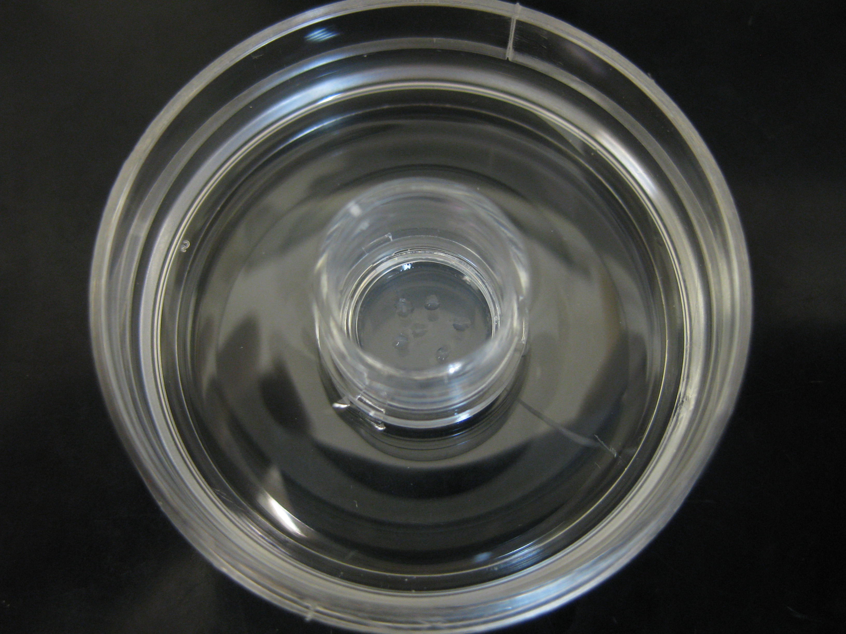
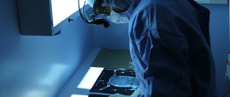
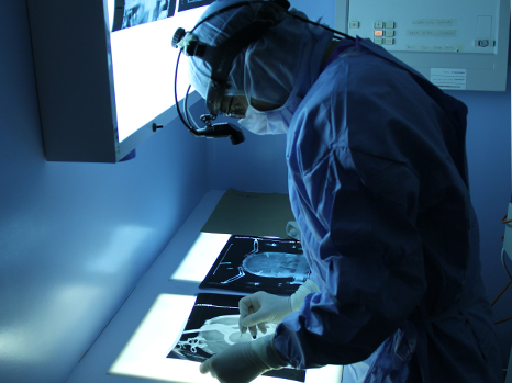
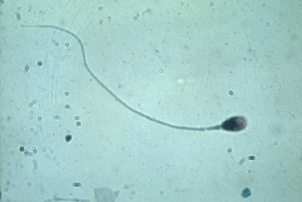
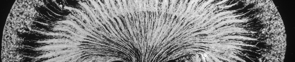
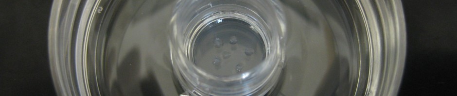
Culture de testicules foetaux humains

R. Habert/Inserm

To assess the harmful effects of endocrine disruptors[3] on the testis development during foetal life, Réné Habert and his colleagues at the Laboratory of Development of the Gonads (UMR 967 – “Stem Cells, Radiation and Genetic Instability” – CEA/Inserm/Paris Diderot University) have been using an original experiment system for several years, which they have developed and named the Fetal Testis Assay (FeTA)[4]. This method allows foetal testes to be kept in a petri dish for several days to assess the effects of the addition of different chemicals on their development and function. Thanks to this method, the researchers provided the first experimental proof that bisphenol A inhibits the production of testosterone in the human foetus. A concentration equal to 2 micrograms per litre of bisphenol A in the culture medium was sufficient to produce these effects This concentration is equal to the average concentration generally found in the blood, urine and amniotic fluid of the population.

Researchers have used the same culture system in this new study, the results of which were published in the Fertility and Sterility journal. They observed that the exposure of human foetal testes to bisphenol S or bisphenol F reduces the production of testosterone that is identical to the reduction caused by bisphenol A. It is the first time bisphenol S and F have been shown to have a harmful effect on physiologic function in humans.

Reminder:

Bisphenol A is a chemical compound that is included in the composition of plastics and resins. It is notably used in the manufacture of food containers and is also found in the protective films used inside food and beverage cans. Several studies have shown that this component has harmful effects on reproduction, metabolism and the brain. The full impact of bisphenol A occurs during foetal life.

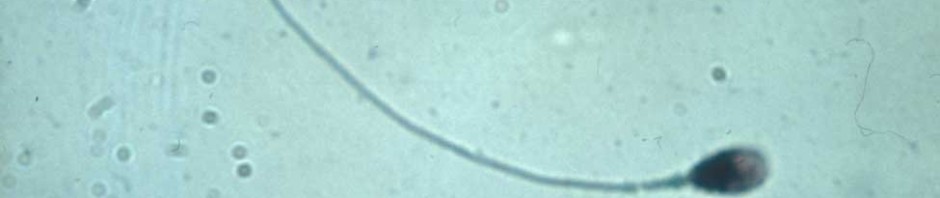
Testosterone produced by the testes in humans and mammals during foetal life is necessary for the masculinisation of internal and external genitalia. Without this hormone, these organs can spontaneously develop in a female sense. A decrease in foetal testosterone production can lead to defects in masculinisation observed at birth (such as hypospadias and cryptorchidism). Furthermore, it is likely that a lack of testosterone produced during foetal life can result in a reduced sperm count in adulthood.

Researchers have also compared the response to bisphenol S and F in human foetal testes to those in the foetal testes of rats and mice. They have observed that humans are far more sensitive to bisphenol S and F than mice.


[1] CEA-IRCM (Institute of Molecular and Cellular Radiobiology), Fontenay-aux-Roses CEA centre.

[2] Press release 17.01.2013 – L’effet néfaste du bisphénol A prouvé expérimentalement (Harmful Effects of Bisphenol A Proven Experimentally) (Plos One, December 2012)

[3] A substance or mixture not produced by the body that alters the function(s) of the endocrine system and consequently causes adverse health effects in an intact organism, or its progeny, or sub-populations.

[4] In collaboration with the Antoine-Béclère Hospital in Clamart.

A new type of heredity described in Paramecia

Considered as an obsolete theory for many years, the transmission of acquired traits has returned to the forefront of debate thanks to the development of epigenetic research(1). In this context, a team from the Institut de biologie at the Ecole normale supérieure (CNRS/ENS/INSERM)(2) has described how in Paramecia, mating types are transmitted from generation to generation through an unexpected mechanism. These types are not determined by the genome sequence but by small RNA sequences transmitted via the maternal cytoplasm, which specifically inactivate certain genes during development. A Paramecium can thus acquire a new mating type that will be inherited by its progeny without any genetic modification being involved. Published in Nature on May 7, 2014, this work highlights a novel mechanism that may be governed by natural selection, thus allowing the evolution of species.

Paramecia, single – cell eukaryote organisms, are hermaphrodite: during their sexual reproduction (or conjugation), the partners exchange their genetic material. Paramecia nevertheless have two “mating types”, called E and O. Conjugation can only occur between different mating types. As early as the 1940s, scientists such as Tracy Sonneborn had noted that mating type was not transmitted to progeny in Mendelian fashion: a new type of trait transmission, not dependent on the chromosomes, had to be involved, but they did not succeed in elucidating it.

Today, the team led by Éric Meyer at the ENS Institut de Biologie 2 has described the mechanism underlying this alternative heredity. To achieve this, they first showed that the difference between the E and O matin g types was due to a transmembrane protein called mtA. Although its encoding gene is present in both types, it is only expressed in E individuals. The scientists then revealed the mechanism by which this gene is inactivated in type O individuals.

Paramecia have two nuclei: a germinal micronucleus that is transmitted during sexual reproduction and a somatic macronucleus – resulting from the latter – where the cell’s genes are expressed. The mechanism
for the transmission of mating types is based on small RNA, called scnARN, which are produced during meiosis. The original function of these RNA is to eliminate from the macronucleus a whole series of genetic sequences called transposable elements, which, like introns (3), have been introduced into the genes during evolution. As a first step, the scnARN scan the maternal macronucleus in order to identify the sequences that were deleted in the previous generation, and then make the same rearrangements in the new macronucleus. However, unexpectedly, this genome “cleaning” mechanism also allows the cell to silence functional genes. In type O individuals of Paramecium tetraurelia , scnARN eliminate the mtA gene promoter, thus deleting its expression. Thus, it is through the scnARN inherited from the maternal cytoplasm, and not from a particular gene sequence, that the mating type of Paramecium is defined.

This silencing process could in principle affect any gene. Thus in theory, Paramecia could transmit to their sexual progeny infinitely variable versions of the macronuclear genome from the same germline. As with
genetic heredity, this mechanism may cause errors that might occasionally endow progeny with a selective advantage. In other words, the somatic macronucleus genome of Paramecium may evolve continuously,
and in certain cases allow a short – term adaptation to changes in environmental conditions. And this can occur without any genetic mutations being involved. This type of Lamarckian heredity (4) may thus offer a
hitherto unsuspected lever for natural selection.

(1) Epigenetics forms part of genetics in its broadest sense; in other words, the study of the mechanisms of heredity. It refers specifically to the study of the hereditary transmission of variable traits that are not dependent on variable DNA sequences.
(2) In collaboration with the Centre de Génétique Moléculaire (CNRS), the Laboratoire Biométrie et biologie évolutive (CNRS/Université Claude Bernard Lyon 1), the Institut Jacques Monod (CNRS/Université Paris Diderot) and CEA (Institut de génomique). Polish, Russian and American teams also collaborated in this work.
(3) Portions of the gene sequence, often non – coding, which must be deleted for the sequence to be functional.
(4) Referring to Jean – Baptiste Lamarck (1744 – 1829) whose theory on the evolution of living beings considered the transmission of acquired traits.

Non-coding genomic regions ameliorate the severity of beta-thalassemia and sickle cell anemia

Beta-thalassaemia and sickle cell anaemia are genetic disorders caused by mutations in a single gene but non-coding genomic regions seem to have a strong influence on disease severity. The teams of Eric Soler, researcher at Inserm unit 967, Fontenay-aux-Roses, France (Inserm / CEA), Swee Lay Thein, Clinical Director of the Red Cell Centre in King’s college London and King’s College Hospital, London, UK, and Frank Grosveld, professor of Cell Biology at the Erasmus Medical center, Rotterdam, unraveled the molecular mechanisms explaining how non-coding genomic variants, located far away from genes, were able to modify the clinical severity of beta thalassaemia and sickle cell anaemia. To reach this goal, the researchers have combined different expertise, including the study of spatial chromosome architecture. This work will be published in The Journal of Clinical Investigation and will be accessible online starting from March 10th 2014.

It is now clear that the vast majority of mutations and genetic variations uncovered by Genome-Wide Association Studies (GWAS), associated with common and rare diseases (diabetes, cardiovascular diseases, cancers) lie within non-coding sequences in the genome, i.e. do not directly hit the structural part of genes. Surprisingly, these genomic variants, found in the human population may be located at considerable distances from genes. This fact strongly complicates their functional analysis, and illustrates the complexity of the human genome organization and its relationship with diseases.

Etude de la drépanocytose. © C Feo/Inserm

Beta-thalassaemias and sickle cell anaemia are among the most common inherited disorders affecting red blood cells. In particular, sickle cell anaemia which affects 300,000 newborns annually, is fast becoming the most common serious genetic disease in UK, France, and the rest of Europe. These disorders are caused by mutations affecting a single gene – the beta globin gene- leading to alterations of the adult haemoglobin. Despite being single gene mutations, both disorders display an extremely variable range of disease severity, many factors modify the disease severity including the ability to produce fetal haemoglobin. Fetal haemoglobin is normally ‘silenced’ in adults, but some adults are able to escape this silencing and continue to produce fetal haemoglobin, and although harmless in healthy adults, fetal haemoglobin  can compensate for the altered adult haemoglobin in patients with beta thalassaemia and sickle cell anaemia, reducing the severity of the anaemia. In 2007, Swee Lay Thein’s lab identified a number of variants in a “gene desert” on chromosome 6q23, at tens of thousands of base-pairs from the closest genes, as affecting the ability to produce fetal haemoglobin in adults. How these genetic variants and the mechanism involved in ‘reactivating’ fetal haemoglobin were the focus of the three labs.

The researchers used a combination of chromosome conformation technologies and high throughput DNA analyses on thalassemia patient samples to elucidate the molecular mechanisms of how non-coding variants exert their action and improve the symptoms of thalassaemia and sickle cell anaemia.

The researchers showed that these variants, in a normal context, physically interact with the MYB gene, located at a distance of more than 80 000 base-pairs, via chromosome folding. In individuals carrying the variants, chromosome folding is diminished leading to a decrease in expression of the MYB gene. 

The decrease of MYB expression in patients carrying the variants leads to a reactivation of fœtal haemoglobin which compensates for the defect of adult haemoglobin and significantly ameliorates sickle cell and beta thalassaemia severity “, the authors say.

These data identify directly and for the first time the MYB gene as the target of chromosome 6q23 non-coding variants. Thus, the MYB gene represents a major therapeutic target for the induction of fœtal haemoglobin, a potential therapeutic approach for beta thalassaemia and sickle cell anaemia, Eric Soler and Swee Lay Thein suggest.

Enhancement of chemotherapy by prevention of tumour cell repair

Chemotherapies are cancer treatments that work by inducing lesions in the DNA of tumour cells in order to inhibit their proliferation. However, the body naturally tries to repair these lesions, and thus reduces the efficacy of chemotherapy. Blocking the mechanisms for DNA repair would help to potentiate chemotherapy by reducing the resistance of cells to treatment. A team of researchers directed by Frédéric Coin, Inserm Research Director at the Institute of Genetics and Molecular and Cellular Biology (IGBMC) in Strasbourg (a Joint Inserm/CNRS/University of Strasbourg Research Unit), has discovered a new drug that inhibits repair: spironolactone, which seems likely to be used in the very short term as an adjuvant to chemotherapy.
Their results are published in
Chemistry & Biology.

UV rays, physical or chemical agents—the human body is constantly subject to environmental insults that cause more or less damage to our DNA. The body has therefore developed a whole system for proofreading and repair. Among these mechanisms, NER (Nucleotide Excision Repair) has been studied for several years by the researchers in a team led by Frédéric Coin and Jean-Marc Egly at IGBMC. This mechanism can thus detect a lesion, and then replace the damaged DNA fragment with an intact fragment.

Cytotoxic chemotherapy is aimed at blocking the division of malignant cells in order to prevent tumour growth. Included among the drugs used to treat many cancers such as colorectal, face and neck, testicular, bladder, ovarian and lung cancers are medications based on platinum. These drugs bind to cellular DNA, induce damage in the latter, and thus prevent its replication. Blocking DNA repair mechanisms, specifically NER activity, would help to potentiate chemotherapy by reducing the resistance of cells to the treatment.

The researchers at IGBMC therefore sought a drug that would inhibit NER activity. They thus tested over 1,200 therapeutic drugs and demonstrated the action of spironolactone, a drug already used for the treatment of hypertension, on NER activity. Specifically, the researchers showed that its action, when combined with that of platinum derivatives, caused a substantial increase in cytotoxicity for malignant colonic and ovarian cells.

Since spironolactone is already in use for other purposes, it does not require a new application for marketing authorisation, and its side-effects are already known. This result thus bodes very well for the rapid development of new chemotherapy protocols that include spironolactone.

illustration coin

© Inserm/ Frédéric Coin

Immunofluorescence labelling, 1 h after treatment, of XPC proteins (in red) and XPB proteins (in green) involved in NER activity. On the right, treatment with spironolactone induces rapid degradation of XPB, which explains its inhibition of NER.

Efficacy of gene therapy demonstrated in canine and murine models of myotubular myopathy

A team of French researchers, led by Dr. Anna Buj-Bello (Genethon/Inserm) and teams at the University of Washington and Harvard Medical School in the United States, have demonstrated the efficacy of gene therapy in models of myotubular myopathy, an extremely severe neuromuscular disease in children. Transfer of the MTM1 gene, which is deficient in the disease, corrected the affected muscles in mice and dogs and prolonged the survival of treated animals. This work, published today in Science Translational Medicine, has been achieved thanks to donations from the French Telethon and the support of the Myotubular Trust.

Discover the images of treated dogs.

ADN

©fotolia

Myotubular myopathy is an X-linked genetic disease affecting 1 in every 50,000 newborn boys. It is caused by mutations in the gene MTM1 encoding myotubularin, a protein involved in the functioning of muscle cells. In its most serious form, it causes hypotonia, generalized muscle weakness and death in the first years of life. There is currently no effective treatment for this severe rare disease.

The study by the French team at Genethon, and the U.S. team at the University of Washington, aimed at evaluating the efficacy of a single intravenous injection of an adeno-associated virus (AAV) expressing myotubularin in the muscles of mice and dogs which carry an MTM1 mutation.

In 2009, the group directed by Dr. Anna Buj-Bello performed the first study of gene therapy on mice with this disease at Genethon. Their success led to the development of a study in dogs which naturally carry this genetic abnormality, in collaboration with U.S. teams from Boston and Seattle. The vectors used for gene therapy have been developed and manufactured at Genethon.

Exceptional results: normalization of muscle strength and respiratory function and prolonged survival

The results of the study indicate an increase in muscle strength and improved respiratory function as well as improved mobility, and prolonged survival.

This normalization is the first demonstration of persistent correction by a single injection of AAV intravenously in a large animal model of neuromuscular disease. A single dose of drug-vector permitted the long-term expression of myotubularin in muscles.

For Dr. Anna Buj Bello, principal investigator at Genethon: “These results are the culmination of four years of research and show how gene therapy is effective for this genetic muscle disease. We finally can envision a clinical trial in patients. These are very promising results. ”

For Dr. Martin Childers from University of Washington: « The implications of the pre-clinical findings are extraordinary for inherited muscular diseases. Two of our dogs treated with AAV-mediated gene therapy appear almost normal with little, if any, evidence, even microscopically, of disease caused by XLMTM. »

For Dr. Alan Beggs, director of the Manton Center for Orphan Disease Research at Boston Children’s Hospital: “Demonstrating that gene therapy is effective in prolonging the lives of these dogs is extremely exciting, providing us with the necessary information to start planning clinical trials in humans.

Fulvio Mavilio, Chief Scientific Officer Genethon and co-author of the study: “These results have a significant impact on the prospect of developing treatments neuromuscular diseases. They are indeed very promising.

Frédéric Revah, CEO Genethon: “For the first time, researchers have obtained a systemic therapeutic effect on neuromuscular disease in dogs with a single intravenous injection: the treatment does not act locally but throughout the body. Genethon is proud to have worked with the best teams in the world and our next goal is working on the implementation of a clinical trial in humans.

Laurence Tiennot-Herment, President of the AFM-Téléthon and Genethon : “This result achieved by our laboratory Genethon, in association with the best American teams, is a major step forward for families who constantly fight the disease. Our determination to defeat the disease is stronger than ever and, thanks to the support of donors from Téléthon, we move step by step toward new victories.

The AFM-Telethon in France, Muscular Dystrophy Association in the United States, Myotubular Trust in Britain, Anderson Family Foundation and Joshua Frase Foundation participated in the financing of this study.

Parkinson’s disease: an immense step forward thanks to gene therapy

A French and English team (AP-HP, Inserm, UPEC, CEA/Mircen, Oxford Biomedica, Cambridge University) has conducted a clinical phase 1/2 gene therapy study among patients suffering from an evolved form of Parkinson’s disease. Fifteen patients were able to benefit from this new treatment, which involves injecting a vector expressing the genes of three enzymes that are essential for the biosynthesis of dopamine, which is lacking in Parkinson’s disease. Thanks to this therapy certain cells in the brain begin to produce and secrete dopamine again. In all the patients, the motor symptoms of the disease were improved for up to 12 months after administration of the treatment. After a period of four years, this study is at this stage demonstrating innocuousness and tolerance of the lentiviral vector used for the first time in human beings. This study was coordinated by Prof. Stéphane Palfi, head of neurosurgery at Henri-Mondor Hospital (AP-HP) within the framework of the neurolocomotor research cluster directed by Prof. Césaro.
It is the subject of a publication in The Lancet

Palfi opération © APHP

Parkinson’s: a common neurodegenerative disease

With about 120,000 patients in France, Parkinson’s disease is the most common neurodegenerative disorder after Alzheimer’s disease. It essentially manifests itself through motor symptoms that steadily grow and become more severe such as trembling, rigidity of the limbs and diminished movement of the body. This pathology is due to the degeneration of neurons that produce dopamine, a neurotransmitter that participates in motor control .

Currently, the treatment of people affected by this disease consists of taking medication that mimics the action of the dopamine missing in the brains of these patients. While this treatment makes it possible to improve motor activity considerably during the first stages of the disease, severe undesirable effects appear at the end of this time such as fluctuations in the effect of the treatment and abnormal involuntary movements, called dyskinaesia.

Developing a new treatment that permits the physiological restoral of missing dopamine

For several years, experts on Parkinson’s disease, researchers and doctors, have held the hypothesis that the intermittent intake of medication during the day alters the functioning of the brain by stimulating neurons in an excessively irregular manner. This phenomenon would constitute the origin of the complications connected with dopaminergic treatment.

The currently most pressing issues in the treatment of Parkinson’s disease thus concern the development of a technology that would make it possible to induce:

  • sustained dopaminergic stimulation;
  • local dopaminergic stimulation in order to induce beneficial motor effects while avoiding the complications that follow stimulation in other regions of the brain not affected by Parkinson’s disease .

This is why researchers today are turning to gene therapy, which consists of causing a therapeutic gene to be expressed directly by brain cells.

Gene therapy consists of introducing therapeutic genes in vivo so that they express directly in the targeted cells.

It rests on the use of viral vectors such as lentiviruses, adenoviruses and AAVs (adeno-associated viruses), which have the ability to introduce their genetic material into the nucleus of host cells.

Some requirements must be absolutely satisfied for a wild virus to be able to be transformed into a vector with the ability to ensure the transfer of genes of therapeutic interest in complete security. These viral envelopes are stripped of their properties for multiplication and rendered non-pathogenic.

Increasing the synthesis of dopamine through gene therapy

In the majority of cases, Parkinson’s disease does not have a genetic origin. However, the biochemical modifications responsible for the symptoms can be corrected by using a gene therapy strategy of the ‘replacement or restoral of function’ type in order to increase the synthesis of dopamine (by expressing genes involved in the biosynthesis of dopamine) and restore the function of dopaminergic cells partially.

It is this approach that was adopted in the phase I/II biomedical study coordinated by Prof. Stéphane Palfi (Henri-Mondor Hospital, AP-HP), the results of which have just been published.

Fifteen patients were operated on by Prof. Palfi, coordinating investigator, in two centres of excellence in neurosurgery – Henri Mondor Hospital (AP-HP) in France and Addenbrookes Hospital in Cambridge, UK.

For the first time in human beings, the team used a lentiviral vector which expresses the genes of three enzymes – AADC (decarboxylase of aromatic amino acids), TH (tyrosine hydroxylase) and CH1 (GTP-cyclohydrolase 1) – essential in the biosynthesis of dopamine. The product was administered in the area of the brain called the striatum during a heavy surgical operation.

Once in the right place, the genes contained in the lentivirus can express themselves and reprogramme cells, which begin to produce and secrete dopamine in the extracellular environment.

Three increasing dosage levels (1×, 2× and 5×) were tested.

‘This biomedical gene therapy study shows innocuousness over the long-term transfer of genes by the lentiviral vector when it is injected directly into the brain of patients suffering from Parkinson’s disease’, explains Prof. Stéphane Palfi. ‘The clinical analysis suggests that the vector used enables a reduction in motor symptoms depending on the vector dose administered, with the strongest dose being the most effective .

The objective of future clinical developments of the vector will be to confirm an improved viral construction that would make it possible to induce an increased release of dopamine (phase 2a). This phase will be followed by a study of the therapeutic effect of ProSavin® by comparing a group of patients receiving the treatment and another group not receiving the treatment (phase 2b). This study, which is pioneering the use in gene therapy of a lentivirus injected in situ, will definitely open up new therapeutic perspectives for diseases of the nervous system.’

Architecture of phase I/II clinical trial

The local and sustained production of dopamine in vivo was restored in 15 patients suffering from an evolved form of this disease. The long-term monitoring of these patients (4 years) evidenced undeniable innocuousness, tolerance and signs of the therapeutic effectiveness of the viral vector depending on the administered dose, with the strongest dose of the vector inducing the most substantial therapeutic effects.

Key figures

15 patients treated

1 lentiviral vector used for the first time in humans

3 dosage levels tested

Research initiated in 2009

This clinical trial follows on from a preclinical study published in 2009, which showed for the first time the effectiveness and innocuousness of the medication in an animal model. Carried out within the framework of the MIRCen translational platform of the CEA, it has opened the door to the clinical study of ProSavin®.

Clinical trial launched to treat Sanfilippo B syndrome using gene therapy

A phase I/II gene therapy clinical trial for children suffering from Sanfilippo B syndrome, a rare genetic disease, enrolled a first patient in October of this year. The trial is being carried out and coordinated by the Institut Pasteur (the trial’s sponsor), Inserm, AFM-Téléthon and Vaincre les Maladies Lysosomales (VML). It is being conducted at Bicêtre Hospital (AP-HP) in Paris. If the treatment is successful it will pave the way towards the development of other gene therapy treatments using the same process.

Asthme enfant

© Inserm / Latron

Sanfilippo syndrome is a rare genetic disease (also referred to as an orphan disease) that affects approximately 1 in 100,000 children. It is caused by a gene mutation that affects lysosomes – organelles that play essential roles in cell functions – including digestion and protein recycling mechanisms. The first symptoms of the disease – hyperactivity, speech disorders – arise at roughly 2 years of age and lead to neurodegeneration, progressive hearing loss, gradual loss of autonomy and premature death, in most cases before the age of 20. There is currently no cure or treatments available to address either the symptoms or the progression of the disease.

This clinical trial is the result of 10 years of collaborative research* carried out by Professor Jean-Michel Heard and his team at the Institut Pasteur (Biotherapies for Neurodegenerative Diseases Unit, Institut Pasteur/Inserm U1115) in partnership with AFM-Téléthon and Vaincre les Maladies Lysosomales (VML). It is based on the development of a viral vector capable of delivering one of the four potentially mutated genes in Sanfilippo patients (corresponding to four essential lysosomal enzymes) to the patient’s brain cells. This trial focuses on the B form of the disease. Cells incorporate the missing gene, provided by the viral vector, into their DNA thus enabling them to produce the missing enzyme.

The treatment consists of several intracerebral vector deposits in several areas of the brain. It was administered to the first patient in October 2013 by Professors Marc Tardieu, from the pediatric neurology department at Bicêtre Hospital (AP-HP), and Michel Zerah, from the pediatric neurosurgery center at Necker Hospital (AP-HP). Scientists and medical professionals consider that the patient’s very young age – two and a half years old –increases the chances of the therapy’s success. Three other children will be enrolled into the trial over the coming months thanks to the cooperation and support of Vaincre les Maladies Lysosomales (VML).

The original construction of the viral vector, produced by the company uniQure, uses innovative technology which enables batches to be manufactured with a high level of purity. Because of this, the process is already compatible for large-scale use. uniQure was chosen as a partner  because it is the first company to receive market approval in Europe for a gene therapy treatment, Glybera®.

Due to the slow progression of Sanfilippo syndrome, benefits of the treatment on the natural progression of the disease will not be appreciated before several years. This trial, if successful, could also open the door to future applications of the viral vector in gene therapy treatments, particularly in the treatment of neurodegenerative diseases.

Gene movements observed in vivo

Certain parts of DNA are highly mobile and their dynamic motion participates in controlling gene expression. The research team working under MariaElena TorresPadilla, an Inserm research director at the Institute of Genetics and Molecular and Cellular Biology (Inserm/CNRS/University of Strasbourg), has just developed a method of observing the organisation and movements of the genome in time and space. The researchers succeeded in marking then monitoring parent genes during cell division. This new method will be a great step forwards to understanding the resulting processes that control gene regulation.

These results were published on October 6, 2013 on the website of the review Nature Structural & Molecular Biology.

In the cell nucleus, DNA is highly dynamic and changes its spatial configuration, in the same way as during the process of cell division. We already know that the spatial configuration of DNA determines whether the genes are active or inactive, in other words whether they are capable of expression. In this study, the researchers attempted to better understand the dynamics of the position of the genome in the nucleus in order to obtain a better overall understanding of the genome and the expression of its genes.

Visualizing gene movements using the “TGV” method

TALE proteins were first discovered in bacteria. They are proteins that bind with “artificial” DNA and are capable of targeting a specific DNA sequence in a cell. In use since 2009, this technology has up till now been used with nucleases, enzymes that are capable of accurately cutting targeted DNA. The work carried out by Maria-Elena Torres-Padilla’s team consisted in using TALE technology to mark a genome sequence and visualize its movement in vivo. The researchers succeeding in merging a green fluorescent protein (mClover) with a TALE protein, which allowed them to observe the localisation of specific DNA sequences inside the nucleus of living cells. This method, known as TGV (TALE-mediated Genome Visualization) gave the expected results and allowed the marked target DNA to be monitored in real-time.

image 1

Figure 1. The green fluorescent protein (GFP) is bound to a TALE protein, which is bound to a DNA sequence. © Yusuke Miyanari


Observing what becomes of male and female genes after fertilization.

All cells in the body contain two complete sets of chromosomes, one from the mother and one from the father.

“We specifically marked chromosomes either from the father or the mother, then using TGV technology, we managed to monitor their location during the subsequent cell divisions,” explains Maria-Elena Torres-Padilla, research director at Inserm and principal author of the study.

image2


Figure 2. The genes from the father were marked with a red fluorescent protein (RFP) while those from the mother were marked with a green fluorescent protein (GFP). This allowed observation in real-time of the positions of the male and female genes during cell division  © Yusuke Miyanari 

“Our observations have opened up important new prospects of finding answers to questions in varied fields of research such as the cell cycle, DNA dynamics and in-depth study of the expression of parent genes, in particular do they behave and are they expressed in the same way,” concludes Maria-Elena Torres-Padilla.

Novel molecules to target the cytoskeleton

The dysfunction of the cytoskeleton, a constituent element of the cell, is often associated with pathologies such as the onset of metastases. For this reason, it is a target of interest in numerous therapies. Teams from CNRS, the Université de Strasbourg and Inserm, led by Daniel Riveline1, Jean-Marie Lehn2 and Marie-France Carlier3, have synthesized molecules capable of causing rapid growth of actin networks, one of the components of the cytoskeleton. This is a breakthrough because, until now, only molecules that stabilize or destroy the cytoskeleton of actin have been available. These compounds with novel properties, whose action has been elucidated both in vitro and in vivo, provide a new tool in pharmacology. This work was published in the journal Nature Communications on 29 July 2013.

The cytoskeleton is mainly composed of actin filaments and microtubules. Made of polymers in dynamic assembly and constantly constructing and deconstructing itself, it affects numerous cellular processes such as intracellular movement, division and transport. It is involved in key steps of embryogenesis and other processes essential to life. Consequently, its malfunctioning can lead to serious pathologies. For example, the onset of certain metastases is revealed by an increased activity of the cytoskeleton. Identifying new molecules that target the cytoskeleton thus represents a major challenge.

Until now, the molecules known and used in pharmacology had the effect of stabilizing or destroying the cytoskeleton of actin. Actin allows vital actions to be performed by assembling and disassembling itself spontaneously, continually and rapidly in the form of filaments that organize themselves and form networks of parallel bundles or intertwined meshes (known as lamellar networks). Derived from supramolecular chemistry[4], the new compounds synthesized by the researchers have original properties: within several minutes, they bring about the growth of lamellar networks of actin filaments. This is the first time that a pharmacological tool induces growth of the actin network — something that living organisms do all the time. In this way, the researchers have shown that the action of these compounds is specific in vivo (on cells). In addition, they have identified the growth mechanism of the actin network by comparative in vivo and in vitro studies in order to ensure the validity of the process.

For cellular or molecular biology, this tool proposes a new mode of possible action on the cytoskeleton and thus opens new research perspectives for deciphering the living world. This finding could lead to the development of new compounds, derived from the same chemistry, and potential candidates for new therapies targeting the cytoskeleton.

[1] Institut de Science et d’Ingénierie Supramoléculaires (CNRS/Université de Strasbourg) and Institut de Génétique et de Biologie Moléculaire et Cellulaire (CNRS/Université de Strasbourg/Inserm).

[2] Institut de Science et d’Ingénierie Supramoléculaires (CNRS/Université de Strasbourg).

[3] Laboratoire d’Enzymologie et Biochimie Structurales of CNRS.

[4] Supramolecular chemistry, the science of self-assembly and self-organization at the molecular scale, focuses on chemical entities resulting from the interactions between molecular objects.

How is the male genome preserved until it reaches the egg?

When the male genome carried in the spermatozoid leaves the male body to reach the egg, it undergoes numerous transformations. A team led by Saadi Khochbin in Mixed Research Unit 823 at the Institut Albert Bonniot Research Centre (Inserm/Joseph Fourier University) in Grenoble has described the molecular mechanisms that enable the transmission of the male genome to the egg. The researchers have revealed the essential role played by a tiny structure  which compact and preserve the genome in the spermatozoid during its journey to the egg. These results were published on July 24th in the journal Genes & Development.

Spermatozoide-khochbin

Spermatozoid – © Inserm / Denise Escalier

One of the challenges of reproduction is to discover how male DNA is carried via the spermatozoids, the highly specialised germinal cells. These are capable of leaving the organism and surviving during their journey from the male to the female body, at which time it is necessary to ensure that the genome it contains is safe in order to preserve it for fertilisation. When spermatozoids leave the male organism and start their journey to the female body, the genome is necessarily secured and preserved until the fertilization. The genome gradually changes its spatial configuration during spermatogenesis. This enables the DNA to be transported in a very compact, and thus very resistant, form. A defect in the compacting process can result in infertility.

Hitherto, although scientists had identified the molecules that contribute to the compaction of the DNA – histones, transition proteins, protamines, the molecular determinants causing these rapid changes in configuration remain obscure.

The “Epigenetics and cell signalling” Inserm Team headed by Saadi Khochbin, CNRS Research Director, described for the first time how the “organising” element in the male germinal cells directs the very accurate and specific compacting of the male genome. It is a special histone called TH2B, which was discovered in 1975, one of the earliest histones to be identified. This tiny protein attaches itself to the DNA during spermatogenesis and gives it the special configuration required for its final compaction. This is how the paternal genome, transported by the spermatozoid, leaves the male body and reaches the egg. The researchers also discovered that, unexpectedly, this histone is also present in the egg and participates in the repackaging of the male genome after fertilisation as soon as it enters the egg.

“We therefore discovered an important element in the transmission of the paternal genetic information that also participates in its packaging for despatch from the male reproductive organ as well as in its receipt by the female cell”, explains Saadi Khochbin, principal author of the study.

The research required the use of several mouse models and approaches involving very sophisticated recent technology for the purpose of exploring the genome as a whole (the genomic and transcriptomic techniques) and understanding new mechanisms on the molecular scale (proteomic approaches and structural modelling).

On a basic level, the research improves knowledge of male genome transmission and the way in which the male genome is transmitted during reproduction; there are also implications in the understanding of infertility and the optimisation of medically assisted procreation.

(French) : Cancers du rein, mélanomes de l’œil et mésothéliomes : un gène de prédisposition en commun

fermer