Menu

Why do the different people’s bodies react differently to a high-fat diet?

A diet rich in greasy foods causes an imbalance in our gut flora. The composition of the gut flora seems to determine the way in which the body develops certain metabolic disorders such as diabetes, regardless of any genetic modification, gender, age or specific diet. This has recently been demonstrated by Rémy Burcelin and Matteo Serino, researchers from the Inserm unit 1048 “Institute of Metabolic and cardiovascular diseases (I2MC)”. It is believed that nutritional additives such as gluco-oligosaccharides and dietary fibers that target the gut microbiota could prevent the development of metabolic disorders. These results have been published in the review Gut of April 2012

Gut flora, otherwise knows as gut microbiota, are the bacteria that live in our digestive tract. There are roughly one thousand different species of bacteria, that are nourished partly by what we eat. Each person has their own specific gut flora and metabolism and these differ according to our dietary habits. Previous studies in mice have shown that a high-fat diet is capable of causing an imbalance in the gut flora, thus causing metabolic diseases such as diabetes or obesity.

Rémy Burcelin’s research team (Inserm unit 1048, Université Toulouse III – Paul Sabatier) spent three months studying how a fatty (1) diet affected the gut flora of male mice of the same age, all with the same genetic background. Most of the mice developed diabetes while remaining thin, whereas some remained thin but did not develop diabetes. Why is this so?

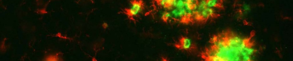
In order to confirm the theory that gut flora affects the way in which our body reacts to a high-fat diet, the research team looked at the microbial profile of different types of mice (thin and diabetic and thin and non-diabetic, which indicates two phenotypes). They showed that there was a difference in the quantities of gut bacteria between diabetic and non-diabetic mice. The thin but diabetic mice presented a flora composed mainly of “bacteroidetes” type bacteria, unlike the thin and non-diabetic mice that presented a flora composed mainly of “firmicutes“ type bacteria.

So is gut flora the cause or the result of metabolic disorders? To find the answer to this question, Rémy Burcelin’s team directly modified the gut flora of a group of mice by adding dietary fibers and gluco-oligosaccharides to their high-fat diet. “By adding these fibers, we modulated most of the physiological characteristics. The metabolism of the mice that we treated with these fibers was similar to that of the thin, non-diabetic mice. But the gut flora of the mice treated with fibers changed greatly compared to that of the other phenotypes observed”, says Matteo Serino.

This project was partly sponsored by the “Florinflam” research program financed by the National Research Agency (ANR) and the FLORINASH research program financed by the EU 7th Framework Programme (FP7). The FLORINASH project (Prevention and treatment of non-alcoholic fatty liver disease) was coordinated by Inserm under the auspices of Rémy Burcelin, Inserm research director, and was contributed to by 6 partners from 4 European countries.

Life expectancy (LE) and Healthy Life Years (HLY) in the European Union, 2008-2010

The HLY (Healthy Life Years) indicates how long people can expect to live without disability. It has been computed annually for each Member State of the European Union since 2005. These figures are released in the framework of the first annual meeting of the European Joint Action on Healthy Life Years (EHLEIS), organized in Paris on April 19, 2012 (ASIEM, 6 rue Albert de Lapparent, from 1:30pm) by the French Ministry of Health. The European Joint Action on Healthy Life Years (EHLEIS) is led by FRANCE, and coordinated by the French National Institute of Health and Medical Research (INSERM).

In 2009 men in the European Union (EU27) could expect 61.3 Healthy Life Years (HLY), representing almost 80% of their life expectancy (LE) at birth of 76.7 years. Women could expect 62 HLY, 75% of their life expectancy (LE) at birth of 82.6 years in 2009.

2010 values for men

In 2010, Sweden has the longest life expectancy at birth (79.6 years) for men in the European Union and Lithuania the shortest (68 years), a gap of almost 12 years. Swedish men also have the most Healthy Life Years (71.7 years) with men in the Slovak Republic having the least (52.3 years), a gap of almost 20 Healthy Life Years. And again Sweden has the highest proportion of years lived without disability (HLY/ LE) in 2010 with 90% of life expectancy without limitations in usual activities. Men in the Slovak Republic on the other hand spend the lowest proportion without disability (73%), a difference of 17 percentage points. This suggests that for men the longer the life expectancy the greater the proportion lived without disability.

In this short period 2008-2010 and despite its low life expectancy and HLY for men, Lithuania experienced the largest gain in HLY, almost three years, whilst the Netherlands saw the largest decline (a loss of 1.3 years). So there is also a tendency for health expectancies in Europe to converge since the gap between Lithuania and the Netherlands fell by more than four HLY in just 3 years. See attached tables for all figures 2008-20010 by Member State.


Better health outcomes for women

In 2010, France and Spain have the longest life expectancy at birth (85.3 years) for women in the European Union and Bulgaria the shortest (77.4 years), a gap of nearly eight years. In 2010 Malta has the highest HLY (71.6 years) for women and the Slovak Republic the lowest (52.1 years), the gap being the same as for men at almost 20 years. However for women in 2010, it is in Bulgaria that the proportion of years lived without disability (HLY/LE) reaches its maximum with 87% of life spent free of activity limitation. This contrasts with the Slovak Republic where women spend the shortest proportion (66%), a difference of 21 points. The results for Bulgaria show that a short life expectancy combined with a low prevalence of activity limitation leads to a large proportion of life expectancy apparently free of disability.

In the short time period 2008-2010, it is again Lithuania that experienced the largest gain in HLY in women (2.4 years), confirming the observation made for men, whilst Finland saw the largest decline (a loss of 1.7 years). As in men, women’s health expectancies show some convergence.

Differences between men and women

Whilst the gap in life expectancy (LE) between men and women is around 6 years (5.9 years) in the European Union in 2009, the gap in HLY is less than one year (0.7 years). Thus the proportion of years lived without disability is 5 percentage points lower for women compared to men (75% vs. 80%).

In 2010, Lithuania has the largest gap in life expectancy (LE) between men and women (10.9 years) and Sweden the smallest (4 years). Lithuania has also the largest gap in HLY (4.6 years) and the Slovak Republic the least (0.2 years). When these two measures are looked at in combination, Portugal has the largest gap in the proportion of years free of disability (HLY/LE) between men and women at almost 9 percentage points and Bulgaria the smallest gap at around about 3 percentage points. But in all European countries women live longer than men and spend a greater proportion of their lives with disabilities. Differences in HLY between men and women are much smaller than differences in life expectancy and in seven cases out of 27 (see figures for 2009), men experience a slightly more HLY than women. This is indeed the case in 2009 for Belgium, Denmark, Italy, the Netherlands, Portugal, Spain and Sweden, a significant number of western European countries.

France, which has the longest female life expectancy in 2009, occupies the 10th place in terms of HLY, illustrating a case where long life does not coincide with a low report of activity limitation. French men occupy respectively the 8th place (out of 27 MS) in terms of life expectancy and 11th place for HLY with respect to 2009 values.

The HLY (Healthy Life Years) is an important European policy indicator and was selected as part of the Lisbon Strategy (2000-2010) to assess the quality of life and functional health status of Europeans. The HLY is also part of the European Community Health Indicators (ECHI) and was set as the overarching target of the first partnership of Innovation Union (research and development component of the new strategy Europe 2020): the Active and Healthy Ageing Partnership, the target being an increase in HLY in the European Union of two years by 2020. HLYs are obtained by applying the prevalence of disability observed in the general population to a standard life table to distribute the years lived into those lived with disability and those lived free of disability. For HLY, the prevalence of disability comes from the annual EU-SILC survey whose implementation is coordinated by EUROSTAT. The prevalence of disability is measured by a general question on activity limitations known as the GALI question: To what extent have you been limited for at least 6 months, due to health problem, in activities people usually do? EUROSTAT calculates and disseminates the HLY as it does for all European policy indicators. The objective is to provide in year t (i.e., 2012) the HLY indicators of year t-2 (i.e., 2010). The Joint Action EHLEIS performs calculations in parallel and compares its results with those of EUROSTAT. The European Joint Action, supported and supervised by the European Commission, disseminates widely the results for all ages (not just birth and age 65 years) through country reports, dedicated websites, Wikipedia, and other media, encourages correct interpretation through training material and an interpretation guide, promotes their use in policies and, lastly and most importantly, produces in-depth analyses of trends and gaps in HLYs and their determinants.

A new hope in the fight against tuberculosis

Researchers from the Institut Pasteur and Inserm, in collaboration with researchers from the University of Pisa, have uncovered the key role played by specific proteins in the virulence of the mycobacterium responsible for tuberculosis, Mycobacterium tuberculosis. They were able to create an attenuated strain of the mycobacterium, which confers a better protection against tuberculosis than the BCG vaccine to mice. This finding represents a major step forward in the scientific quest to develop a new vaccine, more efficient at fighting the disease. This study is being published today in the scientific journal Cell Host & Microbe.

© Institut Pasteur – Photograph of pulmonary tissues infected with Mycobacterium tuberculosis (in pink)

Tuberculosis is one of the most widespread diseases in the world. It is caused by an infection with the mycobacterium Mycobacterium tuberculosis, affecting one third of the world population. According to the World Health Organization (WHO), in 2010, 8.8 million people suffered from tuberculosis, of which 1.4 million died. Thus, M. tuberculosis remains to this day one of the most virulent and dangerous pathogens for man. Despite being effective for children, BCG does not protect adult men and women sufficiently against pulmonary tuberculosis. This type of tuberculosis is extremely contagious; hence, it is essential to create a new vaccine that is more successful at fighting this disease.

A study carried out by researchers from the Institut Pasteur and Inserm, coordinated by Dr Laleh Majlessi (1) and Pr Claude Leclerc (2) (Institut Pasteur/Inserm), in collaboration with Dr Roland Brosch (Institut Pasteur) and Dr Daria Bottai (University of Pisa), shows that a region of M. tuberculosis’ genome can be altered in order to obtain an avirulent strain of the bacterium in mice. This attenuated mycobacterium confers a strong protection against the onset of tuberculosis in a host organism.

The scientists were able to block, in the attenuated strain, the production and transport of certain proteins, called PE/PPE (3), linked to a specific region of the mycobacterium’s genome, the ESX-5 secretion system which can be found in all virulent strains of mycobacteria. They observed that the mice infected with the attenuated strain do not develop tuberculosis. Thus, they showed that the PE/PPE proteins produced by the ESX-5 system play a key role in the virulence of M. tuberculosis.

Furthermore, the scientists observed that the mice immunised with the attenuated strain are efficiently protected against infection by M. tuberculosis. This protection is correlated with the immune response specific to other PE/PPE proteins still present in this strain. Hence, the scientists demonstrated that the mutated strain of M. tuberculosis is a strong vaccine candidate against tuberculosis, triggering a more effective immune response than BCG in mice.

This major finding opens the door to new prospects for the development of a more efficient vaccine against the various pathologies caused by M. tuberculosis, especially against adult pulmonary tuberculosis. Numerous studies will need to be carried out before any of these research findings can be applied to man. The next step for the researchers of the Institut Pasteur, Inserm and the University of Pisa will be the creation of a strain in which other genetic mutations will be introduced in order to render it completely innocuous for man, with the aim to one day conduct clinical trials.

(1) Dr Laleh Majlessi, Immune Regulation and Vaccinology Unit (Institut Pasteur / Inserm U1041)

(2) Pr Claude Leclerc, director of the Immune Regulation and Vaccinology Unit (Institut Pasteur / Inserm U1041)

(3) The genes which encode the PE/PPE proteins belong to two large families that are unique to mycobacteria. They make up 10% of M. tuberculosis’ genome.

Aging of the brain: genetic modifications now identified

The hippocampus is a part of the brain that shrinks as we age, causing memory disorders. An acceleration of this phenomenon is one of the signs of Alzheimer’s disease. Joint international work involving French (1) research teams has shown that genetic mutations are linked to the reduction of the hippocampal volume. These results were obtained from epidemiological studies that analyzed the genomes and MRI scan images of 9232 participants aged from 56 to 84. In France, roughly 2000 MRI scans were performed as part of the study known as the “3-city-study” (2). The results of this work will be published on April 15th 2012 in the review Nature Genetics.

Shrinkage of the hippocampus occurs with age and is caused by the cumulative effect of various factors. Hippocampal atrophy is a recognized biological marker of Alzheimer’s disease, so it is vital that researchers determine the cause of this process.

An international study under the French leadership of Christophe Tzourio looked for genetic variabilities linked to the shrinkage of the hippocampus. To do this, the genomes and MRI scan data of over 9000 persons aged between 56 and 84 were analyzed with a view to detecting a potential link between certain mutations and the reduction in the hippocampal volume. The participants’ data (both subjects presenting dementia and healthy subjects) were extracted from eight cohort studies in Europe and in North America.

The researchers first identified 46 differences in the DNA sequences of the participants, thought to be related to a reduction in the volume of the hippocampus. Eighteen mutations located in different areas of chromosome 12 were found to be strongly linked to shrinkage of the hippocampus. The other links included a mutation on chromosome 2. Then a final mutation on chromosome 9 was also found to be involved in the hippocampal shrinkage in a third and younger sample. These results indicate that “as yet unidentified factors” trigger mutations in precise areas of the genome, causing the reduction of the hippocampal volume.

The hippocampus (shown in red) is a structure located deep down in the brain, that plays an essential role in the memory processes. Its volume decreases with age and this shrinkage occurs more rapidly in cases of Alzheimer’s disease. For this study, computerized analysis of the MRI scan images and calculation of the hippocampal volume had to be developed individually for each participant

© UMR5296 CNRS CEA Université de Bordeaux

The hippocampus (shown in red) is a structure located deep down in the brain, that plays an essential role in the memory processes. Its volume decreases with age and this shrinkage occurs more rapidly in cases of Alzheimer’s disease. For this study, computerized analysis of the MRI scan images and calculation of the hippocampal volume had to be developed individually for each participant.

Once the mutations had been identified, the researchers tried to find out what exactly they modified. They discovered that they changed the structure of genes that were important for numerous functions involved, among others, in cell death (HRK), embryonic development (WIF1), diabetes (DPP), or neuronal migration (ASTN2).

“This study marks a major turning point, since it confirms the fact that genetic factors are linked to a brain structure, the hippocampus, involved in dementia and more generally in the aging of the brain,” explains Christophe Tzourio. This new approach, that studies a targeted area of the brain rather than a disease, will help us to more precisely decipher the mechanisms of Alzheimer’s disease.

The next stages will aim at obtaining a better understanding of how these genetic mutations are actually involved in the overall functioning of Alzheimer’s disease. Although clinical applications are not for the imminent future, these discoveries are a step forward to a better understanding of this disease and of cerebral aging in general.

“This discovery confirms the importance of using sophisticated means such as MRI scanning and genome analysis for the cohort studies. And this is only possible if we have close collaboration between the different disciplines involved,” concludes Christophe Tzourio.

Footnotes

(1) In neuroepidemiology (Inserm U708 – University of Bordeaux, C Tzourio), brain scanning by the neurofunctional imaging group (CNRS/CEA/University of Bordeaux Segalen, B Mazoyer), and in genetics (UMR 744 Inserm University of Lille, P Amouyel).

(2) The 3C study also called the “Three-city-study” denoting the cities of Bordeaux, Dijon and Montpellier, is a large cohort study involving over 9000 persons aged 65, begun in 1999. http://www.three-city-study.com/

(French) Découverte d’une nouvelle règle d’organisation spatiale des chromosomes qui reflète leur fonctionnement

Sorry, this press release is only available in French.

Sport/physical exercise and health: what’s new?

The London Olympic Games kick off on July 27th. With around 100 days to go, Inserm wishes to invite you to look into the research that has been done in the fields of sport and health.

Various research programs over the last few years have shown the benefits of physical exercise on our health, in preventing obesity or in improving the health of patients suffering from chronic, cardiovascular or respiratory diseases in particular.

So what about high-level sports? These also seem to contribute to good health. Are high-level sport and physical exercise both determining factors for our health?

Several research teams in France specializing in epidemiology, sports medicine, physiology and physics are investigating the interactions between sport, physical exercise and health.

Below you will find contacts and detailed information about some of the Inserm teams involved in the fields below, among others:

• The impact of performance on life expectancy and mortality

• The part played by high-level athletes as models for studying respiratory diseases

• The rules of training for competitive sports transposed to physical exercise (notions of gradual build-up, development of different sporting energy systems, regularity, variations in exercises, motricity, assessment, etc.)

• Sport and aging

• Differences in the cardiac system of high-level athletes

A new gene thought to be at cause in early-onset forms of Alzheimer’s disease

A new gene that causes early-onset of Alzheimer’s disease has been discovered by the research team of Dominique Campion at the Insert unit 1079 ”Genetics of cancer and neuropsychiatric diseases” in Rouen. The research scientists showed that in the families of 5 of 14 patients suffering from the disease, mutations were detected on the gene SORL1. This gene regulates the production of a peptide involved in Alzheimer’s disease. The results of this study have been published in the review Molecular Psychiatry issued April 3rd

Precise genetic mutations have been seen to play a part in early-onset forms of Alzheimer’s disease. However, there is a sub-population of patients in whom there is no mutation of these genes. So how can these patients, in whom there are no pre-established mutations, be suffering from early-onset Alzheimer’s?

To reply to this question, the research team working under the leadership of Dominique Campion and Didier Hannequin (Inserm unit 1079 and Centre national de référence malades Alzheimer jeunes, University hospital Rouen), studied the genes from 130 families suffering from early-onset forms of Alzheimer’s disease. These families were identified by 23 French hospital teams within the framework of the “Alzheimer Plan”. Of these families, 116 presented mutations on the already known genes. But in the 14 remaining families, there was no mutation at all observed on these genes.

A study of the genome of the 14 families using new whole DNA sequencing techniques showed evidence of mutations on a new SORL1 gene. The SORL1 gene is a coding gene for a protein involved in the production of the beta-amyloid peptide. This protein is known to affect the functioning of the brain cells (see insert).

Two of the identified mutations are responsible for an under-expression of SORL1, resulting in an increase in the production of the beta-amyloid peptide. “The mutations observed on SORL1 seem to contribute to the development of early-onset Alzheimer’s disease. However, we still need to identify more clearly the way in which these mutations are transmitted on the SORL1 gene within families” states Dominique Campion.

Alzheimer’s disease is one of the main causes of dependency among the elderly. It results from neuron degradation in different areas of the brain. Its symptoms include increased alterations to memory, cognitive functions and behaviour disorders that lead to a progressive loss of independence.

Alzheimer’s disease is characterized by the development of two types of lesion in the brain: amyloid plaques and neurofibrillary degenerescence. Amyloid plaques are caused by extracellular accumulation of a peptide, beta-amyloid peptide (Aβ) in specific areas of the brain. Neurofibrillary degenerescences are intraneuronal lesions caused by abnormal filamentary aggregation of a protein known as a Tau protein.

Asthma: a vaccination that works using intramuscular injection

Asthma is a chronic inflammatory and respiratory disease caused by an abnormal reactivity to allergens in the environment. Of the several avenues of exploration that are currently being developed, vaccination appears to be the most promising approach. In a publication soon to appear in the review Human Gene Therapy, the research scientists at Inserm and CNRS ( Institut du thorax, CNRS/Inserm/University of Nantes) reveal an innovatory vaccine against one of the allergens most frequently encountered in asthma patients. After vaccine was directly injected into the muscle of an asthmatic mouse, a nanovector significantly reduced both the hypersensitivity to the allergen and the associated inflammatory response.

Allergic asthma is a chronic respiratory disease that affects 300 million people throughout the world. The number of people suffering from asthma has doubled over the last ten years and almost 250, 000 people die prematurely from this problem each year. In most cases, asthma is caused by an abnormal reaction to substances in the environment known as allergens. From a physiological point of view, this hypersensitivity results in serious inflammation of the bronchial tubes and the bronchioles in sensitive persons. This alters their ability to breathe correctly.

Current treatment consists in administering corticoids that treat the symptoms and temporarily relieve the disorder, but without curing it. An alternative, long-lasting treatment for allergic asthma is based on a specific immunotherapeutic protocol commonly known as desensitization. Repeated, increased doses of the allergen are administered in order to decrease the hypersensitivity and reduce the symptoms in the event of subsequent exposure. However, the efficiency of this protocol is limited and varies greatly from one patient to another.

Then the research scientists came up with the idea of a vaccination technique using the DNA of the allergenic substance. Rather than administering repeated doses of allergen extract in order to reduce sensitivity, we worked on specific DNA sequences of the allergen responsible for the allergy. “Several studies demonstrated the therapeutic potential of this strategy, but we still had to find techniques that were reliable in human beings”, explains Bruno Pitard, Director of the Biotherapy Innovations team at the Institut du thorax (CNRS/Inserm/University of Nantes). Using these techniques on human subjects meant that the treatment had to be efficient when only a small dose of DNA was injected.

The researchers first tried proving the efficiency of this DNA-based vaccination against the specific allergen Derf1, using a relevant animal model developed by the Bronchial and Allergic Pathologies team led by Antoine Magnan. In Europe,Dermatophagoides farinae 1 (Derf1) is a very common allergen carried by the dust mite Dermatophagoides farinae. More than half of patients presenting allergies to dust mites produce specific IgE type antibodies (Derf1) against this substance that are characteristic of asthma.

In practice, the researchers associated useful genetic sequences of the allergen Derf1 with a nanovector consisting of a synthetic polymer. This DNA sequence, transported by a sort of “molecular taxi” into the muscle cells that ensure protein synthesis of the allergen, modulated the allergic response in asthmatic animals (1).

The vaccine developed in a healthy mouse model was then optimized in a model composed of asthmatic mice. In the asthmatic mice, the vaccine triggers the production of specific anti Derf1 antibodies and a specific cellular response to Derf1, so that the immune system reacts with a protective non-allergizing response when the body comes into contact with the allergen. Two injections were administered at 3 weekly intervals. They significantly reduced the hypersensitivity of the airways and the levels of inflammatory cytokines, that were found in the lungs of asthmatic mice that had not been vaccinated.

These new results validate the whole potential for the use of this new nanovector in DNA vaccination. It is currently undergoing regulatory pre-clinical development with a view to future clinical trials in humans.

Footnote:

(1) Recently, this new class of vector was also used to treat hepatocellular carcinoma (see press release of September 9, 2010Combine a first-rate target with a first-rate vector to build an effective immunotherapy strategy against cancer!”

fermer