Menu

Tracking animal epidemics through road network

Road transport is one of the main vectors of animal epidemics. It is vital to understand how potentially infected animals are transported and exchanged within a country. A French-Italian team, including researchers from the Centre de physique théorique (CNRS/Aix-Marseille Université / Université du Sud Toulon Var) and the Epidémiologie, systèmes d’information, modélisation (Inserm/UPMC)[1] unit, has just presented large-scale numerical simulations to test potential scenarios of a cattle epidemic in Italy. The model is the first to take into account day-to-day variations in the Italian animal transportation network and could lead to new prevention and monitoring strategies. This work has been published on the website of the Journal of the Royal Society Interface.

Transportation of farm animals over long distances is vital for their rearing and for the agri-food industry.  However, it also facilitates the spread of pathogens. This was illustrated by the Foot and Mouth Disease epidemic that hit the UK in 2001 and cost around 8 billion GBP, and the 2006 swine fever epidemic in Germany in 2006, whose indirect costs are estimated at 60 million euros. Furthermore, there is increasing concern that animal diseases could pose a threat to human health, as recently shown by bird flu and the H1N1 virus.

To study how potentially infected animals are exchanged and transported within a country, and how this can affect the spread of an epidemic, the researchers used data from the movement records of 5 million cattle throughout 2007 in Italy. They built a model using tools from the complex network analysis. In mathematics, a network is a series of points (“nodes”) interconnected by communication paths. In the present case, the nodes were farms and the paths represented animal transport routes between them.

This model is novel in that it takes into account changes occurring from one week to another and even from one day to the next in the Italian animal transport network. Traditional models, by comparison, are based on a fixed network, which can lead to inadequate prevention and health screening measures. The researchers built digital simulations to predict how a disease in any Italian farm would propagate by road throughout the country. More importantly, this work could help identify the farms that require priority surveillance once an epidemic breaks out or is suspected. Moreover, in the case of a crisis, the model could be used to trace the path of infection back to the farm of origin. Finally, the research shows that the most important farms to monitor are not only those where animal transport is the busiest, although simpler models that do not account for network dynamics would predict this. Because standard characteristics are insufficient to identify the farms at risk, the researchers are now developing mathematical models to achieve this.

Although this work takes a pure science approach in developing new mathematical models, it could easily serve as a basis for creating a powerful, user-friendly tool for animal health authorities. In addition, the researchers hope to extend their study to the rest of Europe.


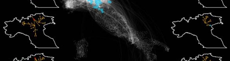
© P.Bajardi, A.Barrat, L.Savini and V.Colizza

Epidemic propagation paths

Each white dot on the main map represents a farm (node), and the lines represent animal transportation. Digital simulations of epidemics can detect groups of farms, defined by the same propagation scenario. Such groups are shown in color on the main map. Farms in a particular group can be located within a single area or be geographically dispersed.

The maps at the sides of the figure show several propagation paths originating in the red group (left) or the blue group (right) from the main map: propagation paths are similar for different origins within the same group.

© P.Bajardi, A.Barrat, L.Savini and V.Colizza

Geographic distribution of groups of farms in the Italian cattle transportation network

Each white dot on the main map represents a farm (node) and the lines represent animal transportation routes. Digital simulations of epidemics can detect groups of farms, defined by the same propagation scenario. Each color shows a different group of farms, illustrating that farms in the same group can be located in a single area or be geographically dispersed.

The networks illustrated at the top of the figure correspond to epidemic propagation paths for each of the groups shown on the map: each group has a different propagation route.


[1] In collaboration with the ISI Foundation (Turin, Italy) and the Istituto Zooprofilattico Sperimentale Abruzzo-Molise (Teramo, Italy).

A fish to detect contaminant endocrinal disruptors

The researchers from INERIS and Inserm (a team managed by Olivier Kah in Inserm unit 1085 “Institute for research into health, environment and work”) have just developed a test in fish that allows us to detect the endocrinal disrupting effects of certain contaminants in the environment. The researchers based their work on a gene that expresses in the brain, and that reacts strongly to certain endocrinal disruptors. In order to make it easier to measure this gene, they used a fluorescent reporter gene. By using the embryos of zebrafish that are transparent, we can see the effects in the brain when the embryos are exposed to disruptor pollutants. The results of these works are published in the review Plos One.

Fluorescence seen in the brain of the fish embryo, induced by the expression of gene cyp19a1b bound to the GFP.

Over the last 20 years, numerous studies have proved the harmful effects of artificial compounds on the reproductive capacity of organisms. Certain pollutants (nonylphenols, bisphenol A, pesticides, pharmaceutical residues, etc.) present in surface water, industrial waste or sediments are capable of mimicking the effects of oestrogens. They thus modify the biological processes that are controlled by oestrogens and are involved in the reproduction and growth functions of organisms, with potentially harmful consequences for the health of living creatures and their progeny. Such substances are known as “endocrinal disruptors” (ED).

Pollutants with oestrogen activity also affect the brain

The originality of the work carried out by the Inserm and INERIS researchers resides in the fact that the study of endocrinal disruptors concentrates on a gene that expresses only in the brain, and demonstrates the sensitivity of the nervous system to pollutants. The results obtained by the researchers confirm that in the fish embryo, a certain number of substances affect the activity of the stem cells in the brain, cells that are vital to the development of the central nervous system. This effect is seen in the expression of a specific gene in the brain that is extremely sensitive to oestrogens: gene cyp19a1b.

A zebrafish model to characterize ODs

Based on these observations, the INERIS and Inserm teams developed a test to detect oestrogen activity in a transgenic fish model. This transgenic zebrafish model (1) helps to identify the effect of pollutants on an enzyme from gene cyp19a1b, aromatase, that is responsible for the synthesis of oestrogens in the body.

The brain of the fish embryos uses a fluorescent reporter known as GFP (Green Fluorescent Protein), that renders it fluorescent when exposed to substances that mimic oestrogens.

21 components (e.g. natural or synthetic oestrogens; alkylphenols, bisphenols) of the 45 tested induced varying degrees of fluorescence. The metabolic capabilities of the model allowed us to detect substances such a certain androgens and certain synthetic progestatives (used in the contraceptive pill).

A test of this type is helpful in evaluating chemical substances, as required by the REACh (2) regulation. This tool is a complement to the existing in-vitro systems, and it has the advantage of integrating what will happen to pollutants in the body and of taking account of their metabolism, something than cannot always be done using tests on cells. Given its sensitivity, it could also be used to monitor aquatic environments.

The INERIS and Inserm researchers have finally opened up new prospects in the field of study of endocrinal disruptors in the central nervous system.

For the researchers themselves, it appears that this simple, robust and sensitive test will have many fields of application in assessing the risks implied in oestrogenic endocrinal disruptors.

Financed by the Ministry in charge of Ecology and the National Research Agency, the research undertaken by INERIS and Insert is aimed at pinpointing the ED potential of these chemicals, that are used in medication, cosmetics, phytosanitory products, plastics, etc.

About INERIS
The mission of the French National Institute for Environmental Technology and Hazards is to help to prevent hazards resulting from economical activities from affecting health, safety of persons and belongings, and the environment. It runs research programs aimed at better understanding the phenomena likely to lead to hazardous situations or dangers for the environment or health and developing its knowledge of prevention. Its scientific and technical know-how are made available to the state authorities, companies and local authorities so as to help them make the best decisions to improve environmental safety. Created in 1990, INERIS is a public commercial and industrial institute placed under the authority of the Ministry of the Ecology, Sustainable Development and Energy. It has a staff of 588 persons, based mainly in Verneuil-en-Halatte, in the Oise ‘département’. Website: www.ineris.fr.

About Inserm
Founded in 1964, the French National Health and Medical Research Institute (Inserm) is a public science and technology institute, jointly supervised by the French Ministry of Higher Education and Research and the Ministry of Health. The mission of its scientists is to study all diseases, from the most common to the most rare, through their work in biological, medical and public health research. Inserm support over 30à laboratories throughout France. In total, the teams include nearly 13,000 researchers, engineers, technicians and administrative staff, etc. Inserm is a member of the French Life Sciences and Healthcare Alliance, founded in April 2009 along with the CNRS (The French National Research Centre), CEA (Atomic Energy and Alternative Energy Commission), Inra (The National Institute for Agricultural Research), Inria (The French National Institute for Research in Computer Science and Control), IRD (The Institute for Research into Development), The Institut Pasteur, the Conference of University Presidents (CPU) and the Conference of Regional and University Hospital CEOs. This alliance forms part of the policy to reform the research system by better coordinating the parts played by those involved and by strengthening the position of French research in this field through a concerted plan.

Footnotes:

(1) In collaboration with Professor B.C. Chung of the Academia Sinica in Taiwan.

(2) Registration, Evaluation, Authorisation and Restriction of Chemical substances: European Parliament rule and directive No. 1907/2006 of December 18, 2006, concerning the registration, evaluation and authorization of chemical substances and the restrictions applicable to these substances.

(French) Progéria – Découverte du mécanisme moléculaire qui préserve les cellules neurales du vieillissement accéléré

Sorry, this press release is only available in French.

(French) ADN tumoral circulant dans le sang : un nouveau biomarqueur du cancer

Sorry, this press release is only available in French.

The neurological bases of anxiety disorders

On June 18th, a one-day scientific symposium in Paris entitled “Neurobiological basis of Anxiety disorders”, got together scientists from 6 of the partners of DEVANX (1) , a European project coordinated by Inserm and launched in 2008. It was an opportunity to take stock of what knowledge has been acquired about the neurological basis of anxiety disorders.

Over the last few years, great progress has been made in our knowledge of brain circuits and the key molecules involved in the manifestations of anxiety. The use of animal models has greatly contributed to our understanding. In mice, it was possible to observe behavioral changes that occurred in situations of emotional conflict, for example how the animal chose between exploring new avenues (curiosity) and withdrawal (fear). It allowed us to describe the way in which the animal acts in a situation of learned fear: how the animal learns to associate a neutral environment with potential danger.

Serotonin and GABA are the 2 main molecules that act as “messengers” (or neurotransmitters) between the neurons involved in states of anxiety. And these are the common targets for “anxiolytic” drugs.

However, the exact role played by these molecules and their interactions with the environment are still unknown. Genetics and the new information gained about brain plasticity need to be integrated into our constantly growing understanding of the mechanisms involved. Patricia Gaspar and Laurence Lanfumey, Research directors at Inserm and coordinators of the DEVANX project, have worked with their colleagues to study the neurobiological bases of anxiety from different angles.

1. Pharmacological aspects

The GABAb receptors present in neurons are targeted by new molecules that work in a completely different way from conventional anxiolytics (benzodiazepines), that act on the GABAa receptors. By finding out more about the structure and the function of GABAb receptors and their interactions with the serotoninergic system, we can propose new therapeutic targets.

A particular example is Benny Bettler’s team, a member of the DEVANX consortium in Switzerland. They demonstrated that GABAb receptors are heterodimers (a combination of two different receptor subunits) that possess partner proteins capable of modifying their binding properties. The pharmacological properties of GABAb receptors vary depending on how the partner proteins are organized. From a therapeutic point of view, positive modulation of these receptors represents a possible strategy for developing new anxiolytics. John Cryan, a partner of the DEVANX consortium in Ireland, has shown that inhibiting GABAb receptors can reduce depressive behavior. Working along the same lines, Laurence Lanfumey’s team in Paris studied the connection between GABAb receptors and the serotoninergic system.

© Gassmann et Bettler, 2012

GABAB receptor subunits: GABAB1a, GABAB1b and GABAB2

These subunits are receptors with seven transmembrane domains bound to the G proteins via the GABAB2 .subunit. The difference between the GABAB1a and GABAB1b subunits is that there are two terminal domains (sushi-domains) on the GABAB1a subunit.

2. The part played by serotonin

In people who suffer from depression, panic attacks, anxiety disorders or phobias, administering drugs that increase the serotonin level reduces these pathologies.

However we know very little about the initial cause of this lack of serotonin that causes the disorders. That is the reason why the researchers need different animal models to discover and analyze the different situations of a brain that is “depleted” of serotonin.

Serotonin is involved in numerous physiological functions: sleep/wake rhythms, impulsivity, appetite, pain, sexual behavior and anxiety. Its action is mediated by around fifteen different receptor sub-types.

The serotoninergic system is a multiple system: it is present in the central nervous system (in the Raphe nuclei of the brain) and in the peripheral nervous system (in the enterochromaffin cells of the digestive tract).

The way in which neurons “specialize” into serotonin neurons is controlled by different molecular factors, depending on their location, and does not all happen at the same time in the course of development.

One of the studies carried out by the genetics specialists as part of the DEVANX project aimed at conditionally targeting the production of serotonin at a given time and in a given location. For example, Dusan Bartsch’s team, a DEVANX partner in Mannheim, produced genetically modified mouse models that allowed them to reduce the serotonin at different times during the life of the mice, by creating models known as inductable models (in which the elimination of a gene could be induced by administering a drug). Patricia Gaspar’s team in Paris characterized mutations in which only part of the serotoninergic neurons was affected (mutation of a Pet-1 transcription factor). In these mice, the team noted that spontaneous anxiety was reduced, but that their fear conditioning was increased. Therefore, the lack of serotonin in the central nervous system could mean that the subjects more easily associate neutral situations with a panic reaction.

3. Other circuits involved: Fear circuits

By linking the research into fear with the latest neurobehavioral findings, it was possible to combine the approaches.

It is becoming more and more evident that it is the normal neuron circuits specialized in dealing with fear that are pathologically affected or amplified in anxiety disorders. So it is very important to understand and analyze how these circuits function in “real situations” in animal models. The end purpose is to find a way of “deconditioning” certain brain circuits that have been abnormally or over-activated.

The new approaches to physiology on the knockout animal, combined with pharmacogenetic research, have made for progress in this field. For example, Agnés Gruart’s laboratory in Sevilla, one of the partners of the DEVANX project, has recorded different neurons from the hippocampal circuits in different fear learning situations and observed the effect of modifying the message conveyed by GABAb and serotonin. Cornelius Gross’s team from EMBL in Rome has shown that we can use serotoninergic receptors (5-HT1A) expressed in different areas of the brain in order to temporarily deactivate highly specific neuron circuits. This allowed them to identify the hippocampal and amygdala circuits involved in the generalization of fear.

Research into anxiety disorders, as in numerous fields of neurosciences, is now using integrated approaches that require multiple fields of knowledge. Molecular studies now need to be integrated into the whole animal context that expresses behavior patterns as similar as possible to physiological situations, while still being strictly controlled as experiments. Genetic tools now provide us with unequalled power for researching into a determined molecule function, or a molecular assembly within a given circuit and a precise time slot. This type of approach will continue to develop in the years to come, with the coming of tools that will allow us to activate or deactivate certain selected neuron circuits.

By solving these intertwined elementary processes step by step, we should at last find the explanation to the mechanisms underlying pathological anxiety disorders.

(1) DEVANX: “Serotonin and GABA-B receptors in anxiety: From developmental risk factors to treatment”, a project funded by the European Commission and launched in 2008. Partners involved: Inserm (coordinator), University College Cork, Ireland, European Molecular Biology Laboratory, Italy/Germany, Central Institute of Mental Health, Mannheim, Germany, Universitaet Basel, Switzerland, Universidad Pablo de Olavide, Spain

(French) Le travail de nuit, un risque pour les femmes ?

Sorry, this press release is only available in French.

Oestrogen and cardiovascular risk in menopausal women

Women are less prone to cardiovascular disease then men; but this difference between the sexes becomes less marked after the menopause. This observation is behind a great deal of received wisdom, where oestrogen is assumed to have a beneficial effect on the heart and blood vessels. Today, new data seems to question these presuppositions. A study has been conducted by a team of Inserm researchers, directed by Pierre-Yves Scarabin (Inserm Unit 1018 “Centre for Epidemiology and Population Health Research”), on 6,000 women aged over 65; its results demonstrate, for the first time, that women with high levels of oestradiol in their blood are exposed to a greater risk of myocardial infarction or strokes. The results are published in The American Heart Association Journal. 

Fotolia

Oestrogen hormones play a key role in sexual development and reproduction in women. Oestradiol is the most active hormone. Its blood levels are particularly high during the active reproductive period. After the menopause, the ovarian function ceases, leading to a significant drop in oestrogen levels in the blood (the adipose tissue then becomes the main source of oestrogen). However, low concentrations of these hormones do continue to circulate and may still exert biological actions.

Throughout their lives, women are less exposed to the risk of cardiovascular disease than men. For many years, this relative immunity displayed by women was attributed to oestrogen undertaking a ‘protector’ role in terms of atherosclerosis and its complications. However, this hypothesis was not confirmed by recent research into the hormonal treatment of the menopause. Oestrogen administration does not prevent ischaemic arterial disease in menopausal women and could even have a harmful effect on women in the highest age bracket.

Until now, no study has been able to clearly identify the link between circulating endogenous sexual hormones and the cardiovascular risk in menopausal women.

Today, this knowledge gap has been reduced by the results of a French cohort study(1) (Three City Study-3C) performed on approximately 6,000 women aged over 65 from among the general public. Oestradiol levels in the blood were measured upon entry into the cohort and, after monitoring performed over a four year period, 150 new cases of cardiovascular disease had appeared.

For the first time, the results demonstrate that high oestrodial levels in the blood lead to an increased risk of myocardial infarction or strokes, although the cause and effect link is not shown. This relation is not influenced by other known factors for cardiovascular risk, namely diabetes and obesity.

Other results show that oestrogen seems to affect some mechanisms involved arterial obstruction, which causes cardiovascular disease. Although the coagulative effect of oestrogen is clearly defined, significant research is now required to establish its role in the inflammatory process, particularly in obese women, where the accumulation of adipose tissues is associated with high oestrogen levels.

This new data questions the beneficial role of oestrogen on the heart and vessels. “Fresh studies must confirm this harmful effect and establish whether these results can be applied to younger

Footnote:

(1) http://www.three-city-study.com/l-etude-des-trois-cites-3c-historique.php

(French) BCG et cancer de la vessie : vers un nouveau protocole pour les patients ?

Sorry, this press release is only available in French.

Preserving sight through enhanced focus on the causes of glaucoma

An article to be published in Plos One describes how Inserm researchers have succeeded in preserving the visual function in rats suffering from glaucoma. This disease is associated with an abnormal increase in intraocular pressure and can lead to blindness. The team of researchers directed by Christophe Baudouin at the Institut de la Vision ((Inserm/CNRS/UPMC) focussed on inflammation-specific molecules: chemokines. Blocking one of the chemokines (CXCR3) led to reduced intraocular pressure, by restoring normal aqueous humour flow, thus protecting the retina and the visual function.

Glaucoma is the second leading cause of blindness across the globe, affecting fifty to sixty million people, 6-7 million of whom are blind. In France, approximately 800,000 people receive treatment for glaucoma. The disease is characterized by the progressive destruction of the optic nerve and an irreversible alteration to the visual function, generally associated with an abnormal increase in intraocular pressure.

This high intraocular pressure is caused by an obstruction to the normal aqueous humour flow on a specific ocular structure: the trabecular meshwork. However, there is little understanding of the degeneration of trabecular tissues, which causes this malfunction.

Current treatment for glaucoma does not directly target the original pathology of the trabecular meshwork.This could partly explain why treatment frequently fails, sometimes leading to blindness even with access to the best possible medical care.

The team of researchers directed by Christophe Baudouin at the Institut de la Vision is dedicated to studying physiopathological mechanisms responsible for glaucomatous trabecular pathology, and, more specifically, the role of specific “chemokine“ molecules in inflammation.

Several researchers from the team have recently demonstrated on the tissues of patients suffering from glaucoma, and on a human trabecular cell line, there is a balance between the CXCL12 chemokine and a truncated form of this molecule, SDF-1(5-67). Whereas the first molecule encourages the viability of trabecular meshwork tissues via the CXCR4 receptor, its other form causes the loss of the trabecular meshwork via the CXCR3 receptor. It would appear that the transition from a “healthy” form to a truncated form is driven by a specific environment and the cytokines and metalloproteases involved in glaucoma.

In a second phase, the researchers used an animal model of glaucoma and observed that blocking CXCR3 reduces intraocular pressure and restores the trabecular filter function, thus preserving the visual function by protecting the retina.

This research has improved understanding of glaucoma. As Alexandre Denoyer, the leading author of the publication explains: “The novel strategy targeting trabecular chemokines could result in the development of an innovative treatment to replace or complement current long-term treatment using eye drops”.

Magnetic stimulation to improve visual perception

Using transcranial magnetic stimulation (TMS), an international team led by researchers from the Centre de Recherche de l’Institut du Cerveau et de la Moelle Epinière (CNRS / Inserm / UPMC) has succeeded in enhancing the visual abilities of a group of healthy subjects. Following stimulation of an area of the brain’s right hemisphere involved in perceptual awareness and in orienting spatial attention, the subjects appeared more likely to perceive a target appearing on a screen. This work, published in the journal PLoS ONE, could lead to the development of novel rehabilitation techniques for certain visual disorders. In addition, it could help improve the performance of individuals whose tasks require very high precision.

TMS is a non-invasive technique that consists in sending a magnetic pulse into a given area of the brain. This results in an activation of the cortical neurons located within the range of the magnetic field, which modifies their activity in a painless and temporary manner. For several years, scientists have been looking at the possibility of using this technique to enhance certain brain functions in healthy subjects.

In this respect, the team led by Antoni Valero-Cabré has carried out research involving the stimulation of a region of the right cerebral hemisphere known as the frontal eye field. Strictly speaking, this is not a primary visual area but it participates in the planning of ocular movements and the orientation of each individual’s attention in the visual space. In a first experiment, a group of healthy subjects tried to distinguish a very low contrast target appearing on a screen for just 30 ms. In some of the tests, the subjects received a magnetic pulse lasting between 80 and 140 ms on this frontal region before the target appeared. The researchers found that the success rate was higher when using TMS. The visual sensitivity of healthy subjects was temporarily increased by around 12%. In a second experiment, the subjects were shown a fleeting visual cue indicating the spot where the target could appear. In this configuration, the enhancement of visual sensitivity, which remained of the same order, was only apparent when the cue indicated the correct location of the target.

Although cerebral functions such as conscious vision are highly optimized in healthy adults, these results show that there is a significant margin for improvement, which can be “enhanced” by TMS. This technique could be tested for the rehabilitation of patients suffering from cortical damage, due for example to a cardiovascular accident, and for that of patients with retinal disorders. The second experiment suggests that rehabilitation based on both TMS and visual cues could be more selective than the use of stimulation alone. The researchers want to further explore this possibility using repetitive TMS, which, in this case, could make it possible to obtain long-lasting modification of cerebral activity.

Furthermore, according to the researchers, TMS could be used in the near future to increase the attentional abilities of individuals performing tasks that require good visual skills.

These experiments benefited from funding from the European ERANET NEURON BEYONDVIS initiative, partly financed by ANR.

fermer