Menu

Auto-immune disease: the viral route is confirmed

Why would our immune system turn against our own cells? This is the question that the combined Inserm/CNRS/ Pierre and Marie Curie University/Association Institut de Myologie  have strived to answer in their “Therapies for diseases of striated muscle”, concentrating in particular on the auto-immune disease known as myasthenia gravis. Through the project known as FIGHT-MG (Fight Myasthenia Gravis), financed by the European Commission and coordinated by Inserm, Sonia Berrih-Aknin and Rozen Le Panse have contributed proof of the concept that a molecule imitating a virus may trigger an inappropriate immune response, causing muscular function to deteriorate. These results have been published in Annals of Neurology, accessible on line.

Myasthenia, a rare auto-immune disease

Myasthenia gravis is a rare auto-immune disease (5,000 to 6,000 patients in France) that produces muscular weakness and exhaustion. It generally affects the facial muscles first, and may then become generalised through the muscles of the limbs or the respiratory muscles, causing respiratory distress.

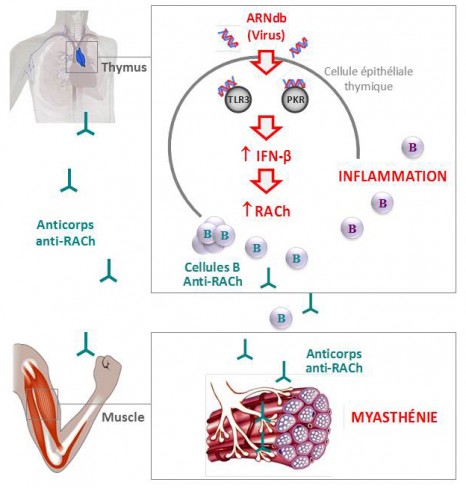
This is due to the production of circulating auto-antibodies that block the acetylcholine receptors (RACh), these neurotransmitters being necessary for transmitting the motor nerve signal to the neuro-muscular junction.

Could a viral infection be the origin of myasthenia?

Myasthenia is a multi-factorial disease in which environmental factors seem to play a key triggering role. Viral infections are suspected but it is hard to prove the role of a virus in triggering the condition. In fact, diagnosis of myasthenia is often made months, or even years, after the actual start of the illness when the virus is no longer detectable, even though the signature left by the virus is visible long after the infection.

Proof of the concept of a viral origin contributed by researchers

Under the European FIGHT-MG project, the team of researchers managed to decode the trigger for the illness by using a molecule that mimics the RNA double viral strand (Poly(I:C)).

To do this, they concentrated on the organ that plays a central role in the disease – the thymus. It is in this gland located in the thorax that the T-lymphocytes mature, these being the key players in immune response that are normally programmed to avoid the development of any auto-immunity.

The researchers were thus able to show in vitro that the Poly(I:C) was capable of specifically inducing an over-expression of RACh through thymal epithelial cells, while activating three proteins (the “toll-like” receptor 3 (TLR3), the protein kinase R (PKR) and interferon-beta (IFN-â)); it is this last that produces inflammation in the thymus.

At the same time, they analysed pathological thymus glands of myasthenia sufferers in whom they observed over-expression of these same three proteins in the immune system, characteristic of a viral infection.

Finally, the researchers also managed to identify the same molecular changes in the thymus glands of mice, after they had been injected with Poly(I:C). After a prolonged injection period, they also observed a proliferation in the mice of B anti-RACh cells, the presence of auto-antibodies blocking the RACh receptors and clinical signs synonymous with the muscular weakness found in myasthenia.

These original results show that molecules that mimic a viral infection are capable of inducing myasthenia in the mouse, something that had never been demonstrated before.

This set of papers published in the Annals of Neurology provides proof of the concept that a viral infection can cause inflammation of the thymus and lead to the development of auto-immune myasthenia.

The next stages of the research will consist in determining which exogenous virus this may be or whether it is a case of the abnormal activation of an anti-viral response by endogenous molecules.

© Inserm / R. Le Panse 

The introduction of a double strand of RNA (Poly(I:C) into the thymal epithelial cell induces the over-expression of the acetylcholine receptors (RACh), via the activation of the “toll-like” receptor 3 (TLR3) and the protein kinase R (PKR),  as well as the production of interferon-beta (IFN-β)). These changes in the thymus gland cause the formation of B anti-RACh cells and the production of circulating auto-antibodies that block the acetylcholine receptors present in the neuromuscular junction.

FIGHT-MG (Fighting Myasthenia Gravis) – a European collaboration making giant leaps forward

The FIGHT-MG project seeks to determine the genetic and environmental risk factors associated with the occurrence of the illness and its development. The project aims also to identify the key immunological molecules associated with its appearance, and to study the pathogenic mechanisms at the neuromuscular junction, establish new diagnostic tests, as well as new treatments (cellular treatments, immuno-regulatory treatments, immuno-absorption of pathogenic auto-antibodies and other pharmacological treatments).

“When one is working on a rare disease, it is essential to work through networking, so as to be able to share our facilities and resources to promote fundamental and clinical research. It is also crucial to communicate permanently with patient associations. It is this combination that enables us to take giant steps in the treatment of rare conditions,” explains Sonia Berrih-Aknin.

FIGHT-MG : http://www.fight-mg.eu/ 

FIGHT-MG started in December 2009 and will last for four years, with a total budget of about six million euros funded by the European Union (FP7). The project involves 12 partners based in seven European countries:

The 12 partners:

Inserm (coordinator), France
Hellenic Pasteur Institute (HPI), Greece
Open University of Israel (OUI), Israel
Fondazione Istituto Neurologico “Carlo Besta” (INNCB), Italy
Oslo University Hospital (OUS), Norway
Hadassah Hebrew University Medical Center (HMO), Israel
Israel Institute of Technology (TECHNION), Israel
University of Paris 6 Pierre and Marie Curie (UPMC), France
University of Basel (UNIBAS), Switzerland
ProteoSys (PSY), Germany
Genopolis Consortium for Functional Genomics (GENOPOLIS), Italy
INSERM TRANSFERT SA (IT), France

The “Myasthenia” team

The “Myasthenia” team, headed by Sonia Berrih-Aknin joined the Institute of Myology directed by Professor T. Voit, just over a year ago in order to get closer to the reference centre for neuromuscular diseases run by Prof B. Eymard, at the Pitié-Salpêtrière Hospital in Paris. The Institute of Myology is an international center of expertise on the muscle and its diseases, a member of the Institute of Biotherapy of rare diseases created by the AFM-Telethon. The Sonia Berrih-Aknin’s team is interested in the etiological and physio-pathological mechanisms of myasthenia and innovative treatments that could improve patients’ quality of life.

Even though winning a European project is very competitive, this team has exceptionally been granted three other projects since 2001, and was responsible for their coordination. Sonia Berrih-Aknin was the coordinator of the “Mechanisms of Myasthenia” project (2001-2005) under FP5, the MYASTAID (2006-2010) project under  le cadre du FP6, as well as the Euromyasthenia Project (2006-2009) through the European Public Health  Directorate. These projects brought a total of more than fifty teams of clinicians, researchers and associations of sufferers in Europe.


The role of the innate immune cells in the development of Type 1 diabetes

Julien Diana and Yannick Simoni of the “Immune Mechanisms in Type 1 Diabetes (Inserm/Université Paris Descartes), directed by Agnès Lehuen, have just published the results of their work on type 1 diabetes in the Nature Medicine journal. This is a disease characterised by the self-destruction of the p pancreatic cells that produce insulin. The researchers reveal the role of the innate immune cells, especially the dendritic cells, that cause the activation of the killer T-lymphocytes whose action is directed against the p pancreatic cells. The results obtained in mice make it possible to consider new ways of regulating the auto-immune reaction generated by the innate immune cells.

Type 1 diabetes, or insulin-dependent diabetes, is an auto-immune disease characterised by the destruction of insulin-producing pancreatic β cells that are present in the Islets of Langerhans which are themselves in the pancreas. The peculiarity of this type of diabetes lies in the fact that the cells are destroyed by T lymphocytes that kill the patient’s immune system. This is an auto-immune reaction. Much of the research has highlighted the role of auto-reactive T lymphocytes in the pancreatic β cells. Yet the mechanisms involved in the initial activation of the immune system that triggers the sequence of events leading to the death of the cells are still ill-defined.

The work of the team working on “Immune mechanisms of Type 1 diabetes” (Inserm/Université de Paris Descartes) in NOD (Non-obese diabetic) mice, the model used for studying Type 1 diabetes, reveals the essential role played by the cells of the innate immune system[1] that were not hitherto considered to be involved in diabetes. These cells were known to play a role in other auto-immune diseases such as lupus and psoriasis. In this study, researchers managed to describe the mechanisms initiating the activation of T lymphocytes attaching themselves to the pancreatic β cells.

The innate immune system is normally activated when an infection occurs. This is when the mobilisation and activation of neutrophils and dendritic cells is observed, constituting the first stages of the immune response. Abnormally in NOD diabetic mice, the natural physiological death of pancreatic β cells, occurring as the process progresses, involves an innate auto-immune response in the pancreas. Researchers have provided details of the introduction of a sequence of activation events in these innate immune cells in the Islets of Langerhans.

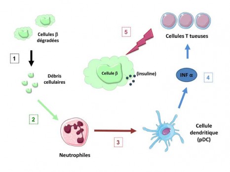
Activation of the immune system leading to death of the p cells

 ©Inserm / Agnès Lehuen – Juliette Hardy

 The natural deterioration of the β cells (1) leaves cell debris in the tissues that abnormally activate the neutrophils (2). These alarm cells in the immune system warn the dendritic cells (pDC) (3) which in turn cause the production of IFN α interferon, an alarm molecule (4). The interferon α then stimulates the T lymphocytes which, by recognising functional pancreatic β cells, cause these cells to die (5).

Innate immune cells and killer T-cells in the Islets of Langerhans

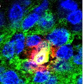
 ©Inserm / Yannick Simoni

A-B) Fluorescent photographs of a pancreatic islet (green) and close-up (B) of a neutrophil  (yellow) producing an activating molecule (red)
C) Photograph of the Islets in untreated mice in which the killer T-cells (white arrows) can be observed.

“We have observed in mice that treatment prevents activation of the innate immune cells, neutrophils and dendritic cells, warning of the onset of diabetes by inhibiting the appearance of auto-immune T responses aimed at the pancreas” explains Agnès Lehuen, head of the Inserm team.

These results show, for the first time, the important role played by innate immune cells in the sequence of events leading to the onset of Type 1 diabetes. Researchers continue to strive to understand how to regulate the auto-immune reaction produced by dendritic cells without compromising the innate immune system, an essential one in cases of infection. Several routes are being taken to attempt to regulate the production of the INF α alarm molecule that precedes activation of the killer T-cells, for example, by specifically targeting certain activation routes for the pDC dendritic cells.

These therapeutic approaches are currently being tested in other auto-immune diseases such as lupus and psoriasis. Such innovative treatments could be useful in the prevention of Type 1 diabetes. It will first be necessary, however, to perform studies in diabetic and pre-diabetic patients to be able to better understand how the innate immune cells function, something that has not been studied until recently in auto-immune diabetes,” concludes Agnès Lehuen.

The research benefited from a grant from LabEx INFLAMEX as part of investment for the future and for the Ile-de-France region.


[1] System present from birth that makes it possible to initiate an immune response to infection, regardless of the infectious agent involved. It is distinguished from the “acquired” or so-called “adaptive” immune system that is a specific response involving recognition of the infectious agent and the memorising of the infectious event.


Le cerveau des parfumeurs se modifie en fonction de leur expérience

A promising clinical trial to reduce the severity of autistic disorders

Yehezkel Ben-Ari, Founder and Honorary Director of INMED (Institut de Neurobiologie de la Méditerranée), INSERM, and Eric Lemonnier, a clinician specialising in autism at the CHRU of Brest, recently published the results of a double-blind clinical trial to evaluate the usefulness of a diuretic in the treatment of autism. Sixty children between 3 and 11 years old with autism or Asperger’s syndrome were treated for 3 months either with a diuretic to reduce their intracellular chloride levels or with a placebo. Although this therapy is not curative, it nevertheless reduced the autistic disorders’ severity in three-quarters of the children. The researchers have filed a request for authorisation to perform a multi-centre European clinical trial in order to determine more precisely the population concerned by this therapy.

Details of this work have been published in the Translational Psychiatry review dated 11 December 2012.

Contribution made by the fundamental research on neuronal chloride

Previous work carried out by the team of researchers led by Yehezkel Ben-Ari in INSERM unit 901, the Institut de Neurobiologie de la Méditerranée (INMED) in Marseille, on intracellular chloride concentrations have demonstrated that they are abnormally high in immature neurons or neurons previously affected by epileptic seizures or other cerebral lesions. Many anxiolytics, analgesics and antiepileptics act by increasing the effects of GABA – the brain’s main chemical mediator – which normally inhibits the neurons. When the cells contain a very high chloride concentration, however, GABA’s effects are reversed. GABA no longer inhibits the neurons; the anxiolytic molecules accentuate these effects instead. These molecules have an excitatory effect, aggravating the disorder rather than alleviating it[1]. This is what has been observed in the case of epilepsy: diazepam, an anxiolytic, actually aggravated the seizures in certain situations. The research team then showed the benefits of a diuretic in mitigating this effect.

From fundamental research to clinical research

Indirect experimental data suggest that the inhibitory transmitter GABA’s action is modified in autism. Eric Lemonnier, a clinician at the CHRU of Brest, pointed out to Yehezkel Ben-Ari that valium is not prescribed to children suffering from autism because their parents say they become more agitated as a result, suggesting that, as in epilepsy and other brain pathologies, their intracellular chloride concentration is increased. This encounter led to the idea of testing a diuretic – in the same way as for epilepsy – to determine whether this could alleviate autistic disorders. A pilot study in 5 children was rapidly set up in 2010 because bumetanide, the diuretic tested, is in common use, particularly in treating high blood pressure. The taking of these molecules can, however, lower the potassium level, meaning that a potassium supplement is required. The researchers then began a randomised double-blind clinical trial in 60 children between 3 and 11 years old with autism or Asperger’s syndrome.

Reduction in the severity of autistic disorders

The children were monitored for 4 months. One group was treated with the diuretic (1 mg of bumetanide) while a placebo was administered to the second group for 3 months. No treatment was administered in the final month. The severity of the children’s autistic disorders was rated at the beginning of the test, the end of the treatment, i.e. after 90 days and one month after the test ended.

After 90 days of treatment, the mean CARS (Childhood Autism Rating Scale) test score of the children treated with bumetanide had significantly improved. The severity of the treated group’s autistic disorders shifted from high (> 36.5) to medium (< 36.5). No significant difference was observed in the score of the group treated with the placebo, however. In total, the clinical diagnosis of 77% of the children who received the treatment improved in the Clinical Global Impressions (CGI) test. When the treatment was terminated, some disorders reappeared. The treatment with bumetanide is therefore reversible.

Various criteria for assessing the severity of disorders: CARS, CGI and ADOS G

The widely-applied Childhood Autism Rating Scale (CARS) behavioural scale was used to rate the severity of the disorders, based on videos of the children’s behaviour during an activity led by a caregiver. The films were analysed with the assistance of their parents. A rating is obtained from the analysis as follows: if the rating is between 30 and 36, the child suffers from a moderate or average disorder; if the rating is higher than 36, the child is severely autistic.

Two other indicators were used to assess the severity of the disorders: the Clinical Global Impressions (CGI) clinical diagnosis, and the Autism Diagnostic Observation Schedule – Generic (ADOS–G) indicator combining assessment criteria such as social interaction and communication.

Dr. Lemonnier explained the case of a 6-year-old boy:

Prior to the treatment, the child presented with low language abilities and little social interaction, was hyperactive and exhibited constantly-combative behaviour. After three months of treatment, his parents, teachers, the hospital nursing staff and his friends at school all said that he was participated more, particularly in the games proposed by the psychologist. His attention and eye contact also improved.”

“Even though it is not curative, the diuretic reduced the severity of most of the children’s autistic disorders. According to the children’s parents, they are more “present””

, added Yehezkel Ben-Ari.

Given the population’s heterogeneity, the researchers assume that the treatment could act differently depending on the severity of the autistic disorders. By forming groups based on severity, the results suggest that the treatment would be more effective in the least seriously-affected children.

As a result, the researchers have filed an authorisation request for a multi-centre European clinical trial in order to determine more precisely the population concerned by this treatment and ultimately obtain a marketing authorisation for this therapy. This test is supervised by a company created by Prof. Ben-Ari and Dr. Lemonnier (Neurochlore). Analyses are also essential in order to assess the long-term effects of taking these molecules and the required dose. Lastly, the researchers stress the need to continue the work on experimental models to determine how chloride is regulated and how it is deregulated in the neural networks of autistic patients.

A patent application has been filed for this work, and a licence has been granted to the Neurochlore start-up. Neurochlore has received funding from the French National Research Agency (ANR) (in the Biomedical Innovation in public-private Research Partnership (BIRP) “Cure Autism” project).


[1] See diagram on p. 3 “Further details”

Adipocyte inflammatoire de l’obèse : une cellule en perte de contrôle

Un diagnostic prénatal des maladies génétiques sur simple prise de sang

Improving chemotherapy effectiveness by acting on the immune system

An Inserm team in Dijon directed by François Ghiringhelli (Inserm unit 866 ‘Lipids, nutrition and cancer’) is to publish an article this week in the Nature Medicine review.  The article suggests that two chemotherapy drugs frequently used to treat digestive and breast cancers may encourage the development of tumours by modulating the anti-tumoural immune response. These results reveal how the immune system can then limit the effectiveness of some cancer chemotherapies. The researchers now intend to block the molecules responsible for negative immune system activation to increase the efficiency of chemotherapy. A clinical trial to test this hypothesis should begin very soon.

Chemotherapy is one of the most frequently used treatments to eliminate cancerous cells. These drugs kill all cells that are multiplying, or block their proliferation (for example, cells responsible for hair growth, explaining the hair loss of treated patients).  In addition to their direct toxic effects, the chemotherapeutic agents also seem to act on the immune system and could make it possible for the body to trigger a direct antitumor immune response in a second phase.

© fotolia

However, this last point is still the subject of hot debate, since some studies suggest, conversely, that chemotherapy eliminates all immune defences.

What now?

The Inserm team directed by Professor François Ghiringhelli (Inserm unit 866 “Lipids, nutrition and cancer”) from the Georges François Leclerc Cancer Research Centre in Dijon observed that two chemotherapeutic agents, 5-fluorouracile and gemcitabine, used to treat colon, breast and pancreas cancers activate a protein complex “inflammasome NLRP3” within some cells in the immune system.

To be more specific, this activation leads to releasing proinflammatory cytokine (interleukin IL-1beta) through these cells. This cytokine “distorts” the immune response related to lymphocytes T and causes the production of another cytokine (cytokine IL-17), which has protumoral properties by encouraging tumour angiogenesis, i.e. vascular irrigation of tumours.

Our results have made it possible to ascertain that the activation of inflammasome limits the effectiveness of chemotherapy. The challenge was then to see whether we could prevent the activation of inflammasome”

explains François Ghiringhelli. The researchers then tested two different strategies:

The first was to test the two drugs on inflammasome NLRP3- or cytokine IL-17-deficient mice.  In these cases, the researchers showed that antitumor activity was not only present, but it actually increased, demonstrating that these two elements (NLRP3 and IL-17) slow down the chemotherapy action.

The second strategy was to treat the mice using an IL-1beta inhibitor. Here again, the effectiveness of chemotherapy was again increased.

These results suggest that targeting the inflammasome and IL-1beta channels, combined with the use of these two chemotherapy agents, can improve the effectiveness of the latter. These tumour cells are eliminated and, in parallel, the damaging immune responses are deleted.

A therapeutic trial combining 5-fluorouracil and IL-1 beta is currently being prepared and should begin soon at the Georges François Leclerc Cancer Research in Dijon.

fermer