Menu

The way the virus continually changes complicates the development of a vaccine against HIV

A team from INSERM Mixed Research Unit 966 Morphogenesis and Antigenicity of HI and Hepatitis Viruses” headed by Martine Braibant and Francis Barin in Tours, has confirmed, with the support of the ANRS, that the AIDS virus has gradually adapted itself to the immune response of the human population during the course of the epidemic. Starting from the time it was isolated in the 1980s to the present day, the virus has become less and less sensitive to neutralising antibodies, complicating the creation of an effective preventive vaccine. Researchers have also shown that the combination of two powerful neutralising antibodies remains capable of neutralising in vitro the most recent HIV variants. Details of the research are published in the journal PLoS Pathogens.


The development of a vaccine that is effective against HIV remains one of the major challenges in the fight against AIDS. Considerable progress has been made in recent years with the identification of human neutralising antibodies capable of blocking in vitro infection caused by the very numerous variants of HIV -1. The scientific community is considering performing clinical trials in humans, using some of these antibodies. One of the issues that remains to be resolved is that of the choice of antibodies and the best combination of antibodies to be used in order to afford the widest possible protection in view of the huge variability of possible forms of HIV -1 currently circulating in the world.

Particules virales du virus de l'immunodéficience humaine (VIH)


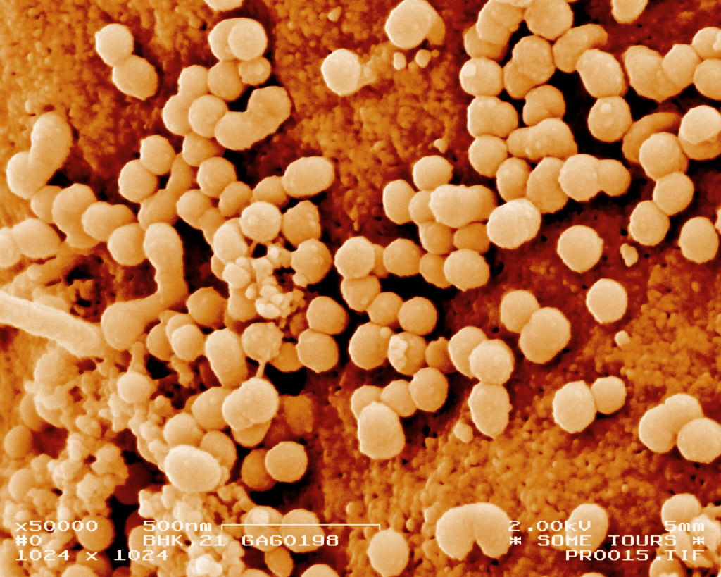
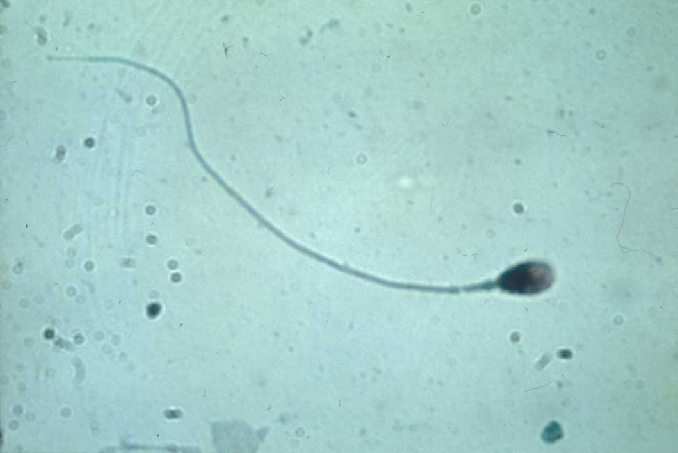
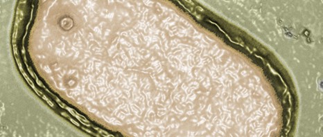
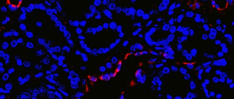
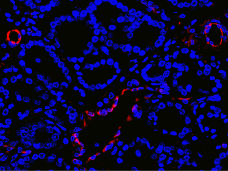
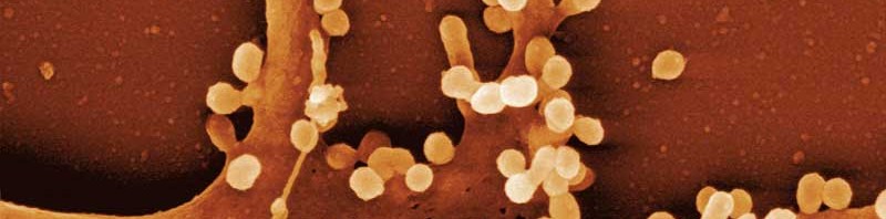
© Inserm /  Philippe Roingeard

The research, headed by Martine Braibant and Francis Barin (INSERM-Université de Tours Mixed Unit) with the support of the ANRS, confirms that during the course of the epidemic HIV-1 has gradually adapted itself to the immune response of the human population by becoming less and less sensitive to certain neutralising antibodies. In order to arrive at these results, the researchers were able to analyse viruses taken from samples in patients infected between the late 1980s and the year 2010[1].

“Although it was known that, at an individual level, the virus was able to adapt and circumvent the individual’s own means of defence, our work confirms that the pressure of selection exercised on the virus has had repercussions on a population-wide scale”, explains Martine Braibant.

Despite this depressing prospect, in the same publication, researchers identify a combination of two powerful neutralising monoclonal antibodies developed by Caltech and the Scripps Research Institute (NIH45-46G54W and PGT128) that still remain capable of neutralising in vitro the most recent variants and do so at a concentration compatible with their use in humans.

This study stresses the need to monitor the development of the sensitivity of HIV -1 variants to the various neutralising antibodies. The work of the researchers opens up interesting prospects for teams involved in researching a vaccine to prevent HIV .

Neutralising antibodies: one of the research pathways for developing an anti-HIV vaccine

Current anti-viral vaccination concepts are largely based on the fact that certain so-called neutralising antibodies, by attaching themselves to viral proteins, inhibit the early stages of HIV infection. When blocked in this way, the virus cannot reproduce and is eliminated.

Scientists have currently identified the immune response to be induced via the identification and characterisation of wide-spectrum neutralisation antibodies. They have not yet succeeded in enabling this type of antibody to be produced in the human body through a standard vaccination. One of the research pathways being pursued is the introduction of gene coding for these antibodies in viral expression vectors so as to cause them to be secreted directly into the circulation. This antibody approach has been tested in France as part of the ANRS/VRI vaccine research programme (Vaccine Research Institute)


[1] PRIMO and SEROCO ANRS cohorts, collaboration with Laurence Meyer and Cécile Goujard, Université Paris-Sud – INSERM U1018.


Novel molecules to target the cytoskeleton

The dysfunction of the cytoskeleton, a constituent element of the cell, is often associated with pathologies such as the onset of metastases. For this reason, it is a target of interest in numerous therapies. Teams from CNRS, the Université de Strasbourg and Inserm, led by Daniel Riveline1, Jean-Marie Lehn2 and Marie-France Carlier3, have synthesized molecules capable of causing rapid growth of actin networks, one of the components of the cytoskeleton. This is a breakthrough because, until now, only molecules that stabilize or destroy the cytoskeleton of actin have been available. These compounds with novel properties, whose action has been elucidated both in vitro and in vivo, provide a new tool in pharmacology. This work was published in the journal Nature Communications on 29 July 2013.

The cytoskeleton is mainly composed of actin filaments and microtubules. Made of polymers in dynamic assembly and constantly constructing and deconstructing itself, it affects numerous cellular processes such as intracellular movement, division and transport. It is involved in key steps of embryogenesis and other processes essential to life. Consequently, its malfunctioning can lead to serious pathologies. For example, the onset of certain metastases is revealed by an increased activity of the cytoskeleton. Identifying new molecules that target the cytoskeleton thus represents a major challenge.

Until now, the molecules known and used in pharmacology had the effect of stabilizing or destroying the cytoskeleton of actin. Actin allows vital actions to be performed by assembling and disassembling itself spontaneously, continually and rapidly in the form of filaments that organize themselves and form networks of parallel bundles or intertwined meshes (known as lamellar networks). Derived from supramolecular chemistry[4], the new compounds synthesized by the researchers have original properties: within several minutes, they bring about the growth of lamellar networks of actin filaments. This is the first time that a pharmacological tool induces growth of the actin network — something that living organisms do all the time. In this way, the researchers have shown that the action of these compounds is specific in vivo (on cells). In addition, they have identified the growth mechanism of the actin network by comparative in vivo and in vitro studies in order to ensure the validity of the process.

For cellular or molecular biology, this tool proposes a new mode of possible action on the cytoskeleton and thus opens new research perspectives for deciphering the living world. This finding could lead to the development of new compounds, derived from the same chemistry, and potential candidates for new therapies targeting the cytoskeleton.

[1] Institut de Science et d’Ingénierie Supramoléculaires (CNRS/Université de Strasbourg) and Institut de Génétique et de Biologie Moléculaire et Cellulaire (CNRS/Université de Strasbourg/Inserm).

[2] Institut de Science et d’Ingénierie Supramoléculaires (CNRS/Université de Strasbourg).

[3] Laboratoire d’Enzymologie et Biochimie Structurales of CNRS.

[4] Supramolecular chemistry, the science of self-assembly and self-organization at the molecular scale, focuses on chemical entities resulting from the interactions between molecular objects.

How is the male genome preserved until it reaches the egg?

When the male genome carried in the spermatozoid leaves the male body to reach the egg, it undergoes numerous transformations. A team led by Saadi Khochbin in Mixed Research Unit 823 at the Institut Albert Bonniot Research Centre (Inserm/Joseph Fourier University) in Grenoble has described the molecular mechanisms that enable the transmission of the male genome to the egg. The researchers have revealed the essential role played by a tiny structure  which compact and preserve the genome in the spermatozoid during its journey to the egg. These results were published on July 24th in the journal Genes & Development.

Spermatozoide-khochbin

Spermatozoid – © Inserm / Denise Escalier

One of the challenges of reproduction is to discover how male DNA is carried via the spermatozoids, the highly specialised germinal cells. These are capable of leaving the organism and surviving during their journey from the male to the female body, at which time it is necessary to ensure that the genome it contains is safe in order to preserve it for fertilisation. When spermatozoids leave the male organism and start their journey to the female body, the genome is necessarily secured and preserved until the fertilization. The genome gradually changes its spatial configuration during spermatogenesis. This enables the DNA to be transported in a very compact, and thus very resistant, form. A defect in the compacting process can result in infertility.

Hitherto, although scientists had identified the molecules that contribute to the compaction of the DNA – histones, transition proteins, protamines, the molecular determinants causing these rapid changes in configuration remain obscure.

The “Epigenetics and cell signalling” Inserm Team headed by Saadi Khochbin, CNRS Research Director, described for the first time how the “organising” element in the male germinal cells directs the very accurate and specific compacting of the male genome. It is a special histone called TH2B, which was discovered in 1975, one of the earliest histones to be identified. This tiny protein attaches itself to the DNA during spermatogenesis and gives it the special configuration required for its final compaction. This is how the paternal genome, transported by the spermatozoid, leaves the male body and reaches the egg. The researchers also discovered that, unexpectedly, this histone is also present in the egg and participates in the repackaging of the male genome after fertilisation as soon as it enters the egg.

“We therefore discovered an important element in the transmission of the paternal genetic information that also participates in its packaging for despatch from the male reproductive organ as well as in its receipt by the female cell”, explains Saadi Khochbin, principal author of the study.

The research required the use of several mouse models and approaches involving very sophisticated recent technology for the purpose of exploring the genome as a whole (the genomic and transcriptomic techniques) and understanding new mechanisms on the molecular scale (proteomic approaches and structural modelling).

On a basic level, the research improves knowledge of male genome transmission and the way in which the male genome is transmitted during reproduction; there are also implications in the understanding of infertility and the optimisation of medically assisted procreation.

Pandoravirus : missing link discovered between viruses and cells

Researchers at IGS, the genomic and structural information laboratory (CNRS/Aix-Marseille University), working in association with the large-scale biology laboratory (CEA/Inserm/Grenoble Alpes University) have just discovered two giant viruses which, in terms of number of genes, are comparable to certain eukaryotes, microorganisms with nucleated cells. The two viruses – called “Pandoravirus” to reflect their amphora shape and mysterious genetic content – are unlike any virus discovered before. This research appeared on the front page of Science on July 19, 2013.

With the discovery of Mimivirus ten years ago and, more recently, Megavirus chilensis[1], researchers thought they had reached the farthest corners of the viral world in terms of size and genetic complexity. With a diameter in the region of a micrometer and a genome incorporating more than 1,100 genes, these giant viruses, which infect amoebas of the Acanthamoeba genus, had already largely encroached on areas previously thought to be the exclusive domain of bacteria. For the sake of comparison, common viruses such as the influenza or AIDS viruses only contain around ten genes each.

In the article published in Science, the researchers announced they had discovered two new giant viruses:
Pandoravirus salinus, on the coast of Chile;
Pandoravirus dulcis, in a freshwater pond in Melbourne, Australia.

Detailed analysis has shown that these first two Pandoraviruses have virtually nothing in common with previously characterized giant viruses. What’s more, only a very small percentage (6%) of proteins encoded by Pandoravirus salinus are similar to those already identified in other viruses or cellular organisms. With a genome of this size, Pandoravirus salinus has just demonstrated that viruses can be more complex than some eukaryotic cells[2]. Another unusual feature of Pandoraviruses is that they have no gene allowing them to build a protein like the capsid protein, which is the basic building block of traditional viruses.

Despite all these novel properties, Pandoraviruses display the essential characteristics of other viruses in that they contain no ribosome, produce no energy and do not divide.

This groundbreaking research included an analysis of the Pandoravirus salinus proteome, which proved that the proteins making it up are consistent with those predicted by the virus’ genome sequence. Pandoraviruses thus use the universal genetic code shared by all living organisms on the planet.

This shows just how much more there is to learn regarding microscopic biodiversity as soon as new environments are considered. The simultaneous discovery of two specimens of this new virus family in sediments located 15,000 km apart indicates that Pandoraviruses, which were completely unknown until now, are very likely not rare. It definitively bridges the gap between viruses and cells — a gap that was proclaimed as dogma at the very outset of modern virology back in the 1950s. It also suggests that cell life could have emerged with a far greater variety of pre-cellular forms than those conventionally considered, as the new giant virus has almost no equivalent among the three recognized domains of cellular life, namely eukaryota (or eukaryotes), eubacteria, and archaea.

Notes
[1] Arslan D, Legendre M, Seltzer V, Abergel C, Claverie JM (2011) “Distant Mimivirus relative with a larger genome highlights the fundamental features of Megaviridae.” PNAS. 108:17486-91
[2] Parasitic microsporidia of the Encephalitozoon genus in particular.

Boosting Muscle capacity

(French) : Découverte d’une nouvelle cible thérapeutique chez E. coli

There is current over-treatment of prostate cancer in France

Research conducted by Cyrille Delpierre (INSERM Unit 1027 “Epidemiology and Public Health Analyses: Risks, Chronic Illness and Handicaps”) in collaboration with the French Cancer Registers Network assessed the proportion of patients potentially suffering from prostate cancer and currently over-diagnosed or over-treated in France. According to a study performed on 1840 patients, current over-treatment involves a considerable number of patients being treated for a cancerous tumour in what is known as Stage T1 (early tumours) and, to a lesser extent, patients suffering from tumours in stage T2 (more advanced tumours). These figures should be taken seriously because over-diagnosis or over-treatment can be associated with undesirable side effects such as impotence or incontinence.

This work has been published in the review Cancer Epidemiology.

photo-cancer-de-la-prostate

© Ghinea N/Inserm Cancer de la prostate (stade précoce)

The generalised use of the PSA[1] test for prostate cancer treatment has led to earlier diagnosis of the condition. This development is an advance in itself, since the early stages are less serious and thus easier to cure. But not all prostate cancers are equally aggressive; like many diagnostic tests, the administration of PSA detects tumours more easily, including those that grow at a slower rate. Consequently, a considerable number of tumours diagnosed from a PSA test develop very slowly, thus exposing patients to the risks of over-diagnosis or over-treatment.

In the absence of markers that make it possible to identify tumours that are specifically aggressive, the main difficulty with prostate cancer diagnosis lies in assessing the benefits for the subject, taking account of the risks of over-diagnosis and over-treatment (which can vary between 30% and 50% according to the literature). It is therefore necessary to compare theoretical life expectancy, and thus to assess such cancers correctly. In practice, if the patient’s life expectancy exceeds 10 years, a period considered necessary for a cancer that has become clinically significant, the treatment is justified.

The purpose of the study conducted by Cyrille Delpierre was to assess the extent of the potential actual over-diagnosis and over-treatment for prostate cancer in France, by taking account of comorbidity rates liable to seriously affect theoretical life expectancy.

The sample consisted of 1840 patients diagnosed in 2001. The proportion of patients over-diagnosed and over-treated was assessed by comparing theoretical life expectancy (taking co-morbidity into account) to life expectancy for those living with the cancer.

It was possible to identify patients in a potential over-treatment situation, i.e. those whose theoretical life expectancy was less than their life expectancy when living with cancer, and among the latter group to discover whether they had actually been treated (either through surgery or through radiotherapy) and thus genuinely over-treated.

Of these, 9.3% to 22.2% of patients with T1 tumours had been over-treated. This represents between 7.7% and 24.4% of patients who had had a prostate ablation and 30.8% and 62.5% of those treated with radiotherapy.

Two per cent of patients with stage T2 tumours had been over-treated, making 2% of patients who had had a prostate ablation and 4.9% who had received radiotherapy.

The presence of a co-morbidity considerably increases these proportions. Patients at stage T1 with more than two comorbidities were in a potential over-treatment situation in almost all cases and were actually over-treated in one third of all cases.

One of the major limits in our study was working with patients diagnosed in 2001. The present situation is different. But the most recent data covering 2008 show inverse proportions of stages T1 and T2. T1 stages are now more frequent than T2. The proportion of minor stages during which a higher risk of over-treatment has been observed is now on the increase”, explains Cyrille Delpierre.

The research highlights that there is a significant proportion of actual over-treatment, mainly observed in patients with co-morbidity.

The main question for researchers around the debate as to the usefulness of PSA, and more generally surrounding all diagnoses, is there for not the test in itself but rather the choice of appropriate treatment.

Diagnosis is a process that starts with the offer of a PSA test, followed in the case of positive results by diagnostic examinations and suitable treatment if disease is detected. “In view of the over-treatment of prostate cancer that has been confirmed, treatment could be restricted, especially for patients with co-morbidities, to supervision that would make it possible to offer the treatment when it becomes appropriate”, proposes Cyrille Delpierre.

The difference observed between potential over-treatment and actual over-treatment is a sign that the risk of over-treatment has been taken into account by urologists, although it needs to be improved. This situation translates the difficulty in suggesting active monitoring and the difficulty of justifying a non-interventionist attitude for patients who are aware that they have cancer.

Over-diagnosis or over-treatment?

Over-diagnosis: the time between diagnosis and the emergence of clinical symptoms and/or death from cancer may be higher than life expectancy, since the cancer will not manifest itself or at least will not kill the person. This situation corresponds to the situation known as over-diagnosis.

Over-treatment: the decision to use an invasive treatment may be the origin of over-treatment when treating a cancer that has not been particularly intrusive during the life of an individual.  It is therefore not always in the patient’s interest to be diagnosed and treated for a tumour that is unlikely to develop much, by using treatment that have undesirable side effects.


[1] Prostate cancer diagnosis is based on detecting a marker in the blood of a specific marker for prostate dysfunction (a specific antigen known as PSA)


When epidemiologists turn to Web 2.0

In the Web 2.0 and Quantified Self” era, scientists are working to improve traditional data collection methods (face-to-face interviews, telephone interviews or paper questionnaires), which are expensive and time-consuming for study participants. Indeed, recruitment, monitoring and data collection tools need to be adapted to fit the habits of the younger generation. The Internet and smartphones offer numerous options for collecting data on participants’ lifestyle, environment and health, in a way involving few constraints, and that can even be fun. In May 2013, the INSERM team headed by Françoise Clavel-Chapelon (Unit 1018, Team 9 Nutrition, Hormones and Women’s Health”) organized an international conference entitled “e-tools and social networks for epidemiology” in Paris. The main objective was to present academic research and ICT (Information and Communication Technology) innovations that can improve epidemiological research.

New data acquisition tools

Carol Boushey, from the University of Hawaii, presented her research on TADA (Technology Assisted Dietary Assessment) technology. TADA is a prototype application for smartphones that collects dietary information easily. The process is simple: you just need to take a picture of your plate before and after a meal. The program’s algorithms do the rest, detecting textures and volumes of foods, and reconstitute the exact composition and dietary intakes.

tada

Carol Boushey demonstrating TADA (Technology Assisted Dietary Assessment)

New techniques to record data on physical activity were also presented. Stephen Intille (NortheasternUniversity, Boston) described

the work done by the Mobile Health Research Group on the use of smartphones and detectors to measure health and physical activity data.

 Suzanne Garland (Royal Women’s Hospital, Melbourne) explained how the SenseWear® Armband is used in the Australian Young Female Health Initiative study. This electronic bracelet collects physiological data (skin temperature, heart rate, etc.) and provides instant information about the number of steps walked, the distance covered, calories burned, sleep cycle, etc.

Furthermore, geolocation techniques are increasingly finding applications in epidemiology. For example, the Veritas tool, presented by Yan Kestens (University of Montreal) based on online cards, can collect spatial information, and the tool presented by John Nuckols (ColoradoStateUniversity) combines recording of geolocation and environmental pollutants data.

Web 2.0 and e-Health

Several talks presented new recruitment techniques, networking, data management and loyalty card schemes run via Internet.

John Wark (Melbourne University) described how Facebook was used to recruit volunteers into the Young Female Health Initiative study.

Finally, several web platforms were presented, such as CleanWEB ePRO®, an integrated electronic management solution for clinical trials, various ad hoc e-health programs rolled out by the BePATIENT company, and the SenseDoc multisensor platform developed by the SPHERE research laboratory (Research Centre of the University of Montreal’s research centre), in collaboration with the RECORD Study team.

capteur

Le SenseWear® Armband de la société BodyMedia®

The major advantages of these web 2.0 platforms are the ability for participants to network, the collection of digital data that can be used very quickly, the connection to e-tools, sensors, smartphone applications that record information automatically, and the ability to obtain individual feedback based on the participant’s profile.

“In addition to saving time and to the improved reliability of data collected through these means, these resources have a fun and interactive component, which is a very important aspect to ensure continued participation of large cohort participants”, explains Françoise Clavel-Chapelon, INSERM Research Director and Principal Investigator of the E3N and E4N cohorts.

This first conference discussed how new tools such as social networks, e-health and spatial technology could have applications in the epidemiological studies of tomorrow. However, before using these innovative methods and tools in epidemiological studies, it is necessary to check their acceptability, reliability and validity. “The E4N cohort (see box below) will offer particularly favourable conditions to carry out this validation and standardization phase”, concludes Guy Fagherazzi, scientific manager of the E4N cohort.

The conference was organised by the INSERM team headed by Françoise Clavel-Chapelon (UMRS 1018, Team 9) who wanted to discuss new ways of collecting epidemiological data for the E4N cohort. The event was run jointly with AFCROs (Association française des CROs), in partnership with Aviesan Santé Publique, the INRIA and three competitive high-tech hubs in the Paris region (Cap Digital, Systematic Paris-Région and Medicen) brought together under the label of “TIC & Santé”. The conference received financial supports from the Mairie de Paris, the Université Paris-Sud and the Ligue contre le Cancer.

E4N: a family cohort

The E4N study is designed to be an extension of the E3N study by following-up the spouses, children and grandchildren of the E3N women. The objective is to better understand the onset of disease, from genetics, family environment and the environment outside the family. This project of a prospective family cohort has been selected by the “Investissements d’Avenir” programme from the ANR (Agence Nationale de la Recherche) for a follow-up over a nine-year period.

For more information: www.e4n.fr

etools

Quels progrès médicaux pour les personnes vivant avec le VIH en France ?

fermer