Menu

Detailed structure of human ribosome revealed

A team at the Institut de Génétique et de Biologie Moléculaire et Cellulaire (IGBMC – CNRS/Université de Strasbourg/Inserm) has evidenced, at the atomic scale, the threedimensional structure of the complete human ribosome and the detailed interactions that occur within it. These findings, obtained using a technology that is unique in France, open the way to further exploring some of the adverse effects of antibiotics, and, in the longer term, to the treatment of diseases related to ribosomal dysfunctions and the deregulation of protein synthesis. This work is published in Nature on 22 April 2015.

Ribosomes are large complexes of proteins and RNA folded together, which — within the cells of all living beings — act as molecular nano-machines in the expression of genes and the biosynthesis of proteins. While the ribosomal structures of different species were already known in detail at the atomic scale, determining the particularly complex structure of the human ribosome remained a major challenge.

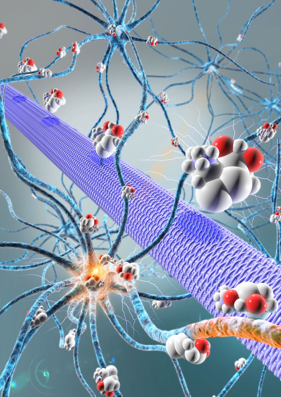
The team led by Bruno Klaholz at IGBMC (CNRS/Université de Strasbourg/Inserm) has now visualized the atomic structure of the complete human ribosome at a resolution greater than 3 angströms (0.3 nanometers). The model obtained represents the 220,000 atoms that make up the two subunits of the ribosome and makes it possible for the first time to explore its detailed arrangement as well as visualize and identify its different amino acids and nucleotides in 3D. The scientists focused in particular on the various binding sites and detailed interactions that occur within this structure. For example, their efforts revealed that after delivering the amino acids they are carrying, transfer RNA continue to interact with the ribosome at a specific site (the tRNA exit site). The team has also shed light on the dynamics of the two ribosomal subunits, which slightly rotate during the protein biosynthesis process, thus heavily remodeling the 3D configuration of the structure at their interface.

These results were achieved using a series of cutting-edge technologies. The samples were highly purified and then frozen before being visualized through cryo-electron microscopy. This method enables scientists to study fixed objects whose orientation does not change and whose structure and biological functions are preserved. A combination of image processing and 3D reconstruction applied to the images obtained by the latest-generation cryo-electron microscope operated by the IGBMC — which is unique in France — made it possible to achieve this rare degree of accuracy.

This detailed knowledge of the structure and dynamics of the complete human ribosome opens the way to further crucial explorations. It is now possible to envisage studying the adverse effects of certain antibiotics designed to fight bacterial ribosomes — and which may target the human ribosome “by mistake”. Listing existing binding sites is the first step towards enhancing the specificity of therapeutic compounds and preventing them from binding to the wrong site. In the longer term, these findings could also help develop treatments for diseases linked to ribosomal dysfunctions and the deregulation of protein synthesis. For example, in the case of cancers, being able to target the ribosomes of diseased cells would make it possible to reduce their protein synthesis rates.

Klaholz 

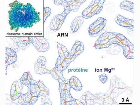
Example of the three-dimensional elements that could be distinguished within the atomic structure of the complete human ribosome (insert). The resolution (of about 1 angström) is able to determine whether or not there are any interactions between the different elements. © H. Khatter, A.G. Myasnikov, S. K. Natchiar & B.P. Klaholz

Saturday 25 April: World Malaria Day

Caused by a parasite of the genus Plasmodium, malaria is transmitted to humans by the bite of a female Anopheles mosquito. It can also be transmitted by blood transfusion, or from mother to foetus at the end of pregnancy.[1]
Paludisme et gène TEP1

Live Plasmodium parasites in the Anopheles mosquito express GFP fluorescence protein, and are visible in green (green arrows). Mosquitoes that express only the sensitive allele of TEP1 are less resistant than those that express only the resistant allele. © Inserm/Lamacchia, Marina

Held on 25 April each year, World Malaria Day is specifically aimed at raising awareness among the general public in order to increase its involvement in controlling this disease. This event is also an opportunity to highlight the advances made in research.

In line with its year-round commitment to malaria research, the team led by Benoît Gamain, Inserm Unit 1134, “Integrated Biology of Red Blood Cell,” is currently working on the PRIMALVAC[2] project, the objective of which is the development of a malaria vaccine for pregnant women (gestational malaria).

This phase I trial is aimed at assessing the vaccine’s safety and tolerance for human subjects, as well as its ability to induce an immune response, enabling pregnant women to be protected against gestational malaria in future.

Research done by Dominique Mazier and her staff at Inserm Unit 1135, “Centre for Immunology and Infectious Diseases,” is focused on the biology and immunochemistry of the hepatic stages of Plasmodium in humans.

A fraction of the parasites located in the liver, known as hypnozoites, may remain “dormant,” before awakening over time, and causing bloodstream infection. This hepatic phase of parasite development, which is specific to the vivax and ovale species, constitutes a double problem for the elimination of malaria: a higher number of patients to be treated, and increased transmission. Unfortunately, primaquine, and a recently developed analogue, tafenoquine, the only drugs capable of killing hypnozoites, have adverse effects that are sometimes serious for patients.

Research done by this team is helping to improve the understanding of this biological phenomenon, and to identify new strategies for an innovative, non-toxic radical cure.

The team led by Stéphanie Blandin, Inserm Unit 963 “Immune Responses in the Malaria Vector Anopheles gambiae,” is seeking to understand how mosquitoes defend themselves from parasites, and to exploit this antiparasitic response to help control transmission of the disease.

[1] For more details, consult the Inserm information pack on this theme (only available in French).

[2] The PRIMALVAC project is coordinated by EVI (European Vaccine Initiative), and has received financial support from the German Federal Ministry of Education and Research (BMBF), Inserm, the French National Institute of Blood Transfusion (INTS), and from Irish Aid via EVI

Natural reparative capacity of teeth elucidated

Researchers at Inserm and Paris Descartes University have just taken an important step in research on stem cells and dental repair. They have managed to isolate dental stem cell lines and to describe the natural mechanism by which they repair lesions in the teeth. This fundamental discovery will make it possible to initiate unprecedented therapeutic strategies to mobilise the resident dental stem cells and magnify their natural capacity for repair.

These results are published in the journal Stem Cells.

The tooth is a mineralised organ, implanted in the mouth by a root. The “living” part of the tooth or dental cavity is the dental pulp (in yellow in the photograph shown opposite) composed of vessels and nerves. Around it is a hard substance, the dentine or ivory, which is in turn covered by an even harder tissue, the enamel. When a dental lesion appears, the dormant stem cells in the pulp awaken and try to repair the tooth by an unknown process.dent1

3D modelling of a teeth. The dental pulp is in yellow. ©Inserm/ Chappard, Daniel

In this study, the researchers from Inserm and Paris Descartes University at Unit 1124, “Toxicology, Pharmacology and Cellular Signaling,” have succeeded in extracting and isolating tooth stem cells by working on the pulp from the mouse molar.

The researchers were thus able to analyse the cells in detail, and identify 5 specific receptors for dopamine and serotonin on their surface, two neurotransmitters that are essential to the body (see schema on page 2).

The presence of these receptors on the surface of these stem cells indicated that they had the ability to respond to the presence of dopamine and serotonin in the event of a lesion. The researchers naturally wondered what cells might be the source of these neurotransmitters, a warning signal. It turns out that the blood platelets, activated by the dental lesion, are responsible for releasing a large quantity of serotonin and dopamine. Once released, these neurotransmitters then recruit the stem cells to repair the tooth by binding to their receptors (see schema on page 2). The research team was able to confirm this result by observing that dental repair was absent in rats with modified platelets that do not produce serotonin or dopamine, i.e. in the absence of the signal.

“In stem cell research, it is unusual to be simultaneously able to isolate cell lines, identify the markers that allow them to be recognised (here the 5 receptors), discover the signal that recruits them (serotonin and dopamine), and discover the source of that signal (blood platelets). In this work, we have been able, unexpectedly, to explore the entire mechanism,” explains Odile Kellermann, leader of the team from Inserm and Paris Descartes University, and the main author of this work.

To take things a stage further, the researchers tried to characterise the different receptors they found. One of the 5 receptors does not seem to affect the repair process. On the other hand, the other 4 turn out to be strongly involved in the repair process. In vivo blocking of just one of them is enough to prevent dental repair.

“Currently, dentists use pulp capping materials (calcium hydroxide) and tricalcium phosphate-based biomaterials to repair the tooth and fill lesions. Our results lead us to imagine unprecedented therapeutic strategies aimed at mobilising the resident pulpal stem cells in order to magnify the natural reparative capacity of teeth without use of replacement materials,” concludes Odile Kellermann.

The foundations have been laid for extending this research done in rodents to stem cells of the human tooth in order to initiate new strategies for repairing teeth.

schéma mécanisme dent EN

© Inserm / Odile Kellermann, Anne Baudry

Inflammatory bowel disease: a gut bacterium with beneficial properties

Several years ago, INRA researchers in Jouy-en-Josas showed that levels of the gut bacterium Faecalibacterium prausnitzii tended to decline in the human gut at the onset of chronic inflammatory bowel disease (IBD). Is this disappearance of F. prausnitzii one of the causes of inflammation, or is its disappearance a consequence of the disease? Today, the same INRA team, in collaboration with an American team (Berkeley), AgroParisTech, lnserm, AP-HP and UPMC, are answering these questions. Not only do their results show that F. prausnitzii plays an active role in protecting against intestinal inflammation, they also propose explanations regarding mechanisms of action. This research is published in the journal mBio on 21 April 2015.

Seven years ago, researchers at INRA (French National Institute for Agricultural Research) drew the attention of scientists and the public to a specific bacterium found in our gut. This bacterium, Faecalibacterium prausnitzii, is abundant in the gut of healthy humans, but as soon as a chronic inflammatory bowel disease (IBD) occurs, it tends to decline. One nagging question has been raised by the scientific community: is the disappearance of F. prausnitzii one of the causes of inflammation, or is its disappearance a consequence of the disease? In other words, is F. prausnitzii a bacterium that might protect our digestive tract from an inflammatory disease?

To find an answer, the researchers used mice harbouring only two types of bacteria in their digestive tract, in contrast to several billion under normal circumstances. Following a treatment that generates inflammation, the presence of F. prausnitzii on its own protects from the development of intestinal inflammation. This demonstrates the anti-inflammatory potential of F. prausnitzii.

The INRA researchers and their colleagues from Berkeley, in collaboration with AgroParisTech, the French National Institute of Health and Medical Research (Inserm), the Paris public hospitals (AP-HP), and Pierre and Marie Curie University (UPMC), also propose new approaches to explaining how this bacterium could protect us. The presence of this bacterium is actually associated with many anti-inflammatory molecules in the gut and bloodstream of animals. F. prausnitzii may be able to provide protection to our digestive tract by a varied arsenal of metabolic activities. For example, salicylic acid, a precursor in the synthesis of drugs used to treat patients with IBD, is found in the gut of mice carrying F. prausnitzii. The bacteria that we harbour may well play an active role in our health via the same strategies that are used in the medical arena.

[exergue]Once there is intestinal inflammation, the decline in the presence of the bacterium F. prausnitzii therefore aggravates the disease. In order to break this vicious cycle leading to chronic inflammation of the digestive tract, the scientists plan to restore the presence of F. prausnitzii using new food supplements containing the bacterium (probiotics) and/or favouring development of the bacterium (prebiotics). [/exergue]This study, which has added to our fundamental knowledge in the area of microbiology, is also at the interface of new industrial and medical applications.ProbioScanning electron micrographs of F. prausnitzii © MIMA 2 Platform, T. Meylheuc)

New gene therapy success in a rare disease of the immune system: Wiskott-Aldrich syndrome

French teams from CIC Biothérapie (AP-HP/Inserm), from pediatric hematology department of Necker Hospital for Children (AP-HP), led by Marina Cavazzana, Salima Hacein Bey Albina and Alain Fischer and from Genethon led by Anne Galy (Genethon/Inserm UMR-S951), and English teams from UCL Institute of Child Health and Great Ormond Street Hospital in London led by Adrian Thrasher and Bobby Gaspar demonstrated the efficacy of gene therapy treatment for Wiskott-Aldrich Syndrome (WAS). Six children that were treated and followed for at least 9 months had their immune system restored and clinical condition improved. This work, which was published today in the Journal of the American Medical Association (JAMA), was carried out with support from the AFM-Telethon.

Wiskott-Aldrich syndrome is a rare congenital immune and platelet deficiency which is X-linked and has an estimated prevalence of 1/250 000. It is caused by mutations in the gene encoding the WAS protein (WASp) expressed in hematopoietic cells. This disease, which primarily affects boys, causes bleeding, severe and recurrent infections, severe eczema and in some patients autoimmune reactions and the development of cancer. The only treatment available today is bone marrow transplantation, which requires a compatible donor and can itself cause serious complications.

The Phase I / II study, with Genethon as the promoter, was launched in December 2010 and conducted in Paris and London to treat severely ill patients without a compatible donor. This study, which is ongoing, assesses the feasibility and efficacy of gene therapy in this indication. The article published in JAMA reports the results for the first six patients, aged 8 months to 16 years, where the monitoring period allowed assessment of the initial effects of the treatment.

The treatment involves collected blood stem cells carrying the genetic anomaly of patients and corrected them in the laboratory by introducing a healthy WAS gene using a lentiviral vector developed and produced by Genethon. The corrected cells were reinjected into patients who in parallel were treated with chemotherapy to suppress their defective stem cells and autoimmune cells to make room for new corrected cells. After reinjection, these cells were then differentiated into the various cell lines that make up the blood (red and white cells, platelets).

To date treated patients showed significant clinical improvement. Severe eczema and severe infection disappeared in all cases. Arthritis was eliminated in one patient and another saw major improvement in vasculitis of the lower limbs and was able to return to normal physical activity without a wheelchair. However, the rate of corrected platelets varies from one patient to another.

Fulvio Mavilio, Chief Scientific Officer Genethon: “We are all very happy and encouraged by the results of this study. It is the first time that a gene therapy based on genetically modified stem cells is tested in a multicenter, international clinical trial that shows a reproducible and robust therapeutic effect in different centers and different countries. For very rare diseases such as WAS, multicenter clinical trials are the only effective way of proving the safety and efficacy of gene therapy and having it rapidly approuved and made available to all patients. We are following the same approach for other rare and less rare blood diseases.

Frédéric Revah, CEO of Genethon, the laboratory of the AFM-Telethon and the trial sponsor, said “These first results of our clinical trial for the treatment of Wiskott Aldrich syndrome are very encouraging. They illustrate not only the ability of Genethon to carry out the upstream research to develop treatments for these rare and complex diseases, but also to construct and conduct international clinical trials, to produce these advanced therapy products, to work with international teams and to manage the regulatory aspects of the trials in France and abroad. These are skills that we implement for other international trials of gene therapy for rare genetic diseases of the immune system, blood, muscle, vision or liver… We will continue the current study with the objective of providing treatment for patients.

Marina Cavazzana: “The results obtained in this multicenter clinical trial constitute an important therapeutic advance (overhang) because they concern a complex pathology which affects almost all of blood cells with dramatic clinical consequences. After transfer of gene, the patients showed a significant clinical improvement due to the reexpression of the protein WASp in the cells of the immune system. The efficiency of the treatment of such a deficit for which a high level of correction of hematopoietic stem cells is required, indicates that it is from now on justifiable to hope to treat other complex genetic diseases as those affecting red blood cells.

Professor Thrasher says: “This is a very powerful example of how gene therapy can offer highly effective treatment for patients with complex and serious genetic disease. It also excitingly demonstrates the potential for treatment of a large number of other diseases for which existing therapies are either unsatisfactory or unavailable.

Caring for blindness: a new protein in sight?

Vasoproliferative ocular diseases are responsible for sight loss in millions of people in the industrialised countries. Many patients do not currently respond to the treatment offered, which targets a specific factor, VEGF. A team of Inserm researchers at the Vision Institute (Inserm/CNRS/Pierre and Marie Curie University), in association with a team from the Yale Cardiovascular Research Center, have demonstrated in an animal model that blocking another protein, Slit2, prevents the pathological blood vessel development that causes these diseases. This work is published in Nature Medicine.

Vasoproliferative ocular diseases are the main cause of blindness in the industrialised countries. Age-related macular degeneration (ARMD), diabetic retinopathy and retinopathy of prematurity (in newborns) are characterised by progressive involvement of the retina, the area of the eye that receives visual information and transmits it to the brain. This damage is caused by abnormal growth of the blood vessels in the retina. These weakened vessels allow leakage of serum—which causes a swelling that lifts the retina—and/or blood, which leads to retinal haemorrhage.

This process involves several proteins required for normal or pathological development of the blood vessels. The action of vascular endothelial growth factor (VEGF) is a particularly decisive factor in this ocular disorder. At present, the main treatments are aimed at blocking its action by injecting inhibitors into the eye. Some patients are or become resistant to these anti-VEGF therapies.

For this reason, the team led by Alain Chédotal, in collaboration with a team led by Anne Eichmann[1],

sought to identify new factors involved in the growth of new blood vessels, angiogenesis. They paid particular attention to Slit2.

Slit2 is a protein already known for its role in the development of neural connections. By acting on its receptors, Robo1 and Robo2, it is also involved in the development of many organs and certain cancers. The researchers therefore formulated the hypothesis that this factor might have a role in the abnormal vascularisation observed in vasoproliferative ocular diseases.

To test this postulate, the scientists inactivated Slit2 in a mouse model. They observed that ramification and growth of the retinal blood vessels were severely reduced, without any change in the stability of the pre-existing blood supply. Surprisingly, they discovered that without this protein, VEGF action was also partly reduced. By simultaneously blocking Robo1 and Robo2, they obtained the same results. They thus demonstrated that Slit2 is essential for angiogenesis in the retina.

“The success of these initial experiments led us to hope that controlling Slit2 might block the chaotic development of blood vessels in ocular diseases,” explains Alain Chédotal, Inserm Research Director.


The team therefore repeated these tests in an animal model for retinopathy of prematurity. As they had suspected, the absence of Slit2 protein prevented abnormal vascularisation of the retina in these young mice.

This work suggests that therapies targeting Slit2 protein and its receptors, Robo1 and Robo2, might be beneficial for patients with vasoproliferative ocular disease, especially those who are resistant to conventional anti-VEGF therapies.

Moreover, it would be interesting to set up other studies to obtain a better understanding of the mechanism of action of Slit2 and its relationship with VEGF. This could open up new avenues for the treatment of tumours.inserm

© Alain Chédotal /Inserm. Retinal blood supply of a one-week-old mouse. Growth is upward. The endothelial cells constituting the vessel walls are shown in blue and their nuclei in red. Green staining indicates the nuclei of proliferating cells, which will give rise to new vessels. Proliferating endothelial cells therefore appear yellow (green added to red).


[1]  Yale Cardiovascular Research Center (Yale University) and the Center for Interdisciplinary Research in Biology (CNRS/INSERM/Collège de France)

Type 2 diabetes: understanding regulation of sugar levels for better treatment

Individuals with type 2 diabetes, who are resistant to insulin, have an excess blood glucose level, which they are now trying to reduce using a new class of diabetes drugs known as the gliflozins. These new drugs lower the sugar level but also produce a paradoxical effect, leading to the secretion of glucagon, a supplementary source of glucose. Joint research units 1190, “Translational Research for Diabetes,” (University of Lille, Inserm and Lille Regional University Hospital), directed by François Pattou, and 1011 “Nuclear Receptors, Cardiovascular Diseases and Diabetes,” directed by Bart Staels[1], describe a new mechanism that controls glucagon secretion in humans, making it possible to elucidate this phenomenon and suggesting a modification of this new type of treatment.

These results, obtained in Lille at the EGID (European Genomic Institute for Diabetes) Laboratory of Excellence, are published in the journal Nature Medicine on 20 April 2015.

The team directed by François Pattou is developing innovative therapies to control the more severe forms of diabetes, a disorder characterised by a high blood sugar levels, i.e. chronic hyperglycaemia. To treat type 1 diabetes, the laboratory is conducting projects based on the production of human islets, which are transplanted into patients. Islet transplantation restores production of insulin, the hormone that controls the level of sugar by storing it when its level in the blood is too high. Analysis of human islets destined for transplantation makes it possible to evaluate the cells and thus improve transplantation. It was in this context that the research team discovered a new mechanism for controlling glucagon secretion in humans, a mechanism that explains a side-effect of a new class of diabetes drugs used to treat type 2 diabetes associated with obesity and characterised by insulin resistance.Îlot de Langherans humain

Human islet of Langerhans (0.3 mm diameter) with alpha cells stained red and beta cells stained green
© Inserm Valery GmyrAccessible on www.serimedis.inserm.fr as soon as the embargo is lifted

When the cells detect a low sugar level (e.g. during fasting), an increase in blood sugar level is required to provide the energy needed by the body. This involves another hormone, glucagon, the role of which is to stimulate sugar production by the liver in order to restore the blood glucose levels to normal as quickly as possible. This hormone, secreted by the alpha cells in the islets of Langerhans in the pancreas, has been somewhat forgotten compared to insulin, which is produced by the beta cells to stimulate storage of sugar. It is, however, an essential part of the physiopathology of diabetes.

In this study, the researchers discovered that a glucose cotransporter, SGLT2, known to reabsorb glucose in the kidney, is present in the alpha cells, and controls glucagon secretion. By measuring the expression of the gene for this transporter in the islets of diabetic donors (type 2), they observed a reduction in SGLT2 expression and an increase in glucagon expression compared with the islets of healthy subjects. This result was confirmed in mice with type 2 diabetes. As they became increasingly obese, expression of the cotransporter declined.

Unexpectedly, by revealing this mechanism, the researchers have explained the paradoxical increase in glucagon level observed in patients using a new class of diabetes drugs, the gliflozins, marketed in the United States and the United Kingdom. This class of drugs targets the glucose transporter located in the kidney, preventing the reabsorption of excess glucose in diabetics and its partial elimination in the urine.

“The diabetes treatment dapagliflozin, by blocking the SGLT2 receptor, stimulates the alpha cells and increases glucagon secretion,” explains François Pattou.

This unexpected effect might at least partially limit the hypoglycaemic effect of this diabetes treatment, and, for the researchers, justifies the simultaneous administration of other drugs that limit glucagon secretion, such as the sulfonylureas or GLP-1 analogues. Before it is marketed in France, which is expected in the next few months, this discovery might enable patients receiving this treatment for type 2 diabetes to optimise its efficacy. 

[1]Université de Lille, Inserm, CHRU Lille, Institut Pasteur de Lille


An electronic micropump to deliver treatments deep within the brain.

Many potentially efficient drugs have been created to treat neurological disorders, but they cannot be used in practice. Typically, for a condition such as epilepsy, it is essential to act at exactly the right time and place in the brain. For this reason, the team of researchers led by Christophe Bernard at Inserm Unit 1106, “Institute of Systems Neuroscience” (INS), with the help of scientists at the École des Mines de Saint-Étienne and Linköping University (Sweden) have developed an organic electronic micropump which, when combined with an anticonvulsant drug, enables localised inhibition of epileptic seizure in brain tissue in vitro. This research is published in the journal Advanced Materials.

Drugs constitute the most widely used approach for treating brain disorders. However, many promising drugs failed during clinical testing for several reasons:

  • they are diluted in potentially toxic solutions,
  • they may themselves be toxic when they reach organs to which they were not initially directed,
  • the blood-brain barrier, which separates the brain from the blood circulation, prevents most drugs from reaching their targets in the brain,
  • drugs that succeed in penetrating the brain will act in a non-specific manner, i.e. on healthy regions of the brain, altering their functions.

Epilepsy is a typical example of a condition for which many drugs could not be commercialised because of their harmful effects, when they might have been effective for treating patients resistant to conventional treatments [1].

During an epileptic seizure, the nerve cells in a specific area of the brain are suddenly activated in an excessive manner. How can this phenomenon be controlled without affecting healthy brain regions? To answer this question, Christophe Bernard’s team, in collaboration with a team led by George Malliaras at the Georges Charpak-Provence Campus of the École des Mines of Saint-Étienne and Swedish scientists led by Magnus Berggren from Linköping University, have developed a biocompatible micropump that makes it possible to deliver therapeutic substances directly to the relevant areas of the brain.

The micropump (20 times thinner than a hair) is composed of a membrane known as “cation exchange,” i.e., it has negative ions attached to its surface. It thus attracts small positively charged molecules, whether these are ions or drugs. When an electrical current is applied to it, the flow of electrons generated projects the molecules of interest toward the target area.

To enable validation of this new technique, the researchers reproduced the hyperexcitability of epileptic neurons in mouse brains in vitro. They then injected GABA, a compound naturally produced in the brain and that inhibits neurons, into this hyperactive region using the micropump. The scientists then observed that the compound not only stopped this abnormal activity in the target region, but, most importantly, did not interfere with the functioning of the neighbouring regions.

This technology may thus resolve all the above-mentioned problems, by allowing very localised action, directly in the brain and without peripheral toxicity.

“By combining electrodes, such as those used to treat Parkinson’s disease, with this micropump, it may be possible to use this technology to treat patients with epilepsy who are resistant to conventional treatments, and those for whom the side-effects are too great,” explains Christophe Bernard, Inserm Research Director.


Based on these initial results, the researchers are now working to move on to an in vivo animal model and the possibility of combining this high-technology system with the microchip they previously developed in 2013. The device could be embedded and autonomous. The chip would be used to detect the imminent occurrence of a seizure, in order to activate the pump to inject the drug at just the right moment. It may therefore be possible to control brain activity where and when it is needed.

In addition to epilepsy, this state-of-the-art technology, combined with existing drugs, offers new opportunities for many brain diseases that remain difficult to treat at this time.


PhotoCP-micropompe

The organic electronic micropump (represented by a purple cylinder) directly releases among the neurons, active molecules (spheres) that will control the activity of these neurons (here they will stop the epileptic activity).

© Adam Williamson, Christophe Bernard, ID Labs, Arab4D (Christophe Bernard: Controlling Epileptiform Activity with Organic Electronic Ion Pumps. DOI: 10.1002/adma.201500482. 2015. Copyright Wiley-VCH Verlag GmbH & Co. KGaA. Reproduced with permission)

[1]Epilepsy in brief

This disease, which affects nearly 50 million people in the world, is the most common neurological disorder after migraine.

The neuronal dysfunctions associated with epilepsy lead to attacks with variable symptoms, from loss of consciousness to disorders of movement, sensation or mood.

Despite advances in medicine, 30% of those affected are resistant to all treatments.

Friday 17 April: World Haemophilia Day

Haemophilia is quite a rare disease, with approximately one in 10,000 people born with haemophilia, and hereditary (i.e. passed on through the parents’ genes)

. It causes a failure of the blood to coagulate. As a result, people with haemophilia do not bleed faster than normal, but bleed for longer.

World Haemophilia Day is on Friday 17 April. On this occasion, the institutes and associations, particularly the World Federation of Haemophilia, work to sensitise and inform the general public and caregivers about hereditary coagulation disorders.

Throughout the year, research is conducted by the Inserm teams to improve replacement therapies.

Bleeding disorders (including haemophilia) are the main concern of the team led by Cécile Denis in Inserm Unit 1176, “Haemostasis–Inflammation–Thrombosis.” The researchers’ work is focused on the development of innovative therapies, such as gene therapy, which could be used to treat this disorder.

The Bettencourt Schueller Foundation reveals the varied and far-reaching list of the winners of its scientific prizes.

The Bettencourt Schueller Foundation reveals the varied and far-reaching list of the winners of its scientific prizes, which, with a total value of €1.9 million, are awarded for high-level biomedical research.

The projects of the top prizewinners are characterised by original themes, enthusiasm, and risk-taking applied to human health and a wide variety of disciplines in biomedical research.

“In research, one needs passion, creativity, work, time… and a little luck,” says Professor Hugues de Thé, the new Chairman of the Foundation’s Scientific Board. As evidenced by the personalities and work of this year’s six top prizewinners, who are involved in the following disciplines:

  • Neurosciences:
    • shedding light on the workings of memory, right down to neuron level, in order to find out what makes the behaviour of every individual unique;
    • exploring the functioning of the brain in vivo, by selective activation of neurons using light;
    • exploring the mechanisms of Huntington’s disease in the depths of neurons.
  • Immunology and Microbiology
    • characterising giant viruses in order to revisit the foundations of virology;
    • drawing the genealogical tree of the cells of the immune system.
  • Cell biology:
    • determining the destination of stem cells by modulating their environment.

The 20 prizewinners on this list were honoured at two separate ceremonies.

On 7 April 2015 at the Institut de France, Françoise Bettencourt Meyers, President of the Foundation, awarded the Coups d’Élan Prizes for French Research, in the presence of the Foundation’s two partners, namely Inserm (French National Institute of Health and Medical Research) and CNRS (French National Centre for Scientific Research), along with eminent representatives from the French scientific community. They were awarded to four prominent public research laboratories, and are aimed at optimising infrastructures and working conditions for researchers, i.e. remodelling, renovation, acquisition of equipment, etc. They also enable laboratories to acquire operational support.

laureats

© Christophe Petit Tesson / CAPA Pictures


Front row, left to right: Valentina Emiliani, Neurophotonic Laboratory, Centre Universitaire des Saints Pères, CNRS / Paris Descartes University, prizewinner; Catherine Jessus, Director, Institute of Biological Sciences (INSB), CNRS; Frédéric Saudou, Grenoble Institute of Neuroscience (GIN), Inserm / Université Joseph Fourier / Grenoble University Hospital, prizewinner;Chantal Abergel, Structural and Genomic Information Laboratory (IGS), Mediterranean Institute of Microbiology (IMM), CNRS / Aix-Marseille University; Yves Lévy, Chairman and Chief Executive Officer of Inserm.

Back row, left to right: Olivier Brault, Chief Executive Officer of the Foundation; Hugues de Thé, Chairman of the Scientific Board of the Foundation; Manuel Théry, University Institute of Haematology (IUH), Saint Louis Hospital, Inserm / CEA, prizewinner.

To date, 50 French laboratories and over 500 researchers have already been awarded the Coups d’Élan Prize for French Research, with an individual value of €250,000.

On 8 April 2015, the Foundation’s other prizes were awarded at a ceremony at the home of Liliane Bettencourt, Honorary President of the Foundation:

Scott Waddell, Professor of Neurobiology at the University of Oxford, received the Liliane Bettencourt Prize for Life Sciences. The ATIP-Avenir Programme grant was awarded to Leïla Perié (Physical Chemistry Curie Unit, CNRS / UPMC / Institut Curie) who, having returned to France, will establish her research team at Institut Curie, Paris. The Prizes for Young Researchers were awarded to 14 young researchers in science and/or medicine to enable them to carry out post-doctoral research in the best laboratories abroad.laureats-2

Front row, left to right: Prof. Nicole Le Douarin, member of the Foundation’s Scientific Board, Fred Etoc, prizewinner, Prof. Alain Pompidou, member of the Scientific Board, Prof. Hugues de Thé, Chairman of the Scientific Board, Prof. Emiliana Borrelli, member of the Scientific Board, Scott Waddell, winner of the Liliane Bettencourt Prize for Life Sciences, Olivier Brault, Chief Executive Officer of the Foundation, Françoise Bettencourt Meyers, President of the Foundation, Jean-Pierre Meyers, Vice-President of the Foundation, Liliane Bettencourt, Honorary President of the Foundation, Nicolas Meyers, member of the Board of Directors of the Foundation.

Back row, left to right: Mandy Muller, Grégory Franck, Hervé Turlier, Paul Monnier, Edouard Hannezo, Denis Jallet, Jean-Rémi King, Paul Blanche, Fanny Langlet, Maël Lebreton, Séverine Martini, Sébastien Paque, winners of the Prize for Young Researchers; Dr Marcel Méchali, Prof. Alain-Jacques Valleron, Prof. Cédric Blanpain, member of the Foundation’s Scientific Board; Armand de Boissière, Secretary General of the Foundation.

Bettencourt Schueller Foundation / Supporting deployment of talent to contribute to the common good

The Bettencourt Schueller Foundation is fulfilling the mission it was given twenty-five years ago by its founders, André and Liliane Bettencourt and their daughter Françoise Bettencourt Meyers, to “give talent wings” to contribute to the success and influence of France.

This mission is expressed in three areas of involvement: life sciences, culture and solidarity. It is driven by the convictions that define a spirit and ways of working, for the greater good, without gain, and with a purpose of social responsibility.

In the last five years, the Foundation has distributed nearly €113 million, including €36 million in 2014. Since 1990, 332 scientific prizes have been awarded to over 5,000 researchers. In total, the Bettencourt Schueller Foundation has awarded, since 1990, a cumulative total of €257 million in donations to support the life sciences.

Further information: www.fondationbs.org

fermer