Menu

Potential of an immunotherapy demonstrated in the treatment of Alzheimer’s disease

CRCNA UMR 892 Centre de Recherche en Cancérologie Nantes-Angers

©Inserm/Latron, Patrice

The involvement of the immune system in neurological diseases suggests that immunotherapy, which has shown its effectiveness in the area of cancer and autoimmune diseases, is also of major interest in the treatment of neurodegenerative diseases. This has been shown by the teams of Nathalie Cartier-Lacave (Inserm Research Director, Inserm/CEA Joint Research Unit 1169, “Gene Therapy, Genetics and Epigenetics in Neurology, Endocrinology, Cardiology and Child Development”) and David Klatzmann (Director of Inserm/Pierre and Marie Curie University Joint Research Unit 959, “Immunology – Immunopathology – Immunotherapy,” and head of the biotherapy department at Pitié-Salpêtrière Hospital, AP-HP), whose work is published today in the journal Brain. The researchers have proven that a molecule called interleukin-2 (IL-2), from the immune system, is able to control inflammation in the brain cells, which is implicated in neurodegenerative diseases such as Alzheimer’s disease, and can restore impaired cognitive functions in the animal model.

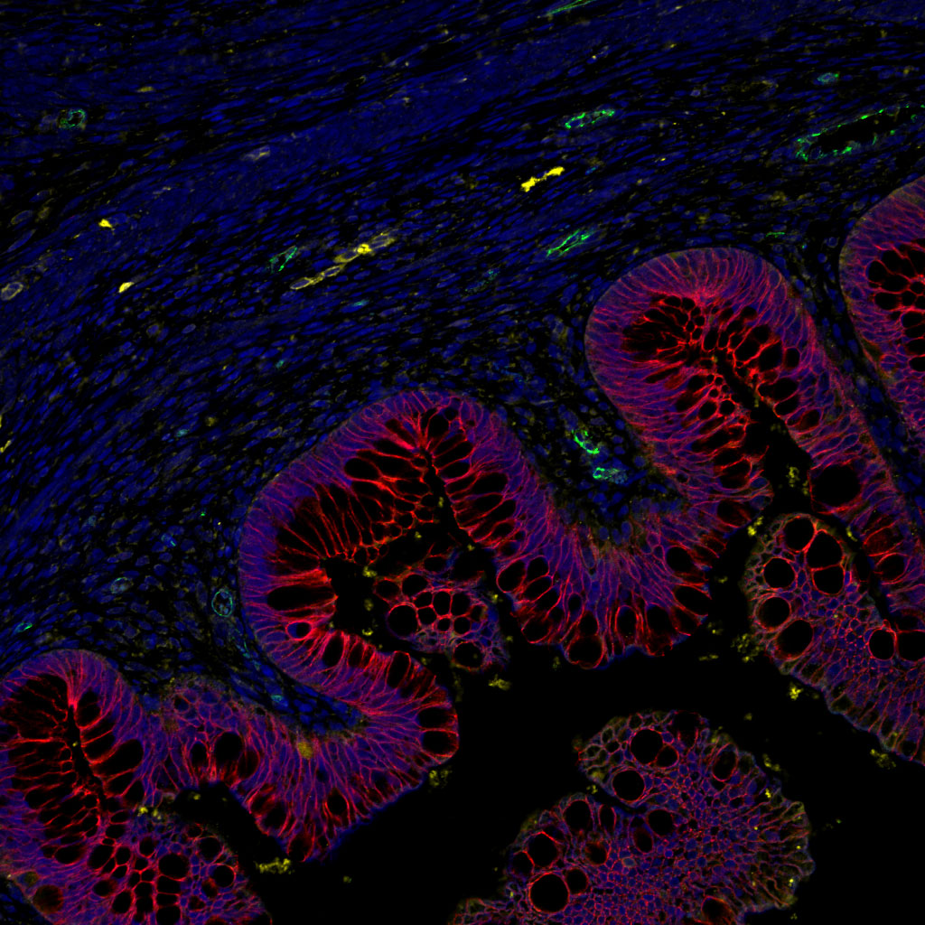
There are many interactions between the central nervous system and the immune system. The cells of the immune system circulate in the brain and can play a role – direct or indirect – in neurological diseases. Thus a direct role has been demonstrated in multiple sclerosis, and a direct role mediated by inflammation has also been found. Neurodegeneration leads to neuroinflammation, which helps to amplify the initial neurodegeneration, generating a vicious circle that aggravates the disease. In Alzheimer’s disease, amyloid peptide β (Aβ) aggregates in the extracellular senile plaques, around which reactive astrocytes and activated microglial cells accumulate. These cells help to dissolve these plaques, and secrete cytokines that regulate the intensity of the brain’s immune response.

Moreover, recent work has shown that mice deficient in IL-2 have diminished faculties for learning and memory, reminiscent of Alzheimer’s disease (AD). Furthermore, IL-2 is currently being evaluated for the treatment for several autoimmune diseases in terms of its ability to stimulate regulatory T lymphocytes (Tregs), the role of which is to control inflammation.

The authors first demonstrated a strong decrease in IL-2 levels in biopsies from patients who had died of Alzheimer’s disease. This led to them to evaluate the therapeutic potential of this molecule in a mouse model of Alzheimer’s disease. The mice were treated at a stage where they already had brain involvement. This long-term treatment caused an expansion and activation of regulatory T lymphocytes in the brain, and led to a reduction in amyloid plaques.

The researchers showed that this reduction in amyloid “load” was accompanied by substantial tissue remodelling marked by an improvement in synaptic structure and function. This improvement is synonymous with recovery of memory deficits.

While untreated mice failed memory tests, treated mice had results comparable with normal mice. These beneficial effects on amyloid plaques and synaptic plasticity are accompanied, in the vicinity of the plaques, by the activation of astrocytes, a type of cell identified as having a protective role in Alzheimer’s disease.

“This work demonstrates the interest of immunotherapies for the treatment of Alzheimer’s disease, and especially the interest of interleukin-2,” the authors believe. “This treatment attacks the consequences of the disease, the synaptic loss and cognitive symptoms that accompany it. Its therapeutic potential now needs to be assessed in humans,” they conclude.

Testosterone for nerve fibre repair

To protect against attack, the body uses natural repair processes. What is involved in the spontaneous regeneration of the myelin sheath surrounding nerve fibres? This is the question addressed by researchers in Unit 1195, “Neuroprotective, Neuroregenerative and Remyelinating Small Molecules” (Inserm/Paris-Sud University). They have discovered, in mice, the unexpected regenerative role of testosterone in this process. This could be a factor in the progression of demyelinating diseases, such as multiple sclerosis, which can present differently in women and men, and heralds new therapeutic opportunities.  

These results are published in PNAS.

The myelin sheath allows the rapid transmission of information between the brain or spinal cord and the rest of the body. Myelin may be targeted by conditions known as demyelinating diseases, such as multiple sclerosis or injuries that lead to its destruction. These diseases disrupt neurotransmission, leading to various symptoms including paralysis. Repair mechanisms then come into play, and bring about myelin regeneration and resolution of symptoms. This regenerative process is erratic, for reasons that are still largely unknown. This question was analysed by the research team “Myelination and Myelin Repair” in Unit 1195, “Neuroprotective, Neuroregenerative and Remyelinating Small Molecules.”

In this study, the researchers present evidence of the unexpected central role of the well-known male sex hormone, testosterone, and of its receptor, the androgen receptor, in spontaneous myelin repair.

“Testosterone promotes the production of myelin by the cells that synthesise it in the central nervous system, in order to repair the sheath, which is essential to the transmission of nerve impulses,” explains Elisabeth Traiffort, Inserm Research Director.

In the absence of testes and hence of the hormone, testosterone, that these organs produce, the spontaneous myelin repair process is disrupted in mice. Indeed, the maturation of cells specialised in myelin synthesis, known as oligodendrocytes, is defective. The researchers also showed that control of this maturation, provided by astrocytes, another type of cell with an important role in repair, is what is compromised.

But why testosterone? Returning to the origins of this hormone, it turns out, surprisingly, that the androgen receptor that enables testosterone to act appeared at the same time as myelin, very late in the evolution of gnathostomes (vertebrates with jaws). According to the researchers, this would explain their very strong association in the myelination process.

“It is perhaps also one of the reasons why progression of demyelinating diseases such as multiple sclerosis often differs between men and women. Our results pave the way for new therapeutic opportunities, and might also benefit research on psychiatric diseases or cognitive ageing,” concludes Elisabeth Traiffort, Inserm Research Director.

La sclérose en plaques

Myelin forms a sheath surrounding neurons © Inserm/Carole Fumat

Storage of umbilical cord blood

Sang cordon ombilical bébé

For the first time in France, a couple has just been given authorisation to entrust a private company with the storage of umbilical cord cells from their unborn child, in view of a potential therapeutic need.

In its Order dated 21 November 2016, the Grasse court authorised the couple to “take and store haematopoietic cells from the umbilical cord and from the placental blood, along with cells from the cord and placenta (…) in light of duly justified therapeutic needs.”

Winner of the 2010 Inserm Honorary Prize, Eliane Gluckman, a haematologist at Saint Louis Hospital, Paris, an Inserm researcher, and a specialist in blood stem cell transplantation, was the first to have performed, in 1987, an umbilical cord blood transplant in a 6 year old child.

See press release from 30 November 2010

Cord blood is currently used to treat patients with blood diseases (leukaemias and lymphomas), and, for certain indications, replaces bone marrow.

Neurons paralyze us during REM sleep

During REM sleep, the brain inhibits the motor system, which makes the sleeper completely immobile. CNRS researchers working in the Centre de Recherche en Neurosciences de Lyon (CNRS/Université Claude Bernard Lyon 1/INSERM/Université Jean Monnet) have identified a population of neurons that is responsible for this transient muscle paralysis. The animal model created will shed light on the origin of some paradoxical sleep disorders, and more particularly the condition that prevents this paralysis. It will also be most useful in the study of Parkinson’s disease, since these pathologies are related. This work was published on December 12, 2016 on the website of the journal Brain.

In spite of being in a deep sleep, the patients talk, move, kick and eventually fall out of bed. They are suffering from a parasomnia called REM Sleep Behavior Disorder[1] (RBD). This disorder usually appears around the age of 50. Muscles are at rest during the REM sleep phase, but in these patients, there is no paralysis, although the reason for this is not known. The sleepers move abnormally, probably reflecting their dream activity.

A team from the Centre de Recherche en Neurosciences de Lyon (CNRS/INSERM/Université Claude Bernard Lyon 1/Université Jean Monnet) has taken one more step towards elucidating this pathology. The researchers identified neurons in the sublaterodorsal nucleus of the brain, ideally located to control motor system paralysis during REM sleep. In rats, they specifically targeted this neuron population, by adding genetically modified viral vectors to it.[2] Once these are in the neural cells, they block the expression of a gene that allows synaptic glutamate secretion. Now incapable of releasing this excitatory neurotransmitter, the neurons can no longer communicate with their neighbors. They are disconnected from the cerebral network necessary for paralysis during REM sleep.

For 50 years, the scientific community has considered that these glutamate neurons generated REM itself. This team’s experience invalidates this hypothesis: despite the absence of activity in this neuron circuit, the rats still experience this stage of sleep. They are fast asleep and disconnected from the outside world, with eyes closed. But these rats are no longer paralyzed. Their behavior is very reminiscent of the clinical profile of patients suffering from RBD. The glutamate neurons targeted in this study play an essential part in REM paralysis during sleep and are reportedly the first neurons affected in this neurological disease.

This research work goes beyond creating a new preclinical model that mimics this parasomnia. It may be of paramount importance in studying some neurodegenerative diseases. Recent clinical research has shown that patients diagnosed with RBD almost always develop the motor symptoms of Parkinson’s disease, on average a decade later. The team is now attempting to develop an animal model that evolves from parasomnia into Parkinson’s disease, in order to understand how neuron degeneration occurs.

2016_12_13_neuroneglutamate

(Left) the glutamate neurons of the sublaterodorsal nucleus emit a spontaneous red fluorescence indicating that the viral vectors used have been successfully added. © Sara Valencia Garcia / Patrice Fort, CNRS

(right) in a normal rat specimen (A and B) the neurons of the sublaterodorsal nucleus (SLD, colored in brown) are glutamate neurons (also colored in black). In rats treated with viral vectors (C and D), neurons are still present (in brown) but are not longer capable of releasing glutamate (absence of black color). © Sara Valencia Garcia / Patrice Fort, CNRS

[1] REM sleep is also called paradoxical sleep.

[2] Provided by researchers at Tsukuba University in Japan

Minimum effort for maximum effect

Ten days after astronaut Thomas Pesquet take-off into space on the Proxima mission, many questions remain about human adaptation to gravity. The research team at Inserm Unit 1093, “Cognition, Motor Activity and Sensorimotor Plasticity” (Inserm/Université de Bourgogne), focuses on the manner in which movements that depend on this parameter are performed. For 30 years, it was thought that the brain, when giving motor commands, continuously compensated for the effects of gravity. In this study, the researchers reveal that it uses gravity to minimise the efforts our muscles have to make. These results have been published in eLife.

Many human and animal activities need our limbs to move in a precise manner. (Such is the case for professional dancers, who have perfect control of their bodies.) For a movement to be performed correctly, the brain has to generate muscular contractions while taking account of environmental factors likely to affect that movement. One of the most important of these is gravity. The brain develops an internal representation of gravity that it can thus use to anticipate its effects on our bodies. But how does that work? Until now, researchers thought that the brain compensated for the effects of gravity at every moment in order to direct a movement. But the Inserm researchers have proposed a new hypothesis. The brain may use the internal representation of gravity to take advantage of it and save energy.

In order to solve this mystery, the research team asked volunteers to perform arm movements under normal gravity and microgravity conditions. Under normal gravity, 15 volunteers performed right arm movements in 17 different directions.

Each movement was made up of two phases, which determined the total duration of the movement: an acceleration phase (e.g.: raising the arm if the initial trajectory is “upwards”) and a deceleration phase (e.g.: stopping the arm on its pathway). This is known as temporal organisation of movement.

If the brain continuously compensated for the effects of gravity, as was thought, the acceleration and deceleration phases would be of constant duration. Under normal gravity, the acceleration or deceleration phase directed by the brain proved to be more or less, depending on the direction of the movement. This observation corroborates the hypothesis that humans have adapted to exploit gravity by modulating the duration of these phases in order to avoid making unnecessary demands on the muscles.

To confirm and validate these results, the researchers simulated weightlessness in an aircraft. The volunteers repeated the arm movements in the 17 directions. While at the beginning of the experiment, the manner of performing the movements was the same as on Earth, the phases of acceleration and deceleration gradually changed in duration.
 

Volunteer performing movements under microgravity conditions in an aircraft travelling in a parabolic arc

© Jérémie Gaveau / Inserm

“This observation clearly shows that our brain captures information from the environment, reprogrammes itself and adapts to new gravity conditions,” explains Jérémie Gaveau, first author of this work. Once the brain has understood, it incorporates the new parameters and sends commands that allow movements to be performed with as little effort as possible.

 “Comparing these results to those of computer simulations shows sophisticated behaviour on the part of the individual. Indeed, our movements are organised to take advantage of the effects of gravity, in order to minimise the efforts that our muscles need to make,” he concludes.

 This is a genuine paradigm shift. This advance might ultimately be used to correctly programme the “brains” of humanoid robots or assist movement in disabled people.

A molecule to regenerate insulin-producing cells in type 1 diabetic patients

illustration-en

Inserm researchers led by Patrick Collombat at Unit 1091,“Institute of Biology Valrose” (Inserm/CNRS/Nice Sophia Antipolis University), show that GABA, a neurotransmitter that is sometimes used as a dietary supplement, can induce the regeneration of insulin-producing cells. This discovery, confirmed in mice and partially validated in humans, gives new hope to patients with type 1 diabetes.

This research is published in the journal Cell.

Type 1 diabetes is a disease characterised by the selective loss of cells that produce insulin, a hormone that lowers blood sugar levels upon sugar intake. These cells are called pancreatic beta cells. Discovering how to regenerate these cells is a major research challenge, as current treatments are not always sufficient in preventing (serious) complications.

Scientists have shown in previous studies that it is possible to regenerate pancreatic beta cells by genetically transforming cells that resemble them: the glucagon-secreting alpha cells. This approach involved the forced activation of the Pax4 gene in all alpha cells. The results also proved that these alpha cells were continuously regenerated and again converted into beta cells, leading to a massive increase in the number of beta cells. “This advance is significant, but it is not possible to carry out this approach in humans,” explains Patrick Collombat, Inserm Research Director. To eventually translate their findings to human, the scientists therefore initiated a search for compounds mimicking the effects of the Pax4 gene.

In this new study, the research team demonstrates that this effect can be induced with no genetic modification using GABA, a neurotransmitter that is naturally present in the body and also available as a dietary supplement.

In mice, GABA induces the continuous, yet controlled, regeneration of pancreatic alpha cells and their transformation into insulin-producing beta cells. The regenerated cells are functional and can cure chemically-induced diabetes multiple times.

With regards to humans, researchers observe that in pancreatic islets (which contain both alpha- and beta-cells) treated with GABA, the number of glucagon-producing alpha-cells is decreased by 37% while a 24% increase in insulin-producing cell count is noted, suggestive of a conversion of alpha-cells into beta-cells.

Finally, by transplanting the equivalent of 500 human islets to mice, the same results are obtained after supplementing the animals for 1 month with GABA (daily). These results are truly encouraging for a putative application in humans. Accordingly, a pilot clinical trial will soon be initiated to determine whether GABA may effectively help patients with type 1 diabetes.

These studies received financial support from ERC and the Juvenile Diabetes Research Foundation.

MAAD Digital: A new website on addiction developed for and by young people

The cause of uncombable hair syndrome identified

Surprised disheveled preschooler girl with long hair

In 1973, the rare syndrome of uncombable hair or ‘pili trianguli et canaliculi‘ was described by a Toulouse dermatologist. More than 40 years later, Michel Simon, Inserm research director his colleagues at the ‘Epidermal Differentiation and Rheumatoid Autoimmunity’ Unit [UDEAR] (Inserm/CNRS/Toulouse III – Paul Sabatier University) have identified its genetic cause. These results are published in The American Journal of Human Genetics.

Uncombable hair syndrome is a rare disease of the hair, the prevalence of which is unknown. It generally begins during childhood between 3 months and 12 years. Dry and unruly, the hair of affected children becomes gradually silver-blond or straw coloured. Hairs stand up on the scalp and grow in all directions. It is impossible to comb it or to flatten it with a comb. In detail, scanning electron microscopy reveals a longitudinal groove running their entire length, with a triangular or kidney-shaped cross-section. However, this syndrome is not disabling and undergoes spontaneous at the end of childhood.


uncombable-hair

he researchers, working with a team from the Human Genetics Institute at Bonn University and dermatologists or geneticists from 7 different countries, have discovered that the disease is due to recessive mutations of a trio of genes that contribute to forming the hair: the gene coding for one of its structural components, trichohyalin (TCHH); or two genes coding for enzymes that take it in turns as target: peptidyl-arginine deiminase 3 (Pad3) and transglutaminase 3 (TGase3).

Furthermore, the researchers have also shown, in mice, that inactivating the Pad3 gene alters the shape of the fur and whiskers of animals, as had already been reported in TGase3-deficient mice.

In conclusion, the absence of TCHH or failure of the biochemical cascade that results in stiffening the hair stem are responsible for the hair formation abnormalities characteristic of uncombable hair syndrome or ‘pili trianguli et canaliculi‘.

These results, as well as describing the molecular cause of the disease and enabling better diagnosis, provide knew knowledge about the hair and the mechanisms of its formation£ concludes Michel Simon, Inserm research director.

For further information

Although extremely rare, the syndrome has long been known. It was brought to public awareness by the famous literary figure ‘Struwwelpeter’ created by children’s author Heinrich Hoffmann in 1845. The book was subsequently translated into English by Mark Twain as ‘Slovenly Peter’. Although he never said so, one might even think that it inspired director Tim Burton to make his film Edward Scissorhands.

Functional human intestine grown from stem cells

photo-mahe

Cross-section of human intestinal organoid with enteric nervous system after transplantation in mice.*

(Copyright CCHMC – Wells/Helmrath labs)

American researchers at the Cincinnati Children’s Hospital Medical Center and French researchers from Inserm (joint research Unit 913 “Neuropathies of Enteric Nervous System and Digestive Diseases”, Nantes) have succeeded in generating a functional human intestine using pluripotent human stem cells. This significant breakthrough was achieved by cultivating human intestinal tissue using nerve cells. Details of their work are published online on 21 November 2016 in Nature Medicine.


The gut is an essential organ of the human body. It is the main interface for exchanges with the external environment and has a surface area equivalent to two tennis courts. Given its importance, the gut has its own nervous system and has been commonly known as “the second brain”. The enteric nervous system controls many functions, which include mixing and propelling food along the digestive tract, hormone secretion and epithelial permeability. Disruptions of this system are the cause of many diseases. Poor gut function can actually affect intestinal muscle contraction. This plays a role in triggering abdominal pain, diarrhoea, constipation and, in serious cases, leads to functional obstruction (intestinal occlusion) that requires surgery.

Researchers have developed an innovative approach to engineering tissue involving the use of stem cells to create a functional human intestine that can be used to study intestinal diseases.

First in vitro studies — finding the right balance

To do this, the scientists successively added a cocktail of molecules designed to promote the differentiation of human pluripotent stem cells into intestinal tissue. The process was virtually the same used in 2010 and 2014 by the same laboratory that succeeded in developing the first generation of human intestinal tissue. However, the intestinal tissue did not possess an enteric nervous system through this approach alone, which is essential for absorbing nutrients and eliminating waste through the digestive tract.

At the same time and in order to develop a functional nervous system, researchers created embryonic nerve cells known as neural crest cells. These cells were manipulated to form cells that are enteric neural cell precursors. “The difficulty with this step was to identify how and when to incorporate the neural crest cells into the developing gut that was previously created in vitro”, explains Maxime Mahé, first co-author of this work and recently recruited as a junior research associate by Inserm.

The co-culture of intestinal tissue and enteric neural cell precursors resulted in human tissue that resembled a developing fetal intestine. This led to the first generation of complex and functional “mini-intestines” (known as intestinal organoids) created entirely from human pluripotent stem cells.


Conclusive in vivo trials

The next challenge was to transplant these functional organoids into a living organism, in this instance, lab mice that lacked an immune system. This step allowed scientists to observe tissue development and function in vivo. Data from this study show that the tissues function and are structured in a way that is remarkably similar to the human intestine. They develop and ensure intestinal functions, such as the treatment of nutrients. Finally, these tissues exhibit motor functions similar to peristalsis, i.e. the series of muscle contractions that move food through the digestive tract.

Researchers then used this technology to study a rare bowel disease — Hirschsprung Disease — a disorder where the rectum and colon do not develop a nervous system, which leads to constipation and intestinal occlusion. A lethal form of Hirschsprung disease is caused by a mutation of the PHOX2B gene. In vitro tests in mice have made it possible for researchers to show that the mutated PHOX2B gene causes significant deleterious changes in the innervated intestinal tissues.

“Our work marks an essential step in understanding human digestive diseases where few models exist. This new technology offers a screening platform for new intestinal treatments. This technology is still in its infancy. However, an approach towards regenerative and personalised medicine is possible, particularly with regards to patient-specific intestinal transplantation”, explains Maxime Mahé.

This discovery yields two significant research possibilities. Firstly, intestinal disorders can be modelled and studied using three-dimensional and functional human tissue involving patient-specific cells. Secondly, new treatments can be tested using this functional human intestine before proposing clinical trials in humans.


In this image we can see the intestinal epithelium in red (E-cadherin) with lumen opening. The tissue underlying the epithelium is rich in blood vessels (green, CD31) and has neurons (yellow, TUBB3) that are also derived from human pluripotent stem cells. The observed tissue is incredibly similar to normal human tissue.

Mitochondria are essential to memory

visuel-marsicano-newsroomMitochondria develop our memory by providing brain cells with energy (c) Charlie Padgett

Numerous studies have shown that using cannabis can lead to short- and long-term memory loss. These effects on memory may be related to the presence of specific receptors on several types of brain cells (glial cells as well as neurons). Inserm researchers led by Giovanni Marsicano (Neurocentre Magendie, U1215) have shown that these effects on memory are related to the presence of these same receptors on the mitochondria, the energy centre of the cell. This is the first time that the direct involvement of mitochondria in higher brain functions, such as learning and memory, has been shown. This work is published in the journal Nature.


Mitochondria are the energy centre of the animal cell. They are present within cells to produce the energy (in the form of ATP) needed for all biochemical processes. To do this, they use oxygen to transform nutrients into ATP. These functions are obviously necessary for the survival of all the cells in the body, but in the brain the impact of mitochondria goes beyond simple cell survival. Although the brain represents only 2% of the weight of the body, it actually consumes up to 25% of its energy. As a result, the energy balance of the brain is highly important for its functions, and is therefore tightly regulated. We know very well that chronic impairment of mitochondrial functions (e.g. in mitochondrial diseases) produces serious neurological and neuropsychiatric symptoms.

However, the direct functional involvement of mitochondria in higher brain functions, such as learning and memory, was not known before now.

In other terms, do we use the mitochondria in our brain when we are learning or remembering something?

This study, which is based on the discovery that the cannabinoid receptor CB1 is also present on the brain mitochondria (where it is known as mtCB1), reveals that this is indeed the case. With the help of innovative tools, the Inserm researchers showed that the active component of cannabis, THC (delta-9-tetrahydrocannabinol), causes amnesia in mice by activating mtCB1 receptors in the hippocampus.

“The impairment in memory induced by cannabis in the mouse requires activation of these hippocampal mtCB1 receptors,” explains Giovanni Marsicano. Conversely, “Genetically deleting them prevents this effect induced by the active drug in cannabis. We therefore think that mitochondria develop our memory by providing the brain cells with energy.”

This study is important not only because it reveals a new mechanism underlying the effects of cannabis on the memory, but also because it shows that mitochondrial activity is an integral part of the functions of the brain.

Jean-Laurent Casanova, specialist in infectious disease genetics, awarded the Inserm Grand Prix 2016

1-Jean-Laurent-Casanova-003-WEB

On Thursday 8 December, at Collège de France, the annual Inserm Awards ceremony will honour eight researchers and engineers whose work has contributed to the Institute’s scientific excellence. The Inserm Grand Prix 2016 will be awarded to Jean-Laurent Casanova, for his work on the human genetics of infectious diseases, in the presence of Marisol Touraine, Minister of Social Affairs and Health, Thierry Mandon, Secretary of State for Higher Education and Research, and Professor Yves Lévy, Chairman and Chief Executive Officer of Inserm.


Jean-Laurent Casanova, Inserm Grand Prix 

In the 19th century, Louis Pasteur demonstrated that microbes are responsible for infectious disease, thus revolutionising medicine. However, when exposed to the same microbe, only a minority of infected children and adults develop a serious illness. When confronted by this enigma, Jean-Laurent Casanova, paediatrician and immunologist, suspected a genetic cause. He then devoted his work to research on infectious diseases of children: tuberculosis, invasive pneumococcal infection, herpetic encephalitis, etc.

Since the 1990s, and thanks to his work, a paradigm shift has occurred. Jean-Laurent Casanova has provided proof that serious infectious diseases are also genetic diseases that make children susceptible to one microbe or another. A pioneer in this area, he has contributed to the identification of several hundred genetic mutations involved in immune deficiencies that predispose to infection.

Today his discoveries make it possible to offer the children affected, and their families, a molecular and genetic diagnosis, together with preventive measures or even therapeutic strategies.

Director of the Laboratory of Human Genetics in Infectious Diseases, which he founded with Laurent Abel, jointly hosted by the Imagine Institute at the Necker Hospital for Sick Children, Paris and The Rockefeller University, New York, Jean-Laurent Casanova, at 53 years of age, is being awarded the Inserm Grand Prix for his entire body of work.

Molecular and genetic diagnosis, made possible by the research carried out by Jean-Laurent Casanova, illustrates the need for an effective implementation of the measures contained in the Aviesan Alliance’s French Plan for Genomic Medicine 2025.


Linda Fried, International Prize

The International Prize honours Linda Fried, Dean and DeLamar Professor at the Columbia University Mailman School of Public Health (New York). Dr Fried is a public health leader in the fields of epidemiology and geriatrics, renowned worldwide for having identified the age-related frailty syndrome. She has also designed an intergenerational tutoring programme, having demonstrated its efficacy in preventing cognitive impairment and dementia.


Catherine Barthélémy, Honorary Prize

The Honorary Prize is awarded to Catherine Barthélémy, Emeritus Professor of the Tours Faculty of Medicine, and former Director of the “Autism” team at Inserm Unit 930, “Imaging and Brain,” for her work on the brain mechanisms involved in autism. With a long history of working toward the early recognition of children with autism, she is currently pursuing her research to enable further advances with these children.


The Opecst-Inserm Prize is awarded to Martine Bungener, researcher, economist and sociologist, President of Inserm’s patients’ association liaison group, GRAM – Groupe de Réflexion avec les Associations de Malades – for her work in support of the dialogue between patients, those close to patients, and researchers. Her work has also contributed to developing the key role of friends and families in the patient pathway.

The Research Prizes are awarded to Rosa Cossart, Research Director at Unit 901, “Mediterranean Institute of Neurobiology” (Inserm/Aix-Marseille University), in recognition of her studies on the synchronisation mechanisms of neural networks, and to Xavier Jouven, researcher, cardiologist and statistician at Unit 970, “Paris – Cardiovascular Research Center” (Inserm/Paris-Descartes University), whose achievements include the discovery of a new form of renal transplant rejection, using mathematical models.

The Innovation Prizes distinguish Benjamin Mathieu, Research Engineer at Unit 1024, “Biology Institute, École Normale Supérieure” (Inserm/CNRS/École Normale Supérieure, Paris), whose work has led to the development of a microscope that allows optical measurement of the activity of the neurons in a living animal, and Céline Tomkiewicz-Raulet, Design Engineer at Unit 1124, “Toxicology, Pharmacology and Cell Signaling” (Inserm/Paris Descartes University), in recognition for her work on the influence of pollutants on breast cancer.

fermer