Menu

The 2018 Inserm Prizes: Spotlight on Health Research

Illustration: ©Flore Avram

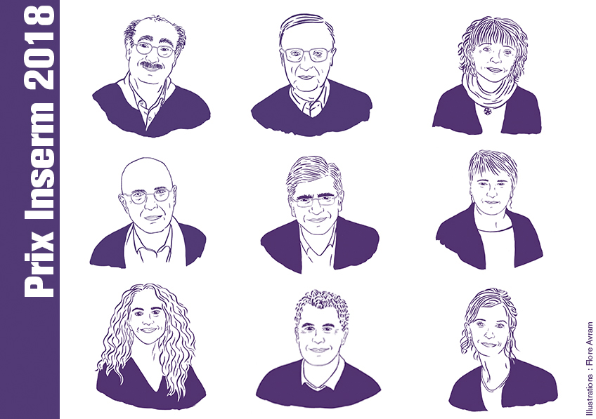
This year’s Inserm Prize ceremony will take place on Tuesday December 11, 2018, at the Collège de France in celebration of nine researchers and engineers whose achievements have helped raise the level of scientific excellence at the Institute, in the service of science for health. “By honoring its talents, Inserm wishes to showcase the diversity and richness of the biomedical research professions, and the creativity and passion of the women and men who drive them on a daily basis”, states Claire Giry, Inserm Director General and acting President. The 2018 Inserm Grand Prize will go to Alain Tedgui for his research on identifying the role of the immune system in atherosclerosis. Two clinical trials directly resulting from his research are currently ongoing.

Alain Tedgui, Inserm Grand Prize

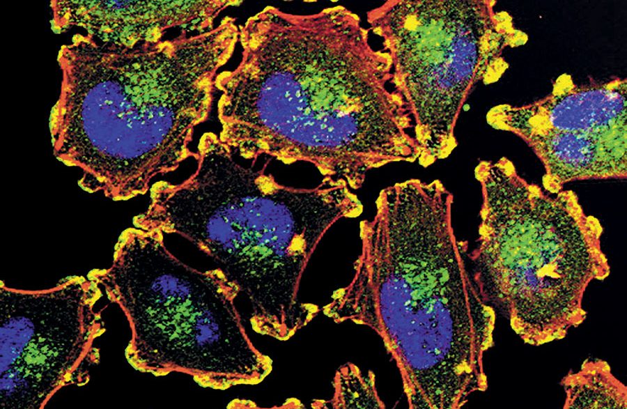
Following a grounding in applied mathematics, Inserm Research Director Alain Tedgui developed a passion for atherosclerosis, one of the primary causes of cardiovascular disease, very early on in his career. Since then, he has gone on to reveal the inflammatory and immune aspects of this cardiovascular condition caused by an accumulation of cholesterol in the arterial walls, and which can lead to myocardial infarction. But is it not actually an auto-immune disease? Tedgui, who leads the Paris-Cardiovascular Research Center at the Hôpital Européen Georges-Pompidou in Paris, has devoted his entire career to this subject, shattering a few medical and scientific dogmas along the way!

Pierre Golstein, Special Prize

Exceptionally this year, Inserm has decided to award a Special Prize to highlight a major discovery made at Inserm upstream of the research being awarded the 2018 Nobel Prize in Physiology or Medicine. The recipient is Pierre Golstein at the Center of Immunology Marseille-Luminy, for having been the first to identify protein CTLA-4, which is now the target of some cancer immunotherapy strategies.

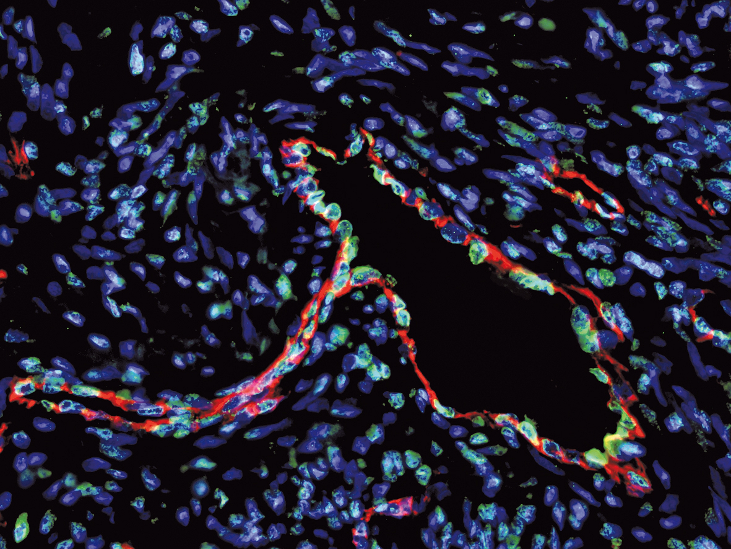
Elisabetta Dejana, International Prize

Inserm also wishes to reward the careers of foreign researchers – careers which tie in with the Institute’s long-standing tradition of international cooperation. This year’s International Prize goes to Elisabetta Dejana, a vascular wall specialist who has had a particularly rich European career so far, spanning Italy, France and Sweden. Experiences which she uses to champion career equality between men and women.

Antoine Triller, Prize of Honor

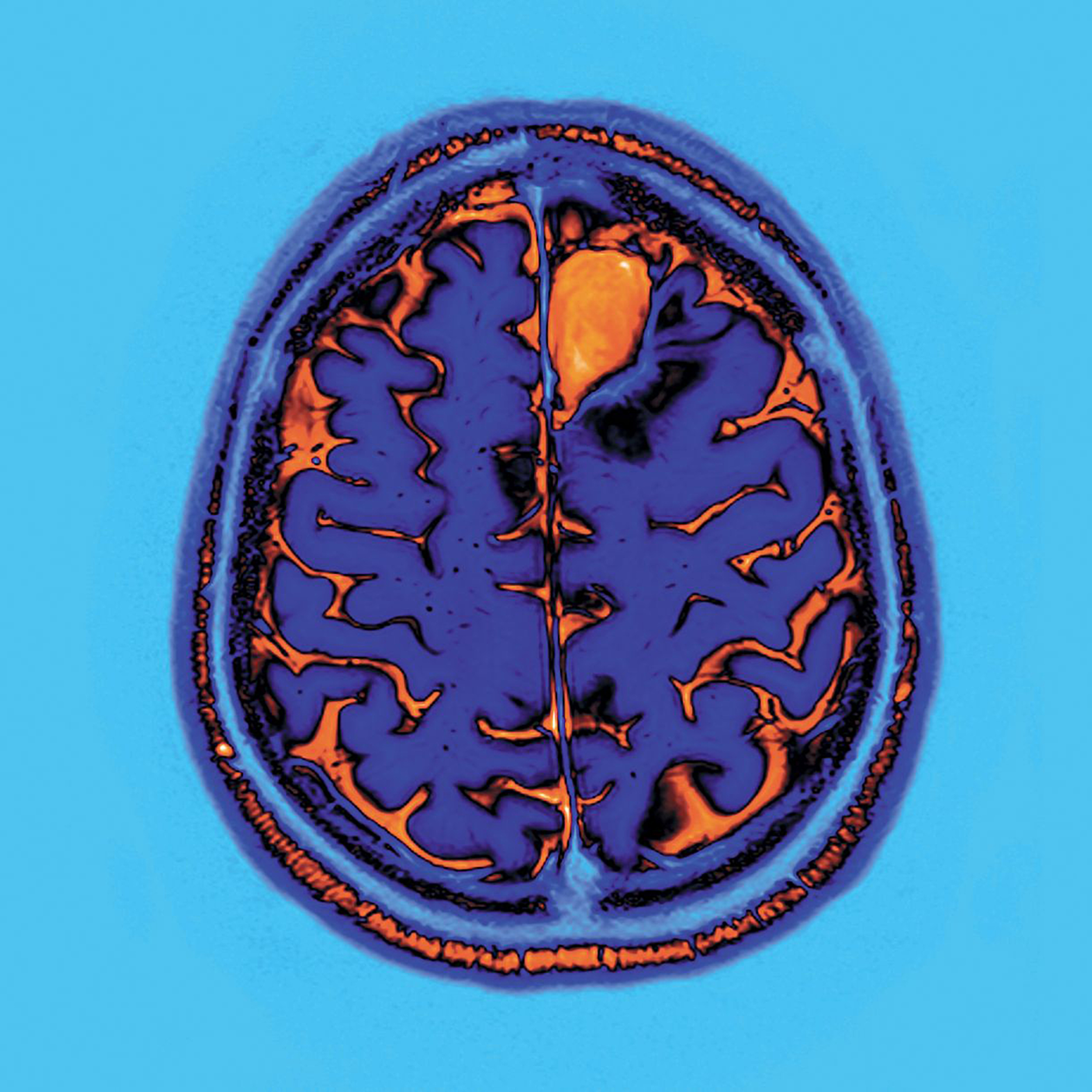
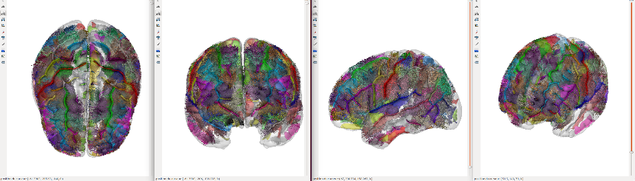
Since 2004, the Prize of Honor acknowledges the career of an eminent scientific figure. This year it goes to Antoine Triller who has devoted his to understanding the cellular and molecular bases of neural communication. He has played a major role in furthering neuroscientific research, most notably through the creation of the Biology Institute of the École Normale Supérieure in which he has always sought to foster the independence and dynamism of the teams.

Robert Barouki, Opecst-Inserm Prize

Since 2013, Inserm has wished to reward efforts to promote research and its ability to be truly in dialogue with societal expectations and citizens’ health questions. This is why the Institute, in partnership with the French Parliamentary Office for the Evaluation of Scientific and Technological Options (Opecst) has created the Opecst-Inserm Prize. Its recipient this year is Robert Barouki, who has always wished, in addition to his research on the exposome, to inform public health decisions.

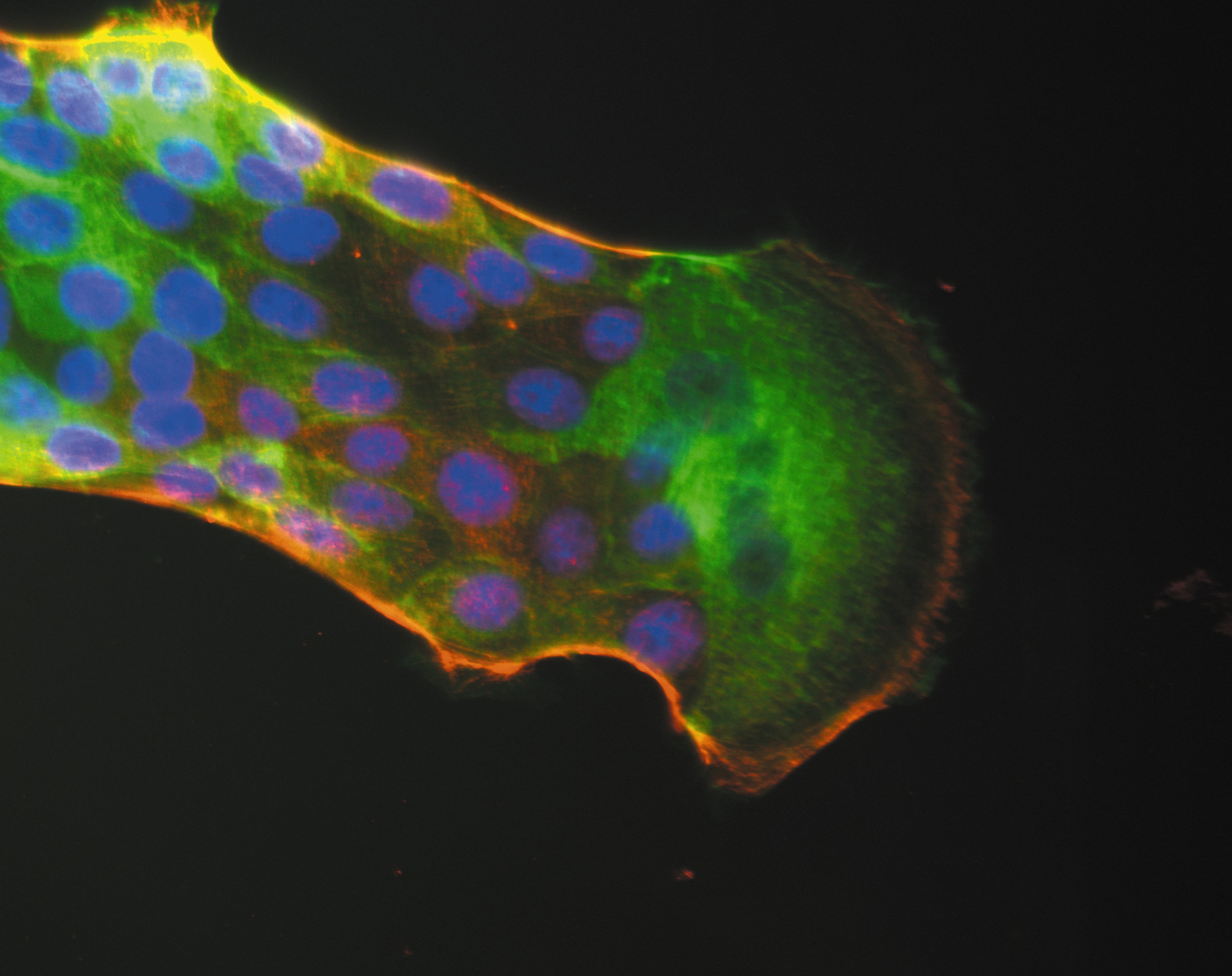
Nathalie Vergnolle and Ana-Maria Lennon-Duménil, Research Prize

The Research Prize honors researchers, lecturer-researchers and clinician-researchers whose work has particularly marked the fields of fundamental research, clinical and therapeutic research, and public health research. Nathalie Vergnolle has worked on the gradual creation of the Digestive Health Research Institute which she currently leads in Toulouse – a specialist structure whose multidisciplinary approach is unique in France. Ana-Maria Lennon-Duménil is a pioneer in the understanding of immune system activation with the prospect of making advances in cancer immunotherapy.

Ahmed Abbas and Nelly Pirot, Innovation Prize

Engineers, technicians and administrative staff also play valuable roles in research: the Innovation Prize is dedicated to them. Ahmed Abbas, responsible for the management of radioactive medicines at the Cyceron platform in Caen, has helped further the development of these products in collaboration with researchers. Nelly Pirot, in charge of the Experimental Histology Network platform of the Institute of Cancer Research of Montpellier, steers the entire technical process. She contributes to reducing the use of animals for research purposes, a primary concern at Inserm.

Gilles Bloch appointed Chairman and Chief Executive Officer of Inserm

Gilles Bloch has been appointed Chairman and Chief Executive Officer of the French National Institute of Health and Medical Research (Inserm) by the Council of Ministers. Gilles Bloch becomes Inserm’s ninth CEO since the Institute was created in 1964. He will assume his post on January 2, 2019.

A graduate of the École Polytechnique, doctor, and researcher specializing in medical imaging, Gilles Bloch spent a large part of his career at the French Atomic Energy Commission (CEA) before taking on major roles within research policy, including heading up the French National Research Agency upon its creation, and then being appointed Head of Research and Innovation at the Ministry of Higher Education and Research.

Gilles Bloch joined the Frédéric Joliot Hospital Unit in Orsay in 1990 to conduct research into nuclear magnetic resonance spectroscopy. He worked as a postdoctoral researcher at Yale University in the United States, and then from 1997 onward held various management positions at the CEA before being appointed Deputy Director of its Life Sciences Division in 2001.

In 2002 he joined the Ministry of Research as an advisor for life sciences, health, and bioethics, then as assistant director. He was entrusted with leading the newly created French National Research Agency (ANR) before becoming Head of Research and Innovation at the Ministry of Higher Education and Research in 2006. In 2009, he was appointed Director of Life Sciences at the CEA.

Since 2015, he has chaired the Université Paris-Saclay group of universities and institutions.

Cancer under pressure: visualizing the activity of the immune system on tumor development

Cancérogenèse : Surexpression de TRF2, marqué en vert, dans les vaisseaux tumoraux, marquage rouge, dans un cancer ovarien. ©Inserm/Wagner, Nicole, 2014

As tumors develop, they evolve genetically. How does the immune system act when faced with tumor cells? How does it exert pressure on the genetic diversity of cancer cells? Scientists from the Institut Pasteur and Inserm used in vivo video techniques and cell-specific staining to visualize the action of immune cells in response to the proliferation of cancer cells. The findings have been published in the journal Science Immunology on November 23, 2018.

Over time, the uncontrolled proliferation of tumor cells results in the accumulation of new mutations and changes to their genome. This gradual process creates significant genetic diversity among the cancer cells in any given patient. And although the cells in the immune system, especially T cells, are potentially able to eliminate these abnormal cells, tumor diversity can have a harmful effect, complicating the action of the immune system and rendering some therapies ineffective. Understanding this frantic race between tumor development and the immune response is key to the success of future immunotherapy techniques.

Scientists in the Dynamics of Immune Responses Unit (Institut Pasteur/Inserm), directed by Philippe Bousso, in collaboration with Ludovic Deriano, Head of the Genome Integrity, Immunity and Cancer Unit (Institut Pasteur), investigated how spontaneous immune responses to tumors influence this tumor heterogeneity. They demonstrated that the immune system can employ mechanisms to significantly reduce tumor diversity, favoring the emergence of more genetically homogeneous tumor cells.

In their study, the scientists marked each cancer cell subclone with a separate color in a mouse model. By monitoring these different colors they were therefore able to characterize the evolution of tumor heterogeneity in time and space. They were also able to observe the contacts between T cells and cancer cells and determine how some tumor cells are destroyed. Their research highlights the drastic impact the immune system can have on tumors by reducing their heterogeneity.


Visualizing the action of stained immune cells.
In this video, the tumor cells are shown in gray. The tumor-specific T-cells, in purple, come into contact with the cancer cells and destroy them. The killed cells are shown in blue. In green, the control cells circulate but do not kill the tumor cells. © Institut Pasteur / Philippe Bousso


Visualizing different clusters of cancer cell clones.
This video illustrates how tumor subclones, each marked by a different color (blue, orange and green), develop in the bone marrow. The vessels are shown in white. © Institut Pasteur / Philippe Bousso

The same impact on the heterogeneity of tumor cells has also been observed in response to immunotherapies that release the brakes on the immune system, an approach which was awarded the Nobel Prize in Physiology or Medicine this year.

This research shows that taking into account the interaction between immunotherapies and tumor heterogeneity could contribute to the development of optimum therapeutic combinations and sequences.

In addition to the organizations mentioned above, this research was funded by the Fondation de France, the French National Cancer Institute (INCa) and the European Research Council (ERC).

The origins of asymmetry: A protein that makes you do the twist

©Inserm/Cochet-Escartin, Olivier, 2014

Asymmetry plays a major role in biology at every scale: think of DNA spirals, the fact that the human heart is positioned on the left, our preference to use our left or right hand … A team from the Institute of biology Valrose (CNRS/Inserm/Université Côte d’Azur), in collaboration with colleagues from the University of Pennsylvania, has shown how a single protein induces a spiral motion in another molecule. Through a domino effect, this causes cells, organs, and indeed the entire body to twist, triggering lateralized behaviour. This research is published in the journal Science on November 23, 2018.

Our world is fundamentally asymmetrical: think of the double helix of DNA, the asymmetrical division of stem cells, or the fact that the human heart is positioned on the left … But how do these asymmetries emerge, and are they linked to one another?

At the Institute of biology Valrose, the team led by the CNRS researcher Stéphane Noselli, which also includes Inserm and Université Cote d’Azur researchers, has been studying right–left asymmetry for several years in order to solve these enigmas. The biologists had identified the first gene controlling asymmetry in the common fruit fly (Drosophila), one of the biologists’ favoured model organisms. More recently, the team showed that this gene plays the same role in vertebrates: the protein that it produces, Myosin 1D,[1] controls the coiling or rotation of organs in the same direction.

In this new study, the researchers induced the production of Myosin 1D in the normally symmetrical organs of Drosophila, such as the respiratory trachea. Quite spectacularly, this was enough to induce asymmetry at all levels: deformed cells, trachea coiling around themselves, the twisting of the whole body, and helicoidal locomotive behavior among fly larvae. Remarkably, these new asymmetries always develop in the same direction.

In order to identify the origin of these cascading effects, biochemists from the University of Pennsylvania contributed to the project too: on a glass coverslip, they brought Myosin 1D into contact with a component of cytoskeleton (the cell’s “backbone”), namely actin. They were able to observe that the interaction between the two proteins caused the actin to spiral.

Besides its role in right–left asymmetry among Drosophila and vertebrates, Myosin 1D appears to be a unique protein that is capable of inducing asymmetry in and of itself at all scales, first at the molecular level, then, through a domino effect, at the cell, tissue, and behavioral level.

These results suggest a possible mechanism for the sudden appearance of new morphological characteristics over the course of evolution, such as, for example, the twisting of snails’ bodies. Myosin 1D thus appears to have all the necessary characteristics for the emergence of this innovation, since its expression alone suffices to induce twisting at all scales.


[1] Myosins are a class of proteins that interact with actin (a constituent of cell skeletons or cytoskeletons). The most well-known of them, muscular myosin, makes muscles contract.

liver cancer and hepatitis C virus to maintain interest in screening programs cirrhotic patients cured of the virus infection with interferon or antiviral direct

Professor Pierre Nahon, the Hepatology Service of Hospital Jean Verdier, AP-HP and Professor Etienne Audureau the Public Health Service of the Henri Mondor Hospital, AP-HP with the University Paris 13, the Inserm and Sorbonne Paris Cité, reported the results of a prospective observational study within the cohort ANRS CO12 CirVir. The latter describes the evolution of patients with compensated cirrhosis due to infection by the virus of hepatitis C, enrolled in liver cancer screening programs or hepatocellular carcinoma (HCC) treated with interferon before 2014 direct antivirals ( DSA) since. The findings, published in the journal Gastroenterology in November 2018, show that if the liver cancer risk is greatly reduced after viral eradication in these patients, it still persists and justified to keep patients with viral cirrhosis C cured in screening programs. This study also confirms the benefits of virologic cure the risk of hepatic carcinogenesis regardless of the type of antiviral treatment.

The direct antiviral (DSA) directed against hepatitis C virus (HCV) have revolutionized the treatment of infected patients since they are made available in 2014 in France. Ensuring virological cure in nearly 100% of patients today at the cost of few side effects, their long-term benefit is still unknown. liver cancer risk or hepatocellular carcinoma (HCC) is the most feared complication in these patients when HCV-induced cirrhosis. Old data obtained before the era of AVD when based on very restrictive interferon treatments allowed to cure that less than 50% of patients have suggested a decreased risk of HCC in case of viral eradication.

The cohort ANRS CO12 CirVir is the oldest prospective cohort of patients infected with the hepatitis B and C supported by the ANRS [1] . Between March 2006 and December 2012, 1353 patients with cirrhosis, uncomplicated and histologically proven originating infection with the hepatitis C virus were enrolled in 39 French centers. All these patients were enrolled in HCC screening programs as recommended, with conducting a liver ultrasound every 6 months. Patients were followed until December 2016, which allows the analysis with a median decline of more than 5 years.

The researchers were able to analyze the incidence of HCC in the two eras successive therapy (interferon and DSA). The analyzes confirm that if the liver cancer risk is greatly reduced after viral eradication regardless of the type of treatment (it is divided by a factor of about 4), it nevertheless persists and justifies maintaining in screening programs patients with viral cirrhosis C cured. Data from the CirVir cohort reported in this study were also allowed to provide answers about the risk of liver cancer in AVD. The latter is indeed not increased compared to interferon era when a number of confounding factors are taken into account in the analyzes.

The cohort ANRS CO12 CirVir, by its prospective longitudinal nature, has in recent years to study the factors associated with the occurrence of major clinical events in patients with cirrhosis. Nearly twenty works based on data collected prospectively in all Hepatology Services of the territory of 10 years have been published in international journals, covering fields as wide as hepatocellular carcinoma, bacterial infections, extrahepatic cancers and cardiovascular diseases.

(1) The cohort ANRS CO12 CirVir multicenter prospective cohort of patients with viral cirrhosis B and / or C uncomplicated, was initiated in 2006 and included 1822 patients. Patient follow-up was completed in 2017 and a large part of them have now been included in the cohort ANRS CO22 HEPATHER .

A new pathway for modulating anti-tumoral immune response

Cellules cancéreuses d’un mélanome.  Inserm/Valencia, Julio C., 2018

Researchers from Inserm, CNRS, Paris-Sud University, Gustave Roussy, and Institut Curie have identified a new agent in regulating PD-L1 gene expression: the eIF4F complex, which plays a role in controlling protein synthesis.

This complex could become a predictive marker for response to immunotherapy. Furthermore, the researchers demonstrate, for the first time, that inhibition of the eIF4F complex gives rise to an anti-tumoral effect related to decreased PD-L1 expression, which therefore elicits immune system intervention.

The researchers hope that eIF4F inhibitors will be able to be used as anti-cancer agents in the future, alone or, more than likely, in combination with other treatments.

Just a few years ago, the immune system, our defense against disease, seemed unequipped to fight cancer. Advances in immunotherapy are making it possible to correct these shortcomings: the immune system can now learn to recognize and destroy cancer cells. Lymphocytes are thus rediscovering their initial ability to fight rather than protect tumors.

The PD-1 (programmed cell death) molecule, expressed on the surface of T cells, binds to another molecule present on the surface of certain tumor or immune cells, PD-L1. This interaction in a way renders the tumor cell invisible to the immune system, by deactivating (or disarming) T cells.

In recent years, immunotherapy targeting the PD-L1/PD-1 interaction has revolutionized the treatment of melanoma and other types of cancer.

However, many patients do not respond to treatment. These agents are highly effective for several months or years, but only in 10 to 20% of patients, for all types of cancer combined.

The development of biomarkers is therefore a key issue in identifying patients liable to respond to treatment,” explains Professor Caroline Robert, Head of the Dermatology Department at Gustave Roussy.

A high PD-L1 level in tumors is a major indicator since this is often associated with a good response to anti-PD1 agents. However, the mechanisms for regulating PD-L1 expression have not been fully elucidated,” points out Stephan Vagner, Inserm Research Director and Head of the RNA Biology Team at Institut Curie.

In this latest publication, the researchers demonstrate, for the first time, that a complex known as eIF4F, involved in initiating the translation of messenger RNA into proteins, regulates PD-L1 expression and that, by targeting eIF4F in tumor cells, anti-tumoral immunity can be stimulated, thus mimicking the effect of immunotherapy.

In this study, the researchers mainly used melanoma as a model, but also conducted tests with lung cancer, breast cancer, and colon cancer cells.

They will now go on to evaluate the findings of the study on the formation of the eIF4F complex, as a predictive marker for response to immunotherapy.

They are, moreover, developing other melanoma treatment models, based on the use of eIF4F complex inhibitors, in combination with other treatments, to increase therapeutic efficacy and fight resistance.

This study was supported by Inserm, CNRS, Gustave Roussy, and Institut Curie. It was also funded by the Ligue Nationale Contre le Cancer (accredited team), French National Cancer Institute, the ‘Ensemble contre le mélanome’ collective, and the ‘Vaincre le Mélanome’ association, SIRIC Socrate, Fondation Bettencourt Schueller, and Fondation ARC for Cancer Research.

Autism Spectrum Disorder: a new brain imaging study appears to challenge the prevailing theoretical model.

©Inserm/Arribarat, Germain

As part of the InFoR-Autism* scientific program, supported by Institut Roche, an MRI neuroimaging study investigated the links between local anatomical connectivity and social cognition in individuals with autism spectrum disorder (ASD). Born out of the partnership between Fondation FondaMental, Inserm researchers, NeuroSpin (CEA Paris-Saclay), and Henri Mondor University Hospital-AP-HP, the results appear to challenge the prevailing theoretical model which suggests that ASD is caused by diminished “long-distance” connections between neurons located throughout the brain, associated with increased “short-distance” neural connectivity between adjacent regions of the brain. Published in Brain, these studies could, if confirmed on a larger scale, pave the way for exploring new therapeutic approaches.

Autism spectrum disorder (ASD) is a neurodevelopmental disorder characterized by communication disorders, diminished social interaction skills, together with sensory and behavioral abnormalities. Genetic and brain imaging studies suggest that abnormal brain development, notably concerning the formation of neural networks and synapse function, could contribute to the onset of ASD.

In recent years, neuroimaging studies have shown that individuals with ASD have functional abnormalities affecting certain areas of the brain known to be responsible for processing emotions, language, and social skills. Studies on brain connectivity in individuals with ASD have notably evidenced diminished “long-distance” connections, in contrast to increased “short-distance” connectivity. These results formed the basis for the development of a theoretical model aiming to shed light on ASD, according to which the deficit in terms of social attention and information processing observed (difficulty in understanding a situation as a whole, attention to certain details) is explained by a saturation of information processed by the brain, linked to the increased neural connectivity between adjacent regions of the brain.

Nonetheless, Professor Josselin Houenou, Professor of Psychiatry at UPEC, Inserm researcher, practitioner at Henri Mondor University Hospital-AP-HP, and last author of the study published in Brain, explains: “This model is based on the study of heterogeneous pediatric populations, comprising autistic children of varying ages and with highly diverse symptoms, and on relatively unspecific neuroimaging methods which do not allow reliable measurement of “short-distance” connectivity.”

In order to test the current model, the authors of this study used an innovation designed by Miguel Gevara, Jean-François Mangin, and Cyril Poupon at NeuroSpin, namely an atlas specifically focusing on the tractography analysis of 63 “short-distance” connections using images obtained by diffusion MRI (dMRI). dMRI evidences white matter bundles in the brain in vivo, by measuring the diffusion of water molecules, notably along the axons. Tractography can then be used to reconstruct, step by step, the nerve fiber bundle paths, represented in tractogram form.

The authors were thus able to study the links between “short-distance” connectivity and social cognition in a homogeneous adult population of individuals with ASD, originating from the InFoR-Autism cohort* (27 patients with ASD, without intellectual deficit, and 31 control subjects), with one of the most extensive databases per patient and per control.

Discovery of diminished “short-distance” brain connectivity associated with diminished social interaction skills and empathy

The strength of the InFoR-Autism cohort* lies in the wealth of data collected for each subject included. We were thus able to establish a link between the neuroimaging results obtained and the social cognition scores, measuring social skills, empathy, social motivation, etc.,” explains Dr. Marc-Antoine d’Albis, Hôpital Henri Mondor, Inserm U955, lead study author.

The results show that subjects suffering from ASD display diminished connectivity in 13 “short-distance” bundles, compared to control subjects. Furthermore, this abnormal “short-distance” bundle connectivity is correlated with a two-dimensional deficit in social cognition (namely social interactions and empathy) in subjects with ASD.

Key: 3D image of “short-distance” bundles, © Miguel Guevara

These preliminary results are well and truly at variance with the current theoretical model according to which diminished social attention and information processing in individuals with ASD is explained by increased neural connectivity between adjacent regions of the brain. These now need to be confirmed by studies in children with ASD, as explained by Professor Josselin Houenou.

According to Professor Josselin Houenou, “These preliminary results nonetheless suggest that these “short-distance” connectivity abnormalities could contribute to certain social cognitive deficits observed in autistic subjects. Similar studies now need to be conducted in children in order to confirm the results obtained in adults. Pediatric cohorts allow children of varying ages – and therefore brain maturation – to be studied, which entails taking into account a much larger population of subjects.

If these initial conclusions were confirmed, this would allow the development of new therapeutic approaches for social cognition deficits to be envisaged. For instance, transcranial magnetic stimulation could be explored since brain connectivity between adjacent regions is located on the surface of the brain.”

*InFoR-Autism

Since late 2012, Fondation FondaMental, Inserm, Inserm Transfert, and Institut Roche have been partners in the InFoR Autism scientific program which aims to monitor clinical, biological and brain imaging variables with a view to studying the stability and progression of ASD. 117 patients and 57 healthy volunteers aged 6 to 56 years in total have been included in the study. This cohort has one of the largest databases (clinical, biological, eye tracking, and imaging) per patient and per control.

“Nested sequences”: an indispensable mechanism for forming memories

©Photo by Annie Spratt on Unsplash

A research team from CNRS, Université PSL, the Collège de France and Inserm has just lifted part of the veil surrounding brain activity during sleep.  Though we know that some neurons are reactivated then to consolidate our memories, we did not know how these cells could “remember” which order to turn on in. The researchers have discovered that reactivating neurons during sleep relies on activation that occurs during the day: “nested” theta sequences. These results were published on November 9, 2018 in Science.

Repetition is the best method for memorization, for neurons themselves. This is the principle behind what neurobiologists call sequence reactivations: during sleep, neurons in the hippocampus related to a task activate very quickly in turn in a precise order, which consolidates the memory of this task. Sequence reactivations are fundamental for long-term memorization and for exchanges between the hippocampus and the rest of the brain. These are only present at rest so they appear after initial neuron activity, which implies that they “memorize” the order they should turn on in. But by which mechanism?

A team of researchers from the Centre interdisciplinaire de recherche en biologie (CNRS/Inserm/Collège de France)1 has answered this question by studying activity sequences in rats’ place cells. These are hippocampal neurons that turn on by following the animal’s position in the environment when it moves. First slowly, while it moves, then very quickly during reactivations of sequences during sleep. But neurobiologists know another type of sequence, called theta sequences, which quickly repeat the activation of the same place cells when the animal moves, in parallel with slow sequences. These theta sequences are therefore called “nested”.

Which of these sequences, slow or nested, is necessary for the appearance of sequence reactivations, and therefore causes the consolidation of memories during sleep? Using an ingenious system, the researchers discovered what deactivates nested sequences, without affecting slow sequences: the animals are transported on an electric train, in a car with a treadmill (see image). When the treadmill is stopped, the nested sequences disappear; they return when the treadmill starts again.

The researchers then observed that after several circuits in the train with the treadmill stopped, place cells in the rats’ hippocampi did not reactivate during sleep in the same order as when awake. On the contrary, after one train circuit with the treadmill on, the sequence reactivations are indeed present. So it is these nested theta sequences during movement that are indispensable for the consolidation of memory during sleep.

The researchers are continuing their work, looking now at the integration of non-spatial information such as objects or textures in nested sequences, and their reactivation during sleep.


  1. Associated member of the Université PSL, since 2009 the Collège de France has been conducting a voluntaristic policy for welcoming independent teams that benefit from pooled technical or scientific services and an exceptional multidisciplinary environment. Twenty-two teams are currently housed in the Centre interdisciplinaire de recherche en biologie and in the Instituts de chimie et de physique du Collège de France. Supported by the CNRS in particular, this is available to both French and foreign researchers. It contributes to making Paris a major player as an attractive place for research.

Bile acid receptor controls hepatitis B virus replication

 Hépatite en voie de guérison : foyers de macrophages contenant de la bile et des pigments. ©Inserm/Hadchouel, Michelle, 1990

Researchers from CIRI – International Center for Research in Infectious Diseases in Lyon (Inserm, CNRS, ENS Lyon and Claude Bernard Lyon 1 University), supported by the ANRS, are demonstrating the link between activation of a bile acid receptor found in liver cells and the reduction in hepatitis B virus replication in mice infected with the virus. This study, coordinated by Professor Patrice André (Claude Bernard Lyon 1 University), was recently published in The FASEB Journal.

Despite the existence of an effective, well-tolerated vaccine, hepatitis B caused by infection with the hepatitis B virus (HBV) is still a global public health problem with 250 million people suffering from chronic infection, at high risk of developing cirrhosis of the liver and cancer. Current direct-acting antivirals significantly reduce the risk of complications, but are unable to control infection via the host’s immune defenses; hence, life-long treatment is required. A team of researchers supported by the ANRS has studied the role of the Farnesoid X receptor (FXR) in controlling HBV infection. FXR is a liver nuclear receptor activated by bile acids. Its main known function is to control the synthesis and excretion of bile acids in bile.

Previous research by the same research team had already suggested interdependence between bile acids and hepatitis B. An initial part of the study, conducted on in vitro cell models, published in October, was able to reveal a mechanism for this interdependence. A second part, conducted in vivo, confirms these results. The researchers were able to provide in vitro evidence that FXR is a host factor favoring HBV replication. This viral multiplication is suppressed following inhibition of FXR expression or during activation thereof via agonist molecules mimicking the action of bile acids.

The researchers then evaluated the effects of treatment with agonists with a view to activating FXR in an HBV-infected mouse model. They then observed that HBV replication was less active in the treated mice compared to the control mice. This result demonstrates, for the first time in vivo, that activation of FXR by an agonist molecule may lead to suppression of HBV replication.

Lastly, scientists also observed that young HBV-infected mice did not respond to treatment, in contrast to adult mice.

These findings suggest that the immaturity of the bile acid structures could lead to an increased risk of chronic HBV infection observed in neonates and young children.

This study introduces a new concept, namely that the role of bile acid metabolism is more far-reaching than expected and extends to the control of at least one viral infection,” points out Patrice André. “FXR agonists could offer an additional therapeutic approach to relieve the burden of life-long treatments for these HBV-injected patients.”

Inserm Transfert has filed a patent for this research. “In order to bring these advances and a new agonist, which we have identified, into the clinical sphere, we have created a start-up, EnyoPharma, which has acquired the patent license,” explains Patrice André, adding: “The results of a phase I trial will be available shortly, and a phase II trial is in the preparatory stages.”

Large Numbers of Older Adults Are Thought to Wear Unsuitable Glasses

©AdobeStock

Researchers from Inserm, Université de Bordeaux and Sorbonne Université have published a study showing that, out of a population of older adults, nearly 40 % have a poorly-corrected vision problem (such as nearsightedness, farsightedness and astigmatism) which could be improved by wearing more suitable glasses. These findings have been published in JAMA Ophthalmology.

Vision problems are common in older adults and associated with negative outcomes in terms of health, quality of life and dependency in activities of daily living. Nearsightedness, farsightedness and astigmatism continue to represent major causes of visual impairment even though they can be corrected simply by wearing the right glasses.

Using data from the Alienor cohort, in which more than 700 people aged 78 or over were evaluated, researchers from Inserm, Université de Bordeaux and Sorbonne Université studied the vision problems related to refractive error (such as nearsightedness, farsightedness and astigmatism) and which were uncorrected or poorly corrected by the right glasses or contact lenses.

In the study, the researchers showed that nearly 40 % of those aged 78 or over were not wearing the right glasses.

There are a number of reasons for this, explains Catherine Helmer, Inserm researcher in charge of the study, which may be related to fatalism with the perception that visual decline is normal with aging or to financial reasons which persist despite the help available.”

In addition, nearly 50 % of those examined at home (due to unwillingness or inability to come to the clinic) and 35 % of those with age-related eye disease (age-related macular degeneration, glaucoma…) were estimated to wear unsuitable glasses.

Beyond the large number of poorly-corrected vision problems in the entire population studied, the yet larger proportion of poor correction in those examined at home should incite prevention strategies aimed specifically at them. Furthermore, the discovery of a large number of poorly-corrected vision problems in people with eye disease – for which they are most often monitored – underlines the need to look out for these unsuitable corrections.” concludes Helmer.

fermer