Menu

Improving Your Sleep Can Protect Your Cardiovascular Health

Scientists have studied the risk of incident cardiovascular disease in relation to five sleep dimensions. © Adobe Stock

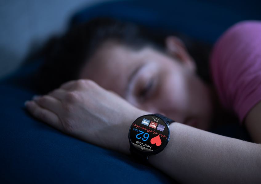
The association between sleep disorders and cardiovascular risk is already well documented. Apnea, sleep deficit, and insomnia are linked to an increased risk of cardiovascular diseases and stroke. A new study published in European Heart Journal has now explored this association in much greater depth. Led by Inserm research director Jean-Philippe Empana and his team at the Paris Cardiovascular Research Center (Inserm/Université Paris Cité) in collaboration with the Centre hospitalier universitaire vaudois (CHUV, Lausanne), the study shows that five aspects of sleep  are almost each equally important to explain the association between sleep and the risk of coronary events and stroke.Improving one of these aspects over time can bring significant benefit. These five aspects are nighttime sleep duration, chronotype (being a morning or evening person), frequency of insomnia, excessive daytime sleepiness, and sleep apnea.

Sleep is essential to health and well-being, and several physiological mechanisms explain this. Poor sleep quality or quantity is therefore associated with a deterioration in health: mood problems, depression, weight gain, infections, diabetes, hypertension, etc. A link has also been established with cardiovascular risk – an issue which Jean-Philippe Empana’s Inserm team at the Paris Cardiovascular Research Center decided to explore further.

To do this, the team studied the risk of cardiovascular accidents (acute coronary syndrome or stroke) in relation to five aspects of sleep: nighttime sleep duration, chronotype (being a morning or evening person), frequency of insomnia, excessive daytime sleepiness, and sleep apnea.

Usually, studies focus on a single sleep dimension, mainly sleep duration or the presence of sleep apnea, but good or healthy sleep encompasses several dimensions,” clarifies Aboubakari Nambiema, first author of this publication and postdoctoral researcher at Inserm.

The researchers incorporated these five patterns of sleep into a single score in order to account for the multifactorial nature of sleep. Each of these patterns was evaluated using a specific questionnaire validated by the scientific community and appropriate for large studies (Pittsburgh sleep quality index-PSQI, Berlin questionnaire or Epworth Sleepiness Scale) and counts for 1 point when it is optimal and 0 points when it is not. The overall score therefore varies from 0 (worst score) to 5 (optimal score corresponding to: 7 to 8 hours of sleep per night, being a morning person, not having insomnia, apnea, or excessive daytime sleepiness).


Monitoring over time

This score was used in two population surveys that study the determinants of cardiovascular health.

One was conducted in Paris and included 10 157 adults aged 50 to 75 years (Paris Prospective Study III, PPS3) and the other in Lausanne, Switzerland, brought together 6 733 participants aged over 35 years (CoLaus|PsyCoLaus study).

The score was calculated at the point of inclusion and then two to five years later. The occurrence of cardiovascular events was then monitored for approximately eight to 10 years.

Although the use of a composite score to study sleep habits had already been tested in the past, this is the first time to our knowledge that a study has investigated its evolution over time and its potential association with cardiovascular diseases,” explains Jean-Philippe Empana.

By combining the data from the two surveys, an initial analysis confirms that the higher the initial (baseline) score, the lower the risk of incident cardiovascular disease. Compared to the participants with a score of 0-1 (10% of the participants), the risk of cardiovascular disease is reduced by 10% for those with a score of 2 (21% of the participants), by 19% for those with a score of 3 (32% or the majority of the participants), by 38% for a score of 4 (27% of the participants) and by 63% for those with the best score of 5 (10% of the participants).

“In other words, almost 60% of incident cardiovascular disease could potentially be avoided if all individuals had an optimal sleep score (score of 5), thus highlighting the potential public health implications of the results,” clarifies Jean-Philippe Empana.

A second analysis that took an unprecedented look at the change in sleep score over time showed that sleep improved in 8% of the participants, deteriorated in 11% and remained stable in the majority of the participants (2/3 of them remained with a score equal to or greater than 3). Importantly, these changes are associated with the occurrence of cardiovascular disease. In particular, this risk decreased by 16% per one point increase in the score over time, regardless of which dimension of the score was improved. “They each seem to be of equal importance,” specifies Nambiema.

The researchers now hope that healthcare professionals and the general population will take advantage of this tool: “the ease and speed of use of the questionnaires as well as the simplicity of calculating the sleep score should encourage their understanding and good adherence,” they predict.

Asleep but Open to the World: We Can Still Respond to External Stimuli

Sleep is generally defined as a period during which the body and mind are at rest, as if disconnected from the world. © Nicolas Decat

When we sleep we are not completely cut off from our environment: we are still able to hear and understand words. These observations, resulting from the close collaboration between researchers from Inserm, CNRS, Sorbonne Université and AP-HP at the Brain Institute and the Department of Sleep Disorders at Pitié-Salpêtrière Hospital in Paris, call into question the very definition of sleep and the clinical criteria that distinguish between its different stages. They are detailed in a new study published in Nature Neuroscience.

Sleep is generally defined as a period during which the body and mind are resting, as if disconnected from the world. However, a new study led by Delphine Oudiette, Inserm researcher, Isabelle Arnulf (Sorbonne Université, AP-HP) and Lionel Naccache (Sorbonne Université, AP-HP) at the Brain Institute, shows that the boundary between wakefulness and sleep is much more porous than it would appear.

The scientists have shown that sleepers with no particular disorders are able to capture verbal information transmitted by a human voice and respond to it by contracting facial muscles. What is more, this astonishing ability manifests itself intermittently during almost all stages of sleep — as if windows to the outside world were temporarily opened.

These new data on sleep behavior suggest that it may eventually be possible to develop standardized protocols for communication with sleepers in order to better understand how mental activity changes during sleep.

On the horizon: a new access route to the cognitive processes that underpin normal and pathological sleep.


A Thousand and One Variations in Consciousness

Even if it seems familiar to us because we do it every night, sleep is a very complex phenomenon. Our research has taught us that wakefulness and sleep are not stable states: both resemble a kaleidoscope of conscious moments… and moments that do not appear to be so,” explains Prof. Lionel Naccache, neurologist at Pitié-Salpêtrière Hospital AP-HP and neuroscience researcher.

It is essential to improve our understanding of the brain mechanisms that underlie these intermediate states between wakefulness and sleep.

When out of sync, they can be associated with disorders such as sleepwalking, sleep paralysis, hallucinations, the feeling of not sleeping at night or, on the contrary, sleeping with the eyes open“, explains Prof. Isabelle Arnulf, head of the Sleep Disorders Department at Pitié-Salpêtrière Hospital AP-HP.

However, in order to distinguish between wakefulness and the different stages of sleep, we have so far used simple and inaccurate physiological indicators, such as specific brain waves made visible through electroencephalography. Such indicators do not capture in detail what is going on inside the heads of sleepers, especially as they are sometimes in contradiction with what the sleepers tell us themselves.

We need more refined physiological measurements that are aligned with the sleeper’s feelings and ability to respond to the outside world; this is to better define their level of vigilance“, adds Delphine Oudiette, Inserm researcher in cognitive neuroscience.


A Game Between Unconsciousness and Lucidity

The research team[1] therefore explored this avenue and recruited 22 people without sleep disorders and 27 narcoleptic patients — i.e. victims of irrepressible sleep episodes.

People with narcolepsy have the particularity of having many lucid dreams, namely in which they are aware of being asleep and can sometimes shape the scenario. In addition, they easily and quickly reach REM sleep (the stage where the lucid dream emerges) during the day, making them good candidates for studying consciousness during sleep under experimental conditions.

One of our previous studies had shown that two-way communication, between the scientist and the dreamer and vice versa, is possible during lucid REM sleep, explains Oudiette. For our latest study, we wanted to know if these findings could be extrapolated to other sleep stages and to individuals who do not have lucid dreams. “

The study participants were asked to take a nap. The researchers had them do a “lexical decision” test in which a human voice uttered a series of words, both real and made up. The participants had to respond by smiling or frowning, in order to place the words in one of the two categories. Throughout the experiment, the participants were monitored using polysomnography—a comprehensive examination to record their brain and heart activity, eye movements, and muscle tone. Finally, upon waking, they had to report whether or not they had a lucid dream during their nap, and whether they remembered interacting with someone.

Most of the participants, whether narcoleptic or not, managed to respond correctly to the verbal stimuli while sleeping. These events were admittedly more frequent during episodes of lucid dreams, characterized by a high level of consciousness; however, we observed them occasionally in both groups, during all sleep phases”, specifies Arnulf.

By combining these physiological and behavioral data with the subjective reports of the participants, the researchers also show that it is possible to predict the opening of these windows of connection with the environment, i.e. the times when the sleepers were able to respond to stimuli. These were heralded by an acceleration of brain activity, and by physiological indicators usually associated with rich cognitive activity.

In people who had a lucid dream during their nap, the ability to dialog with the investigator and talk about this experience on waking was also characterized by a specific electrophysiological signature, adds Naccache. Our data suggest that lucid dreamers have privileged access to their inner world, and that this increased awareness also extends to the outside world.

Further research will be needed to determine whether the increase in these windows is correlated with sleep quality, and whether they could be used to improve certain sleep disorders or promote learning.

More advanced neuroimaging techniques, such as magnetoencephalography and intracranial recording of brain activity, will help us to better understand the brain mechanisms that orchestrate sleep behaviors“, concludes Oudiette.

Finally, these new data could help to revise the definition of sleep, a state that is ultimately very active, perhaps more conscious than we thought, and open to the world and others.

This study was funded by the French National Research Agency and the French Society for Sleep Research and Medicine (SFRMS).

[1] Including PhD students Başak Türker, Esteban Munoz Musat and Emma Chabani, whose participation was essential to the conduct of this study. 

Countering the effects of aging and the occurrence of cancers: new and promising results

By studying immune cells in the lung, researchers from Institut Curie and Inserm have provided new knowledge on the topic.© Fotolia

Cancer and aging are closely linked processes, but the mechanisms underlying this relationship are still not well understood. By studying immune cells in the lung, researchers from Institut Curie and Inserm have provided new knowledge on the topic. They show that targeting ruptures of the nuclear envelope of these cells would represent a new opportunity for therapeutic intervention in age-related diseases, in particular cancer, thus improving the quality of life of the elderly in the long term. Funded by the Fondation ARC, this work has just been published in the journal Nature Aging.

Age is one of the main risk factors for the development of a number of diseases, such as viral or bacterial infections and neuro-degenerative diseases, but also cancers. The economic and societal issues related to the overall aging of the population constitute a major challenge. Furthermore, the notion of “healthy aging” increasingly suggests that targeting aging rather than its consequences is a far better strategy for reducing morbidity among the elderly.

More than two thirds of new cancers diagnosed in France occur in people over the age of 65[1]. The appearance of cancers with age can be explained by the accumulation of genetic alterations during a lifetime, less effective DNA repair mechanisms, and also by an aging immune system with diminished protective functions (immunosenescence). What are the mechanisms that govern this phenomenon? How can we develop strategies to counter immunosenescence?


The nucleus of immune cells sensitive to deformations

It is these questions that the Inserm and Institut Curie researchers attempted to answer. With time, DNA becomes fragile and one of the characteristic markers of cell aging is genome instability. When they patrol through the various tissues within the body, the immune system cells are sensitive to deformations which weaken their nucleus and promote DNA breakage. To maintain the structure of the nucleus and thus the genome’s integrity, the cell relies on a dense network of proteins, which include lamins. Among them lamin A/C is studied in particular since it undergoes alterations over the course of aging. In addition, mutations in the gene coding for this protein are known to cause early aging syndromes.

Repeated ruptures of the nuclear envelope lead to DNA damage. It is vital to understand the processes at work in this regard since they promote not just aging of the body, but also the development of cancers. For example, ruptures of the nucleus make the DNA “visible” by damaging proteins, thus triggering a response from the cell that will promote the development of metastases“, explains Dr. Nicolas Manel, Inserm research director and team leader at Institut Curie.


Observation en microscopie « 2-photons » de la déformation extrême d’un macrophage alvéolaire, lors de son passage entre deux alvéoles.“Two-photon” microscopy of the extreme deformation of an alveolar macrophage, when it passes between two alveoli. During these migrations the nucleus is also deformed, and it is at this point that the DNA can be damaged.


A protein identified in the lung: lamin A/C

At Institut Curie, the Innate Immunity team of Dr. Nicolas Manel, Inserm research director, studied a new experimental model in which the immune system’s cells are deficient in lamin A/C. Researchers looked closely at a population of lung macrophages – alveolar macrophages – which are highly dependent on lamin A/C for their survival. The role of these alveolar macrophages is to constantly monitor the lungs, and they are one of the main entry points for a number of pathogens.

The researchers showed that without lamin A/C, the alveolar macrophages show serious signs of fractures in their nucleus and damage to the DNA, leading to a dramatic drop in their number in the lungs. Furthermore, the surviving alveolar macrophages have many characteristics similar to those of aged alveolar macrophages, and accumulate markers characteristic of aging.

The team also revealed that without lamin A/C in the macrophages, the implantation and growth of lung tumors is a lot faster, encouraged by the malfunction of the aged macrophages.

The loss of lamin A/C would therefore constitute a mechanism for alveolar macrophage aging and a prime study model for understanding how lung cancers develop in the elderly.

Our results open up many new opportunities for studying aging of the immune system, caused by rupture of the nuclear envelope and the decrease in its effectiveness against infections and tumors, in the lungs but also in other organs“, concludes Dr. Nicolas Manel.

These studies are funded in the amount of 2.5 million euros as part of the call for projects “Cancer and Aging” of Fondation ARC for cancer research.

[1] Source INCa : https://www.e-cancer.fr/Professionnels-de-sante/L-organisation-de-l-offre-de-soins/Oncogeriatrie/Epidemiologie

Infection of Certain Neurons With SARS-CoV-2 Could Cause Persistent Symptoms

SRAS-CoV2

Illustration of SARS-CoV-2 infection (immunoreactivity for the S-protein in white) in the olfactory neurons expressing the olfactory marker protein (OMP, in red) in the human nasal epithelium. © Vincent Prévot/Inserm

The brain impacts of infection with SARS-CoV-2, responsible for COVID-19, are increasingly well documented in the scientific literature. Researchers from Inserm, Lille University Hospital and Université de Lille, at the Lille Neuroscience & Cognition unit, in collaboration with their colleagues at Imperial College London, focused more specifically on the impacts of this infection on a population of neurons known for regulating sexual reproduction via the hypothalamus (the neurons that express the GnRH hormone). Their findings suggest that SARS-CoV-2 infection can lead to the death of these neurons and cause certain symptoms that persist over time. The findings of this study have been published in eBioMedicine.

Numerous scientific studies have documented the brain impacts of SARS-CoV-2 infection. One such effect is that a significant proportion of men have low testosterone levels that persist over time. Persistence beyond a period of four weeks is referred to as “long COVID”.

For many years, a research team from Inserm, Lille University Hospital and Université de Lille has been studying the role of certain neurons that express gonadotropin-releasing hormone (GnRH). From the hypothalamus, these neurons control all the processes associated with reproductive function: puberty, acquisition of secondary sexual characteristics, and fertility in adulthood.

These are the same scientists who had, for example, previously revealed that GnRH neuron dysfunction in an animal model of Down syndrome could affect the cognitive function impairment associated with this condition.

In this latest study, the scientists wanted to test the hypothesis that SARS-CoV-2 infection may have harmful consequences on this population of neurons that regulate reproduction.


The Virus Penetrates GnRH Neurons and Alters Their Functions

Following hormone measurements (testosterone and luteinizing hormone) performed three months and then one year after infection in a small group of 47 men[1], the scientists observed that contact with the virus could alter the functions of GnRH neurons, leading to a fall in testosterone levels in certain patients some time after the infectious episode.

The scientists then wanted to verify whether the infection of the GnRH neurons and the subsequently observed hormone abnormalities could be associated with cognitive deficits. To do this, they listed the cognitive symptoms reported by the cohort patients, who underwent extensive testing three months and then one year after the infection.

The outcome was that the proportion of patients reporting memory or attention disorders, regardless of frequency or severity, and also concentration difficulties, tended to be slightly higher in the patients with abnormal hormone measurements, characterized by a decrease in testosterone levels.

“Although these were measurements made on a small sample of only male patients, these findings are very interesting and warrant further exploration in other larger-scale studies,” explains Waljit Dhillo, professor at Imperial College London and co-last author of this study.

To supplement their analyses, the researchers went on to study the cortexes of patients who died as a result of COVID-19. They identified the presence of the virus in the hypothalamus and the death of part of the GnRH neuron population.

“These findings may be worrying on several levels in terms of the role of these neurons in reproduction and their involvement in certain cognitive functions. They point to the necessity to optimize and generalize the medical follow-up of people with persistent symptoms following COVID-19 infection,” concludes Vincent Prévot, Inserm research director and co-last author of this study.

The study also encourages further research into the neurological impacts of long COVID.


[1]These data were collected as part of a larger study evaluating adrenal and thyroid function following Sars-CoV-2 infection: https://pubmed.ncbi.nlm.nih.gov/34008009/

Screens and Child Cognitive Development: Exposure Time is Not the Only Factor to Consider

Enfant déjeunant en regardant l'écran d'un ordinateur portableTo what extent does early or excessive screen exposure affect children’s cognitive development? © AdobeStock

To what extent does early or excessive screen exposure affect children’s cognitive development? This is a question that is currently dividing scientists. A team led by Inserm researcher Jonathan Bernard at the Center for Research in Epidemiology and Statistics (Inserm/INRAE/Université Paris Cité/Université Sorbonne Paris Nord) studied the data of nearly 14,000 children from the French Elfe cohort[1] for each child from the age of 2 to 5.5 years. While this is not the first study to show a negative relationship between exposure time and development, it also highlights that this relationship is not true for all domains of cognition and is much weaker when the family context is properly taken into account. Its findings also confirm[2] a non-negligible negative relationship between exposure to television during family meals and early language development. This research, published in The Journal of Child Psychology and Psychiatry, suggests that while screen time is important, the context of exposure also matters.

In the face of rapid changes in the use of screens and their growing place in everyday life, one question continues to divide scientists: to what extent does too early and/or excessive exposure to screens influence child development? With the majority of studies having focused on language development, less attention has been paid to other cognitive domains. The same applies to the potential influence of the family environment, as well as the child’s daily activities.

The team led by Inserm researcher Jonathan Bernard at the Center for Research in Epidemiology and Statistics (Inserm/INRAE/Université Paris Cité/Université Sorbonne Paris Nord) sought to evaluate the associations between screen use and cognitive development in early childhood, taking into account factors related to social, perinatal, family, and lifestyle conditions. To do this, it looked at data from nearly 14,000 children from the French cohort Elfe, collected for each child from the age of 2 to 5.5 years, between 2013 and 2017.

The parents reported their children’s daily screen time at ages 2, 3.5 and 5.5 years. They were also asked to report whether they had the television on during family meals in their children’s second year. Many factors related to the children’s lifestyle and daily activities also had to be specified. Finally, various cognitive domains were evaluated: language development at age 2, non-verbal reasoning at age 3.5 and overall cognitive development at ages 3.5 and 5.5.

The research team observed that at ages 3.5 and 5.5, screen exposure was associated with lower overall cognitive development scores, particularly in the areas of fine motor, language and autonomy. However, when lifestyle factors likely to influence cognitive development were taken into account in the statistical models, the negative relationship decreased and became of small magnitude.

The results of the study also show that, regardless of exposure time, having the television on during family meals at age 2 (which concerned 41% of children) was associated with lower language development scores at the same age. These children also had poorer overall cognitive development at age 3.5.

“This could be explained by the fact that television, by capturing the attention of family members, interferes with the quality and quantity of interactions between parents and child. Yet such interaction is crucial at this age for the acquisition of language,” explains Shuai Yang, PhD student and first author of the study. He continues: In addition, television adds background noise to family discussions that makes it difficult for the child to decipher sounds and limits verbal understanding and expression. “

These findings suggest that screen time is not the only factor to consider: the context in which screen use takes place could also be an important factor. It is also suggested that not all areas of cognition would be affected in a similar way.

“The first years of life are decisive, not just for cognitive development but also for the establishment of lifestyle habits, adds Bernard. When a child uses a screen excessively, they do so at the expense of other social activities or interactions that are essential for their development. “

The robustness of this study is due to the large number of participants, but also the consideration of factors related to the social profile of the families and the activities of the children.

“While our findings suggest that the harmful effects of screen use in early childhood have a low impact on cognitive development at the individual level and can be compensated for in the years that follow, they do however justify remaining vigilant at population level. When it comes to public health, these little things all add up,” explains Bernard. He added that more long-term studies are needed to evaluate the cumulative impact of these effects from early childhood to adolescence.

The research by his team is continuing through the follow-up of the children in the Elfe cohort in order to answer these questions.


[1]Elfe is France’s first national, longitudinal study dedicated to the follow-up of children from birth to adulthood. More than 18,000 children born in metropolitan France in 2011 were enrolled in it (i.e. one in every 50 children born in 2011). Since the first contact with the families during the maternity stay, the participating parents are regularly interviewed in order to better understand how the environment, family setting and living conditions influence the development, health and socialization of the children. The ELFE study mobilizes around 150 researchers from a variety of scientific disciplines.

[2]See our press release from June 8, 2021: Television During Meals Linked to Poorer Language Development in Young Children

Association Between the Consumption of Food Additive Emulsifiers and the Risk of Cardiovascular Disease

cœurThe scientists used the data reported by 95,442 French adults participating in the NutriNet-Santé study. © Mathilde Touvier/Inserm

Emulsifiers are among the additives most widely used by the food industry, helping to improve the texture of food and extend its shelf life. Researchers from Inserm, INRAE, Université Sorbonne Paris Nord, Université Paris Cité and Cnam, as part of the Nutritional epidemiology research team (EREN-CRESS), studied the impacts on cardiovascular health of the consumption of emulsifiers. They analyzed the health data of 95,442 adults participating in the French NutriNet-Santé cohort study in terms of their overall consumption of this type of food additive. The results suggest an association between the dietary intake of emulsifiers and an increased risk of cardiovascular disease. They have been published in the British Medical Journal.

In Europe and North America, 30 to 60% of dietary energy intake in adults is provided by ultra-processed foods. Recent epidemiological studies have linked high intakes of ultra-processed foods with higher risks of obesity, mortality and chronic diseases (cardiovascular disease, type 2 diabetes, cancer, etc.). Emulsifiers are among the most commonly used additives in industrial foods. They are often added to processed and packaged foods such as certain industrial cakes, cookies and desserts, as well as ice creams, candy bars, breads, margarines and ready meals, in order to improve their appearance, taste and texture and lengthen shelf life. These include celluloses, mono- and diglycerides of fatty acids, modified starches, lecithins, carrageenans, phosphates, gums and pectins.

As with all food additives, the safety of emulsifiers is regularly evaluated on the basis of the scientific evidence available at a given time. However, some recent studies suggest that emulsifiers can disrupt the gut microbiota and increase the risk of inflammation, leading to potentially increased susceptibility to cardiovascular problems.

In order to explore this question in greater depth, French researchers undertook to evaluate the links between exposure to emulsifiers and the risk of cardiovascular disease, including coronary heart- and cerebrovascular disease, i.e. conditions that affect blood circulation and blood vessels in the heart and brain.

Their conclusions are based on the analysis of data from 95,442 French adults (average age 43 years; 79% women) with no history of cardiovascular disease who voluntarily participated in the NutriNet-Santé cohort study (see box below) between 2009 and 2021.

During the first two years of follow-up, the participants completed at least three (and up to 21) days of online dietary records. Each food or drink consumed was then matched against databases to identify the presence and level of food additives, including emulsifiers. Laboratory assays were also performed in order to provide quantitative data.

The participants were asked to declare any major cardiovascular events, such as heart attack or stroke, which were validated by an expert committee following examination of their medical records. Deaths related to cardiovascular disease were also recorded using the French national registry of deaths.

Several well-known risk factors for heart disease, including age, sex, weight (BMI), educational level, family history, smoking and levels of physical activity, as well as overall quality of the diet (e.g. consumption of sugar, salt, energy and alcohol) were taken into account.

After an average follow-up of seven years, the scientists found that the higher intakes of total celluloses (food additive codes E460[1] to E468) were associated with higher risks of cardiovascular disease. In particular, this combination was specifically observed for intakes of E460 (microcrystalline cellulose, powdered cellulose) and E466 (carboxymethylcellulose).

On the other hand, higher intakes of mono- and diglycerides of fatty acids (E471 and E472) were associated with higher risks for all the diseases studied. Among these emulsifiers, lactic ester of mono- and diglycerides of fatty acids (E472b) was associated with a higher risk of cardiovascular and cerebrovascular disease, and citric acid ester of mono- and diglycerides of fatty acids (E472c) was associated with a higher risk of cardiovascular- and coronary heart disease.

High intake of trisodium phosphate (E339) was also associated with an increased risk of coronary heart disease.

No association was detected in this study between the other emulsifiers and the development of cardiovascular diseases.

Given its observational nature, the study cannot therefore establish causality on its own, and the scientists acknowledge it to have certain limitations. For example, the high proportion of women, the higher educational background and overall more health-conscious behaviors among the NutriNet-Santé study participants compared with the general French population, which may limit the generalizability of the results.

That being said, the study sample was large and the authors were able to consider a wide range of potentially confounding factors, while using detailed and unique data on food additives, down to the brand of the products consumed. In addition, the results remained unchanged after multiple sensitivity analyses, thereby strengthening their robustness.

“While these findings need to be replicated in other studies worldwide, they bring new key knowledge to the debate on re-evaluating the regulations around the use of additives in the food industry, in order to better protect consumers,” explains Mathilde Touvier, Research Director at Inserm, and Bernard Srour, Junior Professor at INRAE, principal authors of the study.

NutriNet-Santé is a public health study coordinated by the Nutritional epidemiology research team (EREN-CRESS, Inserm/INRAE/Cnam/Université Sorbonne Paris Nord/Université Paris Cité) which, thanks to the commitment and loyalty of over 170,000 participants (known as Nutrinautes), advances research into the links between nutrition (diet, physical activity, nutritional status) and health. Launched in 2009, the study has already given rise to over 270 international scientific publications. In France, a drive to recruit new participants is still ongoing in order to continue to further research into the relationship between nutrition and health.

By devoting a few minutes per month to answering questionnaires on diet, physical activity and health through the secure online platform etude-nutrinet-sante.fr, participants in France contribute to furthering knowledge of the links between diet and health.

[1] Food additives are identified in the list of ingredients by a European code consisting of the letter “E”, followed by a number making it possible to easily identify the category. For example, E100 for colorants, E200 for preservatives, E400 for emulsifiers and texture agents.

Discovery of an original DNA repair system, bringing new hope for the treatment of breast and ovarian cancer

3D cancer cell © Fotalia

Almost half of breast and ovarian cancers are connected to deficiencies in the biological systems that repair DNA breaks. Researchers from Institut Curie, Inserm and CEA reveal a hitherto unknown DNA repair mechanism involving a protein: Pol, which is able to act during cell division. Published in Nature on 6 September, 2023, their results pave the way for the development of new therapeutic targets for treating cancers, particularly breast and ovarian cancer.

Ultraviolet rays, alcohol, tobacco, genetic predisposition, spontaneous mutations… so many factors constantly damage our genome. Among these lesions, breaks that affect both DNA strands simultaneously are the most harmful. Our body is constantly repairing this damage through several repair systems, including homologous recombination. However, when these mechanisms fail (due to a genetic mutation, for example), they may cause cancer to occur. Also, the proven correlation between these homologous recombination deficiencies and the aggressiveness of cancers or their resistance to current chemotherapies underlines the pressing need for targeted cancer-fighting therapies.


A new major player in DNA repair: Pol

A few years ago, a new player in DNA repair (polymerase theta or PolꝊ) was identified as a therapeutic hope for treating these cancers[1]. The “Alternative DNA Repair Mechanisms in Human Cancers”[2] team headed by Dr. Raphaël Ceccaldi, Inserm researcher at Institut Curie, has just clarified the mechanism for the action of this polymerase and the reason why this enzyme is vital to the development of breast and ovarian cancers.

For the first time scientists have shown that PolꝊ works where other DNA repair pathways do not. While it was believed that DNA repair was impossible during cell division (when DNA is extremely compacted), the team from Institut Curie showed that PolꝊ is active specifically during mitosis when the other contributors to repair were proven ineffective.

“Along with my team at Institut Curie, we looked closely at the mechanisms that the cell puts in place to repair its DNA, enabling cancer cells to survive. It is by understanding such mechanisms that we can build new options to thwart cancer“, explains Dr. Raphaël Ceccaldi, Inserm researcher and team leader at Institut Curie. “Our discoveries on the role and functioning of polymerase theta in maintaining integrity of the genome gives us hope for new therapeutic strategies against cancer, particularly breast and ovarian cancer.“


Genome integrity highly preserved by Pol

Through a collaboration with the team of Dr. Sophie Zinn-Justin, researcher at the CEA (Laboratoire de Biologie Structurale et Radiobiologie), researchers went even further, showing that, in order to repair DNA, PolꝊ had to be activated by an enzyme specifically present during cell division. In addition, the mechanisms that enable this activation of PolꝊ seem to have been extremely well-preserved throughout evolution. This suggests that they play an important role in maintaining the stability of the genome needed for development of eukaryotic organisms.


A therapeutic hope for breast and ovarian cancer

The team of Dr. Raphaël Ceccaldi also discovered that inhibiting PolꝊ during cell division by mitosis prevents the proper repair of DNA and as a result leads to the death of cancer cells. With almost half of breast and ovarian cancers showing DNA repair deficiencies by homologous recombination, this step therefore represents a milestone in the fight against these cancers. Clarifying the molecular mechanisms governing the use and regulation of PolꝊ could ultimately lead to the development of new therapeutic targets for treating these cancers.

CancerPolꝊ (green) marks DNA breaks (gH2AX, red) in the mitotic chromosomes (DAPI, blue) – Scale 5μM


[1] Ceccaldi R, Liu JC, Amunugama R, Hajdu I, Primack B, Petalcorin M, O’Connor KW, Konstantinopoulos PA, Elledge SJ, Boulton SJ, Yusufzai T, D’Andrea AD. Homologous recombination (HR)-deficient tumors are hyper-dependent on POLQ-mediated repair. Nature. 2015 Feb 12;518(7538):258-62.

[2]Cancer, Heterogeneity, Instability and Plasticity unit – CHIP (U830, Institut Curie/Inserm)

First Digital Mapping of the Immune Cells Responsible for Allergies

mastocytesMarking of the different mast cell populations (in green and red), which are major players in allergic responses, on contact with neurons (white) in mouse skin. © Dr. Marie Tauber and Dr. Lilian Basso.

Allergic diseases affect up to one third of the world’s population, and their prevalence is on the increase. In order to develop more targeted and effective therapies, research is mobilizing to better understand the biological and cell mechanisms involved in the onset of allergies. Mast cells – a type of immune cell – is of particular interest to scientists and doctors, but there is little data about them at present. In a new study published in Journal of Experimental Medicine in July 2023, researchers from Inserm, CNRS and Université Toulouse III – Paul-Sabatier, at the Toulouse Institute for Infectious and Inflammatory Diseases (Infinity), broadened our understanding of these cells and created the first digital mapping of mast cells in humans. These findings open up avenues for the adaptation of therapeutic strategies.

Allergic diseases are a major public health problem, to the extent that the World Health Organization (WHO) has classified allergy as being the world’s fourth leading chronic disease. It is currently estimated that 25 to 30% of the population suffers from an allergy, be it food, skin or respiratory allergy, and this proportion could increase to 50% by 2050. A better understanding of the underlying biological mechanisms is a key step if we are to develop more targeted and effective therapies.

This is the goal of Inserm researcher Nicolas Gaudenzio and his team at the Toulouse Institute for Infectious and Inflammatory Diseases. In 2019, the scientists had published a first article in Nature Immunology, showing the crucial role played by immune cells known as “mast cells” in the initiation of eczema. This research has given rise to new therapies that are currently in development.

Mast cells remain poorly understood by scientists. We know that their functions go far beyond problems of allergies and that they can have roles that are either beneficial (such as in fighting bacteria) or not, depending on the pathology. Research has also led to their classification into two large families of mast cells: the CTMCs found mainly in the skin and the MMCs located mainly in the gut mucosa.

However, much remains to be learned about these cells that are complex to study, especially because it is difficult to extract them from tissue.

“If we are to understand how we can act on mast cells and block their harmful action in terms of allergic diseases, we need to improve our knowledge of these cells. This involves determining their location, if there are several types beyond the dichotomy which has traditionally been described, and whether their functions differ according to the tissues in which they are located,” points out Gaudenzio.

In this new study, the research team used more recent technologies to study mast cells more precisely in mice and humans. The scientists used the single-cell sequencing technique: they sequenced the RNA of individual cells from several organs in order to extract their individual “identity card”.

Analyzing human cells with this method reveals a much more complex image than has hitherto been described. Indeed, the cells of over thirty human organs were analyzed thanks to advanced techniques for exploring data banks and bioinformatics. The researchers thus identified not two but seven different subtypes of mast cells, with various characteristics and functions.

From this data, the team was able to create and enable open access to the first “digital mapping” of human mast cells, which allows any scientist to see at a glance which mast cell subtype is associated with which organ and learn more about its function.


mastocytes

This diagram shows, in a simplified way, the distribution of the different mast cell subtypes through different organs of the body.

This approach represents a major paradigm shift since the new mapping makes it possible, just by querying a database, to better understand the natural diversity of mast cells in allergic diseases, and thus open up a process of reflection on the need to adapt therapies to more precisely target the cell subtypes involved.

“This study is the first foundation stone of a vast building that is expected to transform the anti-allergy therapies and move towards a greater personalization of treatments, with more efficacy and fewer side effects. We will continue to supplement this mapping by studying mast cells in different disease settings, in treated and untreated patients alike, so that it is as precise as possible for the scientific and medical community that is working on allergies,” concludes Gaudenzio.  

Omicron BA.1 virus infection in vaccinated patients remodels immune memory

SARS-CoV-2

Cells infected with SARS-CoV-2 © Alberto Domingo Lopez-Munoz, Laboratory of Viral Diseases, NIAID/NIH

Teams from the internal medicine department of the Henri-Mondor AP-HP hospital, the Institut Necker – Enfants Malades, the Mondor Institute for Biomedical Research, the Institut Pasteur, Inserm, and the Paris-Est Créteil University studied immune memory after infection with the Omicron BA.1 variant in patients vaccinated with three doses of the messenger RNA COVID-19 vaccine. The results of this study ( MEMO-VOC) , coordinated by Dr Pascal Chappert and Pr Matthieu Mahévas, in collaboration with Dr Pierre Bruhns and Dr Félix Rey were published on August 4, 2023 in the Immunity review .

The Spike protein of SARS-CoV-2 1 Omicron BA.1 carries 32 mutations compared to the ancestral strain (Hu-1) originally identified. These mutations significantly alter neutralizing antibodies induced by natural SARS-CoV-2 infection and/or vaccination with an encoding mRNA vaccine.

Immune memory is a mechanism that protects individuals against reinfection. This defense strategy of the body, which is the basis of the success of vaccines, includes the production of protective antibodies in the blood (detected by serology) as well as the formation of memory cells (memory B lymphocytes 2 ), capable of quickly reactivate into antibody-producing cells upon re-infection.

The scientific literature has already shown 3,4 that the repertoire of memory B cells generated by two or three doses of mRNA vaccines contains neutralizing clones against all variants of SARS-CoV-2 up to Omicron BA.1.

The research team studied memory B cells after infection with SARS-CoV-2 Omicron BA.1 in 15 individuals previously vaccinated with three doses of the mRNA COVID-19 vaccine encoding the initial Spike protein of the virus. She followed them up to 6 months after infection with Omicron BA.1 to characterize the response of B lymphocytes, from the early immune reaction to the late onset of long-term memory.

This study reveals that infection with the Omicron BA.1 variant mainly mobilizes memory B cells recognizing common proteins between the initial Spike protein and Omicron BA.1 already present in the repertoire formed after vaccination, but few cells directed against specific BA.1 mutations.

Nevertheless, infection with Omicron BA.1 still induces a reorganization in the memory B cell repertoire without altering its diversity, and an improvement in the overall affinity of the memory B repertoire against the common structures of the Spike encoded in the original vaccine (Spike Hu-1) and that of the Omicron BA.1 variant. This reorganization of the memory repertoire is associated with a significant improvement in the ability to neutralize Omicron BA.1.

These results suggest that Omicron BA.1 virus infection in vaccinated patients remodels the memory B cell repertoire and enhances the ability of memory cells to recognize conserved SARS-CoV-2 epitopes and neutralize the virus.

Future vaccine strategies will nevertheless be needed to extend the immune response beyond conserved epitopes to deal with future antigenic variations of SARS-CoV-2.

This study has been labeled a National Research Priority by the ad-hoc national steering committee for therapeutic trials and other research on COVID-19 (CAPNET). The authors thank the ANRS | Emerging Infectious Diseases for its scientific support, the Ministry of Health and Prevention and the Ministry of Higher Education, Research and Innovation for their funding and support.

[1] SARS-CoV-2 protein that allows the coronavirus to enter human cells.
 
[2] Immune cells produced mainly in the lymph nodes and spleen following an infection. They persist for a long time in these regions and retain the memory of the infectious agent. If the body is confronted with them again, these cells are immediately mobilized and quickly reactivate the immune system for effective protection of the individual.
 
[3] Sokal, A., Broketa, M., Barba-Spaeth, G., Meola, A., Ferna´ ndez, I., Fourati, S., Azzaoui, I., de La Selle, A., Vandenberghe, A., Roeser, A., et al. (2022). Analysis of mRNA vaccination-elicited RBD-specific memory B cells re- veals strong but incomplete immune escape of the SARS-CoV-2 Omicron variant. Immunity 55, 1096–1104.e4. https://doi.org/10.1016/j. immuni.2022.04.002.
 
[4] Goel, R.R., Painter, M.M., Lundgreen, K.A., Apostolidis, S.A., Baxter, A.E., Giles, J.R., Mathew, D., Pattekar, A., Reynaldi, A., Khoury, D.S., et al. (2022). Efficient recall of Omicron-reactive B cell memory after a third dose of SARS-CoV-2 mRNA vaccine. Cell 185, 1875–1887.e8. https:// doi.org/10.1016/j.cell.2022.04.009.
 
[5] Part of a molecule capable of stimulating the production of an antibody.

Remission from HIV-1 infection: discovery of broadly neutralizing antibodies that contribute to virus control

Antibody fragments of EPCT112 bNAb (blue) discovered at the Institut Pasteur by Hugo Mouquet’s team, here forming a complex with the HIV-1 envelope protein (Env) (shown in yellow and orange

Some HIV-1 carriers who have received an early antiretroviral treatment during several years are able to control the virus for a long term after treatment interruption. However, the mechanisms enabling this post-treatment control have not been fully elucidated. For the first time, teams of scientists from the Institut Pasteur, Inserm and the Paris Public Hospital Network (AP-HP), supported by ANRS | Emerging Infectious Diseases, have investigated and revealed how neutralizing antibodies, including those described as broadly neutralizing, contribute to virus control. These key findings were published in the journal Cell Host & Microbe on July 10, 2023. A clinical trial involving the use of broadly neutralizing antibodies should begin in France before the end of 2023.

“Post-treatment controllers” is the term used to describe the rare HIV-1 carriers who, having initiated treatment early and maintained it for several years, are able to control the virus for years after that the treatment has been discontinued. These individuals were identified several years ago in part through the VISCONTI[1] study, which assembled the largest cohort of long-term post-treatment controllers in France. Although the mechanisms of viral control enabling the long-term remission from HIV-1 infection without antiretroviral therapy have not been fully elucidated, the identification of these cases provides a unique opportunity to refine our understanding of the factors associated to HIV-1 infection control.

A study conducted by the Institut Pasteur’s Humoral Immunology Unit led by Dr. Hugo Mouquet in collaboration with the team led by Dr. Asier Sáez-Cirión, Head of the Institut Pasteur’s Viral Reservoirs and Immune Control Unit, is now contributing to efforts to describe these mechanisms in more detail.

Asier Saéz-Cirión explains: “Our investigation published in 2020 on the immune response in post-treatment controllers marked a major first step in demonstrating an effective and robust antibody response to HIV-1 in some of these individuals, which may contribute to this control[2].This knowledge has now been further advanced by our new study. By investigating the role of antibodies in a specific “post-treatment controller” case with particularly high serum levels of broadly neutralizing antibodies, we discovered that remission was probably linked to the activity of this type of antibodies.

Hugo Mouquet describes the discovery: “Our study describes for the first time in a post-treatment controller a family of broadly neutralizing antibodies (bNAbs) targeting the HIV-1 envelope protein, of which the antibody EPTC112 is one of the most active member.

The antibody EPTC112 neutralizes about a third of the 200 viral variants of HIV-1[3] tested in vitro and is able to induce the elimination of infected cells in the presence of natural killer (NK) cells, the immune cells eliminating abnormal cells in the body.

This study therefore provides important insights on how neutralizing antibodies modify the course of HIV-1 infection in this individual from the VISCONTI cohort. Although the HIV-1 virus circulating in this subject was found to be resistant to EPTC112 neutralization due to mutations in the region targeted by this antibody, it was effectively neutralized by other antibody populations isolated from the blood of the individual. Hence, the study suggests that neutralizing antibodies from the EPTC112 family impose a selective pressure on the HIV-1 virus. Although the virus escaped the action of these bNAbs, it remained susceptible to the neutralization by other anti-HIV-1 antibodies produced in this individual. This observation suggests the existence of a cooperation between the various populations of neutralizing antibodies.

The fact that we discovered a potential link between the production of neutralizing antibodies, including bNAbs, and the HIV-1 control is exciting to better understand the underlying mechanisms of viral control, particularly by studying additional post-treatment controllers with similar profiles. Indeed, we wish to continue investigating on a short term whether the antibody responses in other ‘post-treatment’ controllers also contribute to long-term remission from the infection,” explains Hugo Mouquet.

This discovery paves the way for new avenues of HIV-1 therapy and fuels hopes of therapeutic approaches for increasing the chances of remission without antiretroviral treatment through the use of broadly neutralizing antibodies. To this end, a clinical trial involving the administration of broadly neutralizing antibodies[4] should begin in France before the end of 2023.

This Phase II trial conducted by the ANRS RHIVIERA consortium through a partnership between the Institut Pasteur, AP-HP, Inserm and the Rockefeller University in New York, will investigate the combination of an antiretroviral therapy in the primary infection phase with two long-acting HIV-1 bNAbs versus placebo to determine whether these antibodies contribute to establishing viral remission after antiretroviral treatment discontinuation. 69 patients in the primary HIV-1 infection[5] phase are planned to be enrolled. They will first receive a short-term antiretroviral treatment, followed by a therapy with the two bNAbs targeting two different regions of the virus envelope protein. It will be possible to stop therapy after a year of close monitoring based on a detailed set of criteria. This trial will enable us to determine whether this therapeutic strategy is able to induce a sufficient immune response to control the infection after the discontinuation of antiretroviral therapy,” concludes Hugo Mouquet.

VIH

Antibody fragments of EPCT112 bNAb (blue) discovered at the Institut Pasteur by Hugo Mouquet’s team, here forming a complex with the HIV-1 envelope protein (Env) (shown in yellow and orange) © Institut Pasteur


[1] HIV: The Antibodies of “Post-treatment Controllers”

[2] Transient viral exposure drives functionally coordinated humoral immune responses in HIV-1 post-treatment controllers study, Nature Communication, 11 avril 2022

[3] There are two types of HIV: HIV-1 and HIV-2 that differ from each other at molecular level. Variants occurring within these two types exhibit different levels of transmissibility, virulence and immunogenicity due to the various mutations associated with them.

[4] https://rhiviera.com/project/anrs-rhiviera-02/

[5] Primary infection: early phase of HIV-1 infection during which the viral load is high. The HIV virus invades the body, attacking the immune system and destroying its CD4 lymphocyte reservoirs.

Air Pollution Accelerates Eye Ageing

© Freepik© Freepik

Numerous studies are now reporting the harmful effects of air pollution on the central nervous system (neurodegenerative diseases in adults, neurodevelopmental disorders in children). Glaucoma, the second leading cause of blindness worldwide, is a neurodegenerative disease of the optic nerve whose principal characteristic is thinning of the retinal nerve fiber layer. In a study of a cohort of 683 elderly Bordeaux residents followed up over 10 years, researchers from Inserm and Université de Bordeaux at the Bordeaux Population Health research center showed accelerated thinning of the retinal nerve fiber layer in the participants more exposed to air pollution, especially those who had a greater exposure to PM2.5 (fine particulate matter of a diameter of 2.5 microns or less). This study therefore suggests a possible increased glaucoma risk for the inhabitants of areas with fine particulate pollution, even at levels below the current European Union regulatory thresholds (25 micrograms/cubic meter). The results have been published in Environmental Research.

Air pollution is a global public health issue. The harmful effects of air pollutants on respiratory and cardiovascular functions have been widely documented in the scientific literature. It is also increasingly evident that chronic exposure to air pollution has adverse effects on the central nervous system, with an increased risk of neurodegenerative diseases in adults and neurodevelopmental disorders in children.

The retinal nerve fiber layer (RNFL) is part of the central nervous system, and its thinning represents the main characteristic of glaucoma[1], an eye disease associated with the progressive destruction of the optic nerve, most often caused by excessive pressure inside the eye. This disease is the second leading cause of blindness in the developed countries.

Researchers from Inserm and Université de Bordeaux studied the effect of exposure to higher concentrations of air pollutants (fine particulate matter[2] and nitrogen dioxide) on the ocular neurodegenerative processes. To do that, they followed up for a 10-year period a population of 683 people aged 75 years or over who were resident in Bordeaux at the time of their inclusion in the Aliénor[3] cohort. This is the first prospective study on this subject.

As part of this study, the volunteers underwent eye exams every two years between 2009 and 2020 in order to measure changes in RNFL thickness.

In addition, their exposure to air pollution over the previous 10 years was determined using their home address, with the help of annual exposure maps for each pollutant. These detailed maps, with a resolution of 100 meters, were produced using air quality control station measurements and meteorological and geographical characteristics (proximity of a road, population density, distance from the sea, altitude, etc.)[4].

According to the results of this study, people having been exposed to higher concentrations of fine particulate matter had more rapid thinning of the retinal nerve layer over time.

These results are represented in the figure above which shows that the participants exposed to a 25 µg/m3 concentration of PM2.5 had a faster reduction in the thickness of this layer compared to those exposed to a 20 µg/m3 concentration.

These findings suggest that exposure to a high concentration of pollutants over time could increase the risk of glaucoma.

As regards PM2.5, the 10-year average exposure estimates were below the annual regulatory threshold of the European Union (established at a maximum of 25 μg/m3) for all participants, but above the limit values recommended by the WHO in 2005 (10 μg/m3) and further reduced in 2021 (5 μg/m3).

“The results of this study confirm the previous observations on the effects of air pollution on neurodegenerative processes – at ocular level in our case. They constitute an additional argument in favor of lowering the European regulatory thresholds[1], as recommended by the WHO, as well as lowering the effective exposure of the French population, which continues to exceed current regulatory thresholds in some places,” explains Laure Gayraud, PhD student in epidemiology and first author of the study.

“More generally, our study documents the effects of air pollutants on neurological aging. Taking the example of ocular aging, it suggests that exposure to high concentrations of pollutants over time could lead to an acceleration of neurological ageing, as was observed in studies on brain aging,” explains Cécile Delcourt, Inserm research director and last author of this research.

The aim is now for the scientists to expand the field of study to national level, using data from other French cohorts in order to learn more about the effects of pollutants on eye aging.


Diagram of the eye and retina

© Adobe stock


[1]To learn more, consult our in-depth article on glaucoma (only available in French).

[2]PM2.5 corresponds to fine particulate matter of a diameter of 2.5 microns or less.

[3]Alienor is an epidemiological study in the general elderly population that explores the relationships between age-related eye diseases and other major determinants of these diseases (genetic, nutritional, environmental, or vascular factors).

[4]These maps were previously produced by the team of Kees de Hoogh and Danielle Vienneau from the Swiss Tropical and Public Health Institute (co-authors of the publication).

[5]The Members of the European Parliament’s Environment Committee recently voted in favor of lowering the thresholds for several pollutants by 2030, including fine particulate matter.

fermer