Menu

A global overview of antibiotic resistance determinants

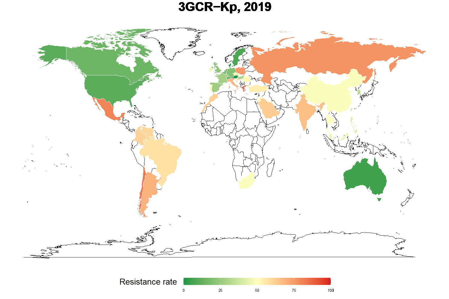
Proportion of third-generation cephalosporin resistance in Klebsiella pneumoniae, for blood infections, 2019 (data from ATLAS, Pfizer) © Institut Pasteur, Eve Rahbé

To understand the main determinants behind worldwide antibiotic resistance dynamics, scientists from the Institut Pasteur, Inserm, Université de Versailles Saint-Quentin-en-Yvelines and Université Paris-Saclay developed a statistical model based on a large-scale spatial-temporal analysis. Using the ATLAS antimicrobial resistance surveillance database, the model revealed significant differences in trends and associated factors depending on bacterial species and resistance to certain antibiotics. For example, countries with high quality health systems were associated with low levels of antibiotic resistance among all the gram-negative bacteria1 investigated, while high temperatures were associated with high levels of antibiotic resistance in Enterobacteriaceae. Surprisingly, national antibiotic consumption levels were not correlated with resistance for the majority of the bacteria tested. The results suggest that antibiotic resistance control measures need to be adapted to the local context and to targeted bacteria-antibiotic combinations. The results of the study were published in the journal The Lancet Planetary Health on July 10, 2023.

Antibiotic resistance (ABR) is currently one of the most urgent threats to global health. It is a natural phenomenon, but improper use of antibiotics is contributing to it by selecting resistance and complicating bacterial infection-control strategies.

Worldwide surveillance of antibiotic resistance, especially under the aegis of WHO has been set up, and several databases have been created to record ABR worldwide, with the long-term aim of improving understanding of the causes to help tackle the phenomenon.

Antibiotic resistance varies considerably depending on the bacterial species, but a recent study2 estimated that in 2019, 1.27 million deaths worldwide were attributable globally to ABR and ABR was associated with 4.95 million deaths.

To identify the main factors associated with worldwide antibiotic resistance dynamics, a multidisciplinary research team at the Institut Pasteur developed a statistical model and analyzed antibiotic resistance data from the ATLAS database, which contains data collected since 2004 in more than 60 countries on every continent. The scientists analyzed the data by testing a large number of determinants to reveal the main factors of antibiotic resistance and understand how they relate to the dynamics observed worldwide.

Research teams study how antibiotic resistance emerges in a bacterium in a Petri dish or in an individual, but we are currently lacking a population-level, global overview that can be used to investigate links between resistance and specific factors like national health system quality for different species of pathogenic bacteria. To understand the dynamics of antibiotic resistance, it needs to be studied at every level. That is what this study sets out to do,” explains Eve Rahbé, a PhD research student in the Institut Pasteur’s Epidemiology and Modeling of Bacterial Escape to Antimicrobials Unit and first author of the study.

The first stage of the study was to select relevant factors that could influence antibiotic resistance dynamics.

“Although some biological factors are known, it was also important for us to investigate hypotheses associated with socioeconomic and climate factors,” continues the scientist.

A total of eleven independent factors were selected, including health system quality (based on the GHS index3), antibiotic consumption and national wealth (GDP per capita), as well as data on travel and climate variables. Statistical models were then developed to study potential associations between the ATLAS data and the selected factors.

The analysis of global data for the period 2006-2019 initially revealed an increase in resistance to carbapenems for several species, although global trends were stable for other resistances. The study also demonstrated that the dynamics and factors associated with antibiotic resistance depend on bacteria-antibiotic combinations. Surprisingly, however, national antibiotic consumption was not significantly associated with resistance for the majority of bacteria tested (except for quinolone consumption for fluoroquinolone-resistant Escherichia coli and Pseudomonas aeruginosa and carbapenem consumption for carbapenem-resistant Acinetobacter baumannii).

Conversely, high health system quality was associated with low levels of antibiotic resistance in all the gram-negative bacteria1 tested. High temperatures were associated with high levels of antibiotic resistance, but only for Enterobacteriaceae (Escherichia coli and Klebsiella pneumoniae).

This study reveals the wide range of factors leading to antibiotic resistance among different pathogenic bacteria at global level, and the need to adapt resistance control approaches to the local context (country, transmission context) and the specific bacteria-antibiotic combination,” concludes Philippe Glaser, Head of the Institut Pasteur’s Ecology and Evolution of Antibiotic Resistance Unit and co-last author of the study.

“Our statistical model can be applied to other databases, such as the WHO database. Improving understanding of resistance determinants, which differ from one country to the next and probably even vary among regions in the same country, is crucial and will be useful in adapting public health measures,” concludes Lulla Opatowski, a Professor at Université de Versailles Saint-Quentin-en-Yvelines, scientist in the Epidemiology and Modeling of Bacterial Escape to Antimicrobials Unit and co-last author of the study.

This research was funded by the research organizations cited above, the LabEx IBEID and an independent research Pfizer Global Medical Grant. 

See the fact sheet about antibiotic resistance at pasteur.fr

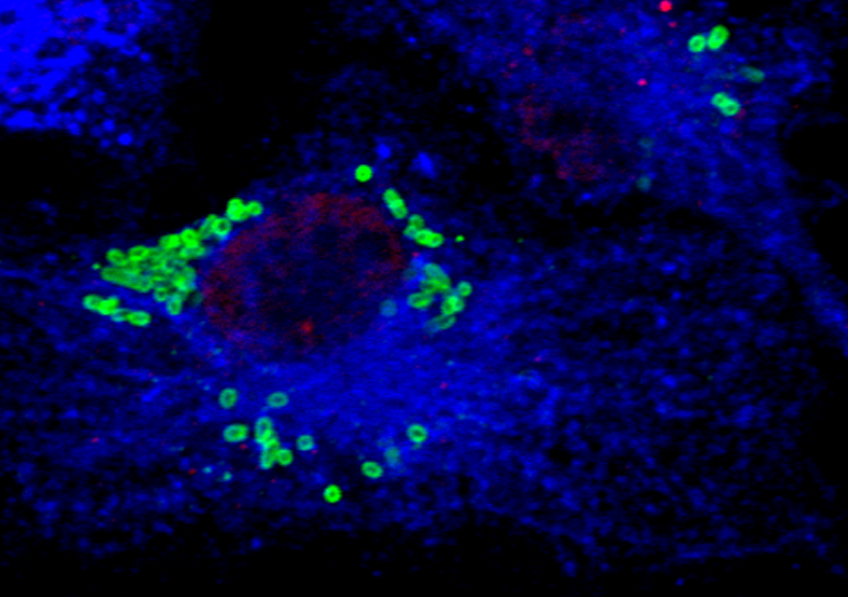
1 Gram-negative bacteria are bacteria with two membranes that are more resistant to antibiotics because of the low permeability of their outer membrane.

2 Lancet, https ://doi.org/10.1016/S0140-6736(21)02724-0

3 GHS: Global Health Security Index

The Record Heat of Summer 2022 Caused Over 61,000 Deaths in Europe, With Nearly 5,000 in France

Europe is the continent experiencing the greatest warming, up to 1°C above the global average. © Adobe Stock

The summer of 2022 was the hottest ever recorded in Europe, characterized by an intense series of heatwaves that beat records of temperature, drought, and forest fires. Although Eurostat had already reported unusually high excess mortality for these dates, the fraction of mortality that could be attributed to the heat had not until now been quantified. And this is precisely what a study by scientists from Inserm and the Barcelona Institute for Global Health (ISGlobal) set out to do. Their analysis estimates that between May 30 and September 4, 2022 there had been 61,672 heat-related deaths in Europe. These findings suggest that our current strategies for adapting to heatwaves may still be insufficient. The full study has been published in Nature Medicine.

In 2003, Europe experienced one of the most intense heatwaves in its history, causing over 70,000 deaths. Since then, adaptation strategies have been developed in an attempt to respond quickly to intense heat and protect the most vulnerable populations. Given the continuous increase in heatwave episodes in recent years and the estimations that they could double in number by 2050, it is crucial to better characterize the mortality associated with them and evaluate the efficacy of the strategies put in place.

With the summer of 2022 being the hottest ever recorded in Europe, scientists from Inserm at the France Cohorts JSU[1] and the Barcelona Institute for Global Health looked at the number of heat-related deaths for this period.

The research team obtained temperature and mortality data for the 2015-2022 period in 823 regions of 35 European countries, representing a total population of over 543 million people. These data were used to estimate epidemiological models to predict heat-related mortality for each region and week of the summer period.

The records first revealed that the temperatures were above average throughout all the weeks of the summer period. The largest heat-related abnormality was recorded between July 11 and August 14, during which, according to the researchers, 38,881 deaths were caused by the heat. In this period, there was a particularly intense pan-European heatwave between July 18 and 24, for which a total of 11,637 deaths were attributed to the excess heat. All in all, the analysis reveals that between May 30 and September 4, 2022 there would have been 61,672 heat-related deaths in Europe.

France is the country that recorded the greatest increase in temperature compared with the seasonal averages, with +2.43ºC above the average values for 1991-2020, followed by Switzerland (+2.30ºC), Italy (+2.28°C), Hungary (+2.13ºC) and Spain (+2.11ºC).


Analyses by Country, Age and Sex

Based on the epidemiological models they built, incorporating data on temperature and number of deaths[2], the scientists were also able to propose a country-specific analysis. They showed in absolute terms that the country with the largest number of heat-related deaths throughout the summer of 2022 was Italy, with a total of 18,010 deaths, followed by Spain (11,324), and Germany (8,173). France comes in 4th place with 4,807 heat-related deaths[3].

Beyond the estimated number of heat-related deaths, the study also included an analysis based on age and sex. At European level, the results highlight a very marked increase in mortality in the upper age groups, with the vast majority of deaths concentrated in the age range of 80 and over.

For the sex-based analysis, the data show that heat-related premature mortality was 63% higher in women than in men. This increased vulnerability of women to heat is observed in the general population and above all among the over-80s, where the mortality rate is 27% higher than that of men. In contrast, the male mortality rate is 41% higher among those under 65 and 13% higher between the ages of 65 and 79.

For the research team, these findings should encourage us to take action to implement more robust preventive and protective measures. Indeed, the fact that many countries already had active prevention plans, unlike in 2003, suggests that the adaptation strategies available to us today may still be insufficient.

“The acceleration of warming observed over the past ten years underlines the urgency of in-depth re-evaluation and reinforcement of prevention plans, paying particular attention to the differences between European countries and regions, as well as between age and sex, which currently mark the differences in vulnerability to heat,” explains Hicham Achebak, Inserm researcher and last author of the study.

Europe is the continent with the highest level of warming, up to 1°C more than the world average. In this context, the team’s estimates also suggest that in the absence of an effective adaptive response, the continent will face an average of over 68,000 excess deaths each summer by 2030 and over 94,000 by 2040.


[1] The France Cohorts Joint Service Unit (JSU) brings together several academic supervisory bodies, including Inserm, Ined, Université Paris-Saclay, Université Paris Cité, and Sorbonne Université. Its long-term objective is to offer a range of services for epidemiological cohorts.

[2] The mortality data were obtained via Eurostat and the temperature data via ERA5-land reanalysis.

[3] This study focuses on heat-related mortality during the summer of 2022 using weekly data and estimated nearly 5,000 deaths in France. On the other hand, Santé Publique France has recently used daily data for the same period and estimated nearly 7,000 deaths. This difference is due to the fact that the weekly data generally underestimate the impact of heat on mortality.

Neurodevelopmental Disorders in Children: A New Gene Called Into Question

ADN© Double helix DNA – National Human Genome Research Institute, National Institutes of Health.

In the face of childhood neurodevelopmental disorders, how can we get out of the therapeutic “dead end”? The answer could well be found in the genes of the proteasome – an intracellular mechanism that is responsible for removing defective proteins from the cell. A research team from Inserm, CNRS, Nantes Université and Nantes University Hospital, at the Thorax Institute and in collaboration with international teams, studied the genome of 23 children with neurodevelopmental disorders. What they found were fifteen mutations in the PSMC3 gene of the proteasome, which may be involved in their disease. This research, published in Science Translational Medicine, opens up new research perspectives in order to better understand these diseases and identify treatments.

The origin of neurodevelopmental disorders in children remains difficult to identify, with patients and their families often having to wait several years for a diagnosis.

A research team from the Thorax Institute (Inserm/CNRS/Nantes Université/Nantes University Hospital), led by Stéphane Bézieau, Head of the Medical Genetics Department at Nantes University Hospital, has been working on the genetics of neurodevelopmental disorders in children for several years. In particular, its research has led to the identification of the role of a gene called PSMD12 in a childhood neurodevelopmental disease. This gene is expressed in a large complex of proteins located in the cells, which is called the proteasome.

The proteasome acts as a kind of “garbage collector” within the cell. By eliminating the defective proteins it contains, the proteasome plays a decisive role in a large number of cell processes. Alterations that may appear on some of its constituent genes are likely to affect its ability to break down defective proteins. Their accumulation results in the development of a wide variety of pathologies.

In new research[1] in collaboration with international teams, the team continued to explore the links between proteasome gene mutations and neurodevelopmental diseases. This time it was more specifically interested in the proteasome PSMC3 gene and its involvement in the neurodevelopmental disorders of 23 young European, U.S. and Australian patients with neurological symptoms (delayed speech, intellectual disability, or behavioral problems) frequently associated with abnormalities of the face and malformations of the skeleton, heart and other organs.

Thanks to the full sequencing of the genome of these patients, the researchers have revealed fifteen mutations in the PSMC3 gene likely to explain the origin of the symptoms.

“It quickly became apparent that the cells of patients with a defective PSMC3 gene were literally overloaded with unnecessary and toxic proteins,” explains Frédéric Ebstein, Inserm researcher and first author of the study.

He compares this phenomenon to that observed in some age-related neurodegenerative diseases, such as Alzheimer’s or Parkinson’s.

“The discovery of the involvement of a second gene in childhood neurodevelopmental disorders provides unprecedented insight into this group of rare diseases that had been unknown until recently, clarifies researcher Sébastien Küry, an engineer at Nantes University Hospital, who co-signed this research. This research, combined with the team’s recent discovery of other genes involved [but not published as yet, ed.], opens up major perspectives in the understanding of this group of neurodevelopmental diseases as well as prospects for their treatment,” he concludes.


[1]This research is supported by the French National Research Agency (ANR), the European Union (European Joint Programme on Rare Diseases), and the insurance company AXA.

A Surprising Discovery About the Pulse

pouls

The pulse wave is used in everyday life to check heart rate. © Adobe Stock


We are all familiar with taking our pulse to check our heart rate. This signal is due to the propagation of a wave caused by the arteries dilating under the surge of blood from the heart. While we thought we knew the pulse well, the latest research by an international team led by Inserm researcher Stefan Catheline at the Laboratory of Therapeutic Applications of Ultrasound (Inserm/Université Claude Bernard Lyon 1/Centre Léon Bérard) shows that this was not the case. Their findings, published in Science Advances, show that the arteries not only dilate but also twist under the effect of the blood flow. This phenomenon generates a second “flexural” wave which propagates much more slowly. While it ultimately provides information on the same parameters – heart rate and arterial elasticity – the unprecedented measurement of this wave adds to our knowledge of the pulse.

Since 1820, pulse wave has been used in everyday life to check the heart rate of an athlete or inanimate person, or to assess arterial health, for example. It corresponds to the dilatation of the arterial wall following the surge of blood caused by the heart’s contractions, which propagates in an undulating manner along the arteries throughout the body.

An international research team led by Stefan Catheline, Inserm researcher at the Laboratory of Therapeutic Applications of Ultrasound (Inserm/Université Claude Bernard Lyon 1/Centre Léon Bérard), has just shown that in reality there is not one pulse wave but two. In addition to the principal wave, which is well-known and felt when touching the carotid artery or base of the wrist, there is a second one, which is more discreet but easily observable on ultrasound: the “flexural wave”, which had never been described until now.


A Chance Finding

It was somewhat by chance that Catheline’s team made this discovery. Specializing in waves and ultrasound therapies, it had been asked to test an innovative tool to analyze the retina: laser Doppler holography. This consists of photographing the organ at high speed and in very fine resolution to observe what is happening, and particularly to follow the arteries in motion. The researchers who developed this tool wanted to know if it could be used to calculate the speed of propagation of the pulse wave in the retina. Catheline’s team not only managed to measure this wave – which circulates at around one meter per second – but also detected a second wave signal nearly one thousand times slower.

The principles of fundamental physics on wave circulation in tubes are what enabled the scientists to better understand this phenomenon. Along the arteries, the two wave types actually propagate in two ways under the effect of the passage of the blood. The first is symmetrical to the central axis of the vessel and is when the arterial walls dilate and increase in diameter. The second is asymmetrical and results from the tube twisting in a so-called “sinusoidal” manner.

“Imagine a snake that swallows a prey which slides down the digestive tract – with the snake undulating away at the same time,” explains Catheline.

Following this discovery, the research team performed new ultrasound pulse measurements along the carotid artery of individuals and found both waves.

“It took us less than one afternoon to confirm the finding. This second wave, called a ‘flexural wave’, is present on all the recordings and is not difficult to observe. If it has never been described, it is simply because no-one had been looking for it,” explains Catheline.


schéma onde de pouls

The most well-known pulse wave (dilatation wave) is caused by the walls symmetrically separating outwards from the central arterial axis under the effect of the blood surge. And the newly-discovered flexural wave is caused by the artery twisting from side to side of this axis. © Stefan Catheline


And the Clinical Applications?

The principal pulse wave is widely used in medicine and reflects an individual’s cardiovascular health. Its speed of propagation depends on the condition of the artery walls: the younger and more supple they are, the slower the speed – and vice versa with age, with rigid arteries being a risk factor for cardiovascular events. However, given the high propagation speed of this wave, it is necessary to measure it over several centimeters to obtain a reliable value.

“With the flexural wave that we are describing here, whose slow speed ranges from one tenth to one thousandth of a meter per second depending on the arterial diameter, it is easier to study the signal on very short fragments and with other types of equipment than ultrasound, especially X-ray and MRI, explains Catheline. One millimeter is sufficient to obtain an accurate value, for example, to assess the state of the arteries in the retina,” he explains.

The researcher sees a second advantage in using this flexural wave in the clinic: by continuing to propagate in the veins there where the principal pulse wave is no longer detectable due to the distance from the heart, it would also provide information on the rigidity of the venous wall. He specifies, however, that in order to make it a clinical tool, research is needed in humans in order to correlate propagation speed and wall elasticity, as had been done previously for the principal dilatation wave.

How Blood Stem Cells Detect Pathogens and Guide Immune Response

Cellule du système immunitaire infectée par BrucellaImmune system cell infected with Brucella (green); endocytosis compartment (blue). © CIML

Correct immune system function depends on the continuous supply of white blood cells derived from stem cells that reside in the bone marrow.  These are known as blood stem cells or hematopoietic stem cells. Researchers from Inserm, CNRS and Université d’Aix-Marseille at the Center of Immunology Marseille-Luminy have now discovered a new role played by these cells in immune response. In their article published in Journal of Experimental Medicine, they describe how they are able to recognize and directly interact with a bacterium called Brucella in the bone marrow, thanks to a receptor present on their surface. This is the first demonstration of the direct recognition of a living pathogen by the blood stem cells, which attests to their very early contribution to the immune response.

Blood stem cells, otherwise known as hematopoietic stem cells, are stem cells that reside in the bone marrow. They multiply and give rise to all blood cells, namely the red cells that transport oxygen and the white cells that participate in the immune response.

With regard to immune response, the blood stem cells had until now only been seen as the cells from which the white cells originate. However, a growing body of evidence suggests that they can also contribute directly and actively to the immune response. For example, recent data have shown that they can directly detect cytokines, which are proteins released during infection or inflammation.

In a new publication, a research team from Inserm, CNRS and Université d’Aix-Marseille led by Michael Sieweke and Jean-Pierre Gorvel[1] wanted to further the scientific knowledge in this area. The researchers succeeded in describing the mechanisms at work during the encounter between the blood stem cell and a specific pathogen: the Brucella bacterium, which is a mandatory reportable microorganism/toxin (MOT)[2].

Brucella causes an infectious disease called brucellosis (also known as Malta fever or Mediterranean fever), which is one of the most widespread zoonoses posing a significant threat to human health worldwide[3]. Brucella is an intriguing pathogen and very interesting for the scientists to study because of its ability to establish persistent and chronic infections and evade the immune response of its host[4].

The scientists found that the blood stem cells present in the bone marrow were able to detect Brucella. Their observations show that CD150, a specific receptor on the surface of the blood stem cells, interacts with Omp25, a protein present on the surface of Brucella.

bactérie BrucellaGraphic Summary of the Discovery. Thanks to the CD150 receptor on their surface, the blood stem cells in the bone marrow are able to detect Brucella. After recognizing it, they begin to produce more white blood cells. © CIML


“Our study reveals the mechanisms by which these blood cells are able to detect bacteria via a special receptor. We can consider this as a direct ‘handshake’ between the stem cell and the bacterium. Never had anyone imagined that the blood stem cell could recognize a living bacterium,” explains Sandrine Sarrazin, Inserm researcher and co-last author of the study.

The scientists then showed that this “handshake” leads to a rapid response by the stem cells, whereby they begin to produce more white blood cells. This is the first demonstration of the direct recognition of a living pathogen by blood stem cells and attests to a very early and unexpected contribution of these cells to the immune response.


How Brucella Uses Stem Cells to “Hack” the Immune System

The scientists then wondered whether this mechanism was more beneficial to the host or to the bacterium.

Thanks to meticulous observations, they found that Brucella directs the stem cells to produce the white blood cells it favors for infection. The bacterium is able to invade the white blood cells produced by the blood stem cells and use them to multiply and establish itself in the body. In this particular case, the stem cells therefore contribute to the spread of the bacterium.

“This research sheds new light on the sophisticated mechanisms that pathogens use to evade the immune system’s defenses. While the increased production of white blood cells would be beneficial if they could effectively fight infection, Brucella is able to exploit them in order to multiply,” explains Gorvel, one of the co-last authors of the study.

“This mechanism can be seen as an evasion strategy used by the bacterium to advance the infection,” summarizes Sieweke, another co-last author of the study.

The publication of this study marks an important step in understanding the complex dance between Brucella and the hematopoietic stem cells. It not only provides crucial information on the pathogenesis of brucellosis, but also opens up new avenues for the development of targeted therapeutic interventions.

“In addition to improving knowledge about how the immune response works, our study ultimately allows us to envisage the development of a targeted therapy capable of preventing the interactions between Brucella and the blood stem cell, preventing the spread of the bacteria in the body and helping patients with brucellosis,” concludes Gorvel.


[1] This research is the result of a collaboration between two research teams at the Center of Immunology Marseille-Luminy (CIML, CNRS/Inserm/Aix-Marseille Université): Michael Sieweke’s Stem cell and macrophage biology team and Jean-Pierre Gorvel’s Immunology and cell biology of pathogen/host cell interactions team.

[2] The Brucella experiment was therefore conducted at the Center for Immunophenomics (CIPHE) under biosafety level 3 conditions.

[3] The World Health Organization (WHO) has identified brucellosis as being one of the seven most neglected zoonoses, contributing to poverty, hindering development, and causing substantial economic losses in developing countries.

[4] Previous studies conducted at Gorvel’s laboratory had enabled crucial discoveries with the aim of elucidating the mechanisms underlying these phenomena.

When We Always Benefit From Focusing on Health: Inserm Unveils Its New Advertising Campaign

A still from the Inserm commercial. © Directed by: Ugo Mangin, production: Yohannes Cousy

One year after its large-scale poster campaign in France’s railway stations and Paris subway, Inserm uses its slogan On gagne tous les jours à s’intéresser à la santé [We always benefit from focusing on health] in a 30-second TV commercial broadcast from June 7 to July 30.

Directed by Ugo Mangin with the support of the Insign agency and commissioned by Inserm’s Communications Department, this commercial illustrates just how many of our daily actions are – without our knowing – the result of research conducted at the Institute.

Throughout the 30-second sequence, the viewer is immersed in the thoughts of a woman, who is out and about and comments on the everyday scenes she sees around her. This is how we find out that a saxophonist has preserved his breathing thanks to specific proteins, that the Nutri-Score enables a young woman to eat a healthy sandwich, and that a middle-aged man continues to skateboard thanks to his chondrocytes.

In keeping with the 2022 poster campaign, the commitment to integrating surprising and complex science into an everyday scene was maintained in order to engage viewers and encourage them to visit Inserm’s website to find out more.

“Inserm is a strong brand, a label of quality in terms of health science that French people trust. Nevertheless, we need to be even more visible and anchored in everyone’s daily lives in order to communicate on our research as close as possible to where the needs are and show the findings of that research. This campaign is the link between the biomedical science produced at Inserm and its concrete translation for the health of us all,” points out the Communications Department.

“The COVID-19 crisis has shown just how medical research is essential to our health. Through this commercial, we wanted to make the impact of our research visible to everyone. It is essential for the general public to realize that excellence in research is conducive to quality medicine. It is our role to find more promising and effective new treatments as our knowledge advances,” explains Prof. Didier Samuel, Chairman and Chief Executive Officer of Inserm.

Immunotherapy for blood cancer: remote destruction of tumor cells demonstrated

Attaque d’une tumeur par des cellules CAR TCAR T cells attacking a tumor, visualized using intravital imaging. Live tumor cells are shown in white, apoptotic tumor cells in blue and CAR T cells in green. © Morgane Boulch, Philippe Bousso / Institut Pasteur

The aim of immunotherapy strategies is to use cells in the patient’s own immune system to destroy tumor cells. CAR T cell therapy is an immunotherapy that is effective in treating blood cancer. Around 35,000 people are affected by blood cancer each year in France, with 1.24 million cases worldwide. By closely investigating some of the immune cells generated during this therapy, known as CD4 T cells, scientists from the Institut Pasteur and Inserm, in collaboration with clinicians from the Paris Public Hospital Network (AP-HP), discovered that these cells are capable of remotely neutralizing tumor cells by producing interferon gamma (IFN-γ). This study raises new hopes for blood cancer patients with an incomplete response to CAR T cell therapy and for cancers sensitive to IFN-γ. The results were published on May 29, 2023 in the journal Nature Cancer.

CAR T cell therapy is an immunotherapy that has produced remarkable results in treating certain types of leukemia or lymphoma. But some patients who receive this treatment relapse because their tumor cells evade the therapy. A multidisciplinary team of scientists from the Institut Pasteur and Inserm and clinicians from the Paris Public Hospital Network (AP-HP) sought to shed light on how the therapy works with the aim of obtaining even more effective responses.

The principle of CAR T cell therapy is to isolate the patient’s T cells, genetically modify them so that they specifically target tumor cells, and multiply them before injecting them back into the patient in large numbers. This army of killer CAR T cells is composed of CD4 T cells and CD8 T cells in varying proportions from one patient to the next. While we know that CD8 killer T cells need to come into direct contact with tumor cells to destroy them, the mechanism of action of CD4 T cells had not previously been fully explored.

The research team studied these CD4 CAR T cells more closely and revealed a very interesting property: their ability to kill tumor cells remotely by secreting a molecule involved in the immune response, interferon gamma (IFN-γ).

For some types of cancer that are sensitive to IFN-γ, this destruction mechanism is highly efficient. We also observed that, among patients with a high quantity of CD4 T cells, those that produce a large amount of IFN-γ respond better to treatment,” explains Philippe Bousso, Head of the Institut Pasteur’s Dynamics of Immune Responses Unit (Inserm 1223) and last author of the study.

To reveal the novel mechanism of action of these remote killer cells, the scientists began by exploring preclinical models to analyze the mechanism in detail, in particular using in vivo imaging techniques, then they checked the relevance of the results on samples from patients.

CAR T cells attacking a tumor, visualized using intravital imaging. Live tumor cells are shown in white, apoptotic tumor cells in blue and CAR T cells in green. The white circles indicate the destruction of tumor cells when they come into direct contact with CAR T cells, and the red circles show the remote destruction of tumor cells. © Morgane Boulch, Philippe Bousso / Institut Pasteur


This discovery opens new avenues for adjusting treatments to prevent tumor cells evading CAR T therapy. It also raises new therapeutic hopes for patients, offering the possibility of a more personalized treatment approach whereby a larger quantity of CD4 CAR T cells can be used to activate IFN-γ depending on tumor cell sensitivity,” comments Philippe Bousso.

By developing a better understanding of how CD4 T killer cells work, it may also be possible to extend the scope of this therapy to other solid tumor cancers that are sensitive to IFN-γ. The clinical data will be confirmed on other cohorts.


This research was funded by the institutes cited above, and also by the French National Cancer Institute (INCa) and the European Research Council.

A Major Advance in the Genetics and Risk Factors of a Form of Infarction That Mainly Affects Women

SCAD is a form of infarction that mainly affects women. © Fotalia


Spontaneous coronary artery dissection, more commonly known under the acronym SCAD, is a cause of infarction of which 9 out of 10 of its victims are women in their forties in apparent good health. Still poorly understood, it is often underdiagnosed, which complicates treatment despite the fact that it could represent up to one third of infarction cases in women under 60 years of age. In order to understand its genetic causes and biological mechanisms, a new international study led by Inserm Research Director Nabila Bouatia-Naji at the Paris-Cardiovascular Research Center – PARCC (Inserm/Université Paris Cité) was set up. Its findings show the genetic causes that define the risk of SCAD to be very numerous and distributed across the entire patient genome. The study has identified 16 genomic regions associated with a higher risk of SCAD, paving the way for a better understanding of the biological mechanisms that underlie this disease. The study was published on May 29, 2023 in Nature Genetics.

Unlike the majority of cardiovascular diseases, such as myocardial infarction, which mainly affect older and/or overweight men, spontaneous coronary artery dissection (SCAD) is a form of infarction that affects women in 9 out of 10 cases. Although these women are often in their forties, the disease can occur earlier – in the year after giving birth, or later – during the transition to menopause. Despite being increasingly recognized as a major form of infarction within this population, SCAD remains quite poorly documented due to a lack of data and a lack of knowledge of its specific risk factors – particularly genetic.

Over the past 20 years, considerable progress has been made in detailing the mechanisms of development of coronary diseases such as atherosclerosis and the very rare and syndromic forms of cardiovascular diseases. Such knowledge is essential in order to better understand these diseases and devise improved and personalized strategies for their prevention and treatment.

Nevertheless, research has lagged far behind in the understanding of diseases such as SCAD that affect women at key stages of their lives. It is therefore essential to now focus on this understudied cardiovascular disease and its own specific genetic risk.

The team of Inserm geneticist Nabila Bouatia-Najia conducted a large-scale study on the subject, coordinating a meta-analysis of 8 genome-wide association studies (GWAS)[1].  By comparing the genetic data of over 1900 patients with around 9300 healthy individuals, the scientists identified 16 genomic regions (or loci) of genetic predisposition to SCAD.


Towards a Better Understanding of the Biological Mechanisms

This study began by showing that the genetic variations most commonly found in patients having survived SCAD play a role in the composition of the “cement” that surrounds the coronary artery cells.

However, one of the genes identified is F3 and it encodes the tissue coagulation factor. Normally, the tissue factor initiates coagulation at cell level in order to resorb any hematomas. The results of the study suggest that a lack of F3 expression is often found in patients, constituting a potential cause of poor artery repair, which can lead to tearing. Poor hematoma resorption would therefore be a hitherto unknown genetic cause.

One of the other objectives of this study was to position SCAD in relation to other cardiovascular diseases in order to better understand its epidemiological particularities. Using the data that determine genetic cardiovascular risk factors and clever statistical methods, the scientists revealed a robust link between high blood pressure and risk of SCAD, while confirming that high cholesterol, overweight, and type 2 diabetes had no impact on this risk.

“This finding could therefore be clinically interesting in the longer term, to encourage doctors to closely monitor blood pressure changes in patients at increased genetic risk of SCAD,” explains Bouatia-Naji, Inserm Research Director and last author of the study.

Finally, this study reveals a genetic link between SCAD-related infarction and atherosclerosis-related infarction. Indeed, the researchers have shown that a large number of genomic regions predisposing to SCAD are shared with those of atherosclerosis-related infarction. However, even if these were the same genetic variants, the alleles[2] that are more common in SCAD patients are routinely described as being less common in subjects with atherosclerosis-related infarction.

“This finding is very surprising because they show that, depending on whether you are faced with a young woman with no risk factors, or an older man with risk factors, the genetic causes and biological mechanisms associated with their infarction can be opposed.  Our findings highlight the need to better understand the particularities of cardiovascular diseases in young women in order to improve their follow-up, which is currently identical to that of atherosclerosis-related infarction,” concludes Bouatia-Naji.

Building on these findings, the team is now working to develop new cell and animal models that better account for the genetic factors involved in the disease, particularly in order to better study their impact on the condition of the arteries. Always with a longer-term objective in mind: to shed the spotlight on a cardiovascular disease that is essentially female and all too often neglected, and to improve how it is understood and treated.


[1] Genome-wide association study, widely performed for several years now, which consists of analyzing the entire genome of thousands of healthy and sick individuals in order to identify the genomic regions which contain the genes influencing the vulnerability of people to the condition in question

[2] An allele is a version of a genetic variant resulting from a change in the DNA sequence. Any DNA sequence can have several alleles, which often determine the appearance of different hereditary characteristics.

InScience 2023: The Festival That Does Your Health Good

affiche Inscience 2023

June 1 to 15, 2023 will mark the third edition of InScience – a festival held by Inserm for visitors to discover and discuss questions of health and medical research. With its rich program deployed across France, InScience is particularly intended for young people, enabling everyone to learn more about Inserm’s work and become actively involved in their own health.  With meetups with scientists, podcasts, videos, virtual reality games, comic books… there is something for all tastes.

For the past three years, InScience has aimed to break taboos surrounding health, counter stereotypes of the world of research, and bring the scientists and general public closer together – enabling the two worlds to meet directly. This festival reflects Inserm’s desire to democratize scientific research and make it more transparent, giving each individual the tools to become actively involved in their own health.

Support from the French National Research Agency (ANR)[1] means that this year the festival is bigger than ever, offering a richer program than previous editions. Original forms of scientific popularization, including podcasts, an original comic, escape games, and a partnership with Tik Tok influencer Tangleroux complete the offering. Places that are very popular with the general public, such as Ground Control in Paris, will host scientific mediation activities.


carte lieux InScience 2023

The highly comprehensive program (see below), with events that can be attended in person or online, seeks to make medical research advances accessible to all citizens, regardless of where they live. The events will be hosted in no fewer than 24 places in 12 French cities, and are open to everyone free of charge.


[1] Inserm’s various regional offices had responded together and won the ANR call for proposals on the theme of Science with and for society (SwafS)

Discover the Program

The program can be found on the Inserm website, as well as the dedicated Twitter and Tik Tok accounts.

Check out the teaser.


Focus on Original Formats

InScience is also about original ways to popularize science, for example through podcasts and a comic.

Through a series of 10 podcasts available on all platforms, InScience sets out to discover women and men who work for the health of us all: researchers. The objective being to talk about research professions and create vocations among young people.

In addition, a collaboration between Inserm and comic book author Maité Robert has led to the creation of a comic narrating the research projects of ten 2018-2019 ANR winners. The idea is to show that medical research is maybe not so complicated when explained with words and images that are accessible to everyone.

This comic can be obtained free of charge at InScience events.

Women and Parkinson’s Disease: Physical Activity Found to Be Beneficial More Than Twenty Years Before Diagnosis

marche sportive retraitée© AdobeStock

Although physical activity is regularly discussed as being one of the avenues for preventing Parkinson’s disease (PD), studies so far had been unable to eliminate certain biases or had not focused on its specific role in women. Researchers from Inserm, Université Paris-Saclay and Université de Versailles Saint-Quentin-en-Yvelines (UVSQ) with the Gustave Roussy Institute studied the impact of physical activity on the development of PD in nearly 100,000 women from the French cohort E3N followed up over 29 years. They observed that the more the participants had been active in their lives, the lower was their risk of developing the disease – with such a benefit already present more than 20 years before diagnosis. The researchers have also shown that in women with PD there had been a significant decrease in physical activity in the 10 years prior to diagnosis, probably due to bothersome precursor symptoms. These findings, to be published in Neurology, call for consideration to be given to the rollout of preventive physical activity-based programs in people at risk of PD.

Parkinson’s disease (PD) is a progressive neurodegenerative disease characterized by the destruction of certain neurons in the brain and the accumulation of proteins toxic to them. It is the second most common neurodegenerative disease in France and there is still no cure; therefore it is essential to identify avenues for prevention.

Previous studies had shown a reduced risk of developing PD in people with who engage in high levels of physical activity. However, the limited number of such studies involving women did not make it possible to confirm this association in that population. In addition, these studies generally had a relatively short participant follow-up period with only one evaluation of physical activity, which did not make it possible to eliminate certain biases – particularly that of reverse causation.

Reverse causation is expressed as follows: precursor symptoms of PD (constipation, sleep disorders, smell disorders, subtle motor signs, etc.) may be present several years before the disease is diagnosed. Their effects could lead people to modify their behavior (such as how much physical activity they do) prior to diagnosis, which is liable to distort the statistical analyses that study the relationship between these behaviors and the risk of developing the disease.

A team led by Alexis Elbaz, Inserm researcher at the Epidemiology and Population Health Research Center (Inserm/Université Paris-Saclay/UVSQ), studied the impact of physical activity on the onset of PD in women from the E3N cohort over a follow-up period of 29 years. E3N is a French cohort of nearly 100,000 women who have been followed-up since 1990. With nearly 1,200 women with PD identified in 2018[1], it is currently the world’s largest female prospective cohort for this disease.

The changes in the physical activity[2] of each participant throughout the 29 years of follow-up were estimated based on the information collected via six individual questionnaires completed at different stages of the follow-up. The data obtained compared the practice of physical activity prior to diagnosis in the participants with PD with those of the participants of the same age without PD.

In order to reduce the risk of a reverse causation bias resulting from the potential influence of the precursor symptoms on physical activity in the years prior to diagnosis, the scientists examined the impact of physical activity evaluated more than 5, 10, 15 and 20 years prior to diagnosis on the risk of developing PD.

What they observed was that the more physically active the participants, the less likely they were to develop PD, even when the physical activity had been evaluated more than 20 years before diagnosis. The most active women therefore had a 25% less risk of developing the disease compared with the least active.

Overall, the participants with PD had been less active than the others throughout the follow-up period, including more than 20 years before diagnosis. This gap between women with and without PD increased further in the 10 years before diagnosis, suggesting that the precursor symptoms occurring in this interval could indeed be responsible for a decrease in physical activity in the women who will go on to develop the disease but who have not yet been diagnosed.


“These findings support the protective effect in women of physical activity against the risk of developing Parkinson’s disease, even over the very long term, specifies Berta Portugal, PhD student and first author of this research. They support the utility of implementing physical activity programs to prevent Parkinson’s disease in people at risk and call for conducting other studies in order to understand what activity type and level of intensity are the most beneficial,” she adds.


“This study demonstrates the importance of studies with a lengthy follow-up within large-scale cohorts to better account for the precursor symptoms and the statistical bias they cause in evaluating the benefit of physical activity on the development of Parkinson’s disease,” concludes Elbaz.


[1]The percentage of women with PD in the cohort is similar to that of the general population in Europe over the same period of time.

[2]The concept of “physical activity” goes beyond sport alone. In this study, it is quantified by the distance walked daily, the number of flights of stairs climbed, journeys taken by bicycle, time spent doing light and heavy household activities, time spent doing recreational activities, whether light (gardening, sports of moderate intensity) or vigorous (intensive sports).

Eating Broccoli to Limit Skin Allergies

brocoli

The scientists specifically focused on dietary compounds naturally present in cruciferous vegetables, such as broccoli. © Unsplash

The severity of skin allergies can vary depending on many environmental factors, including diet. However, the role of specific nutrients had not been well documented until now. In a new study, researchers from Inserm and Institut Curie at the Immunity and Cancer[1] unit have shown that the absence in the diet of compounds found in certain vegetables, particularly broccoli and cabbage, could worsen skin allergies in animal models. These findings, published in Elife, highlight the importance of a balanced diet alongside therapeutic interventions offered to patients.

Skin allergies are caused by an inappropriate immune response to compounds present in the environment, with the extent of their severity varying according to many factors, including diet. However, the impact of the different compounds present in food is still poorly understood, which complicates the use of nutrition-based strategies to alleviate patient symptoms.

In this research, the scientists focused on dietary compounds that act on a molecule present in the body called the aryl hydrocarbon receptor (AhR). Such nutrients are naturally present in cruciferous vegetables, such as broccoli.

While previous studies had already shown an association between these dietary compounds and the worsening of inflammatory bowel disease and neuroinflammation, their effect on allergic immune reactions had not been documented until now.


Worsening Allergies

The research team used a mouse model of skin allergy. Some of the animals were fed a diet containing no AhR-activating compounds in order to evaluate its potential impact on the severity of their allergy.

The researchers showed that the absence of these nutrients is associated with an increase in the skin’s inflammatory state and a worsening of the skin allergy, which did not occur in the mice whose diet contained those compounds.

The scientists wished to go further in order to understand the biological mechanisms of action of these nutrients. When the nutrients were absent, they observed the overproduction of a molecule called TGF-beta in the epidermis of the mice. This overproduction disrupts the normal functioning of a population of immune cells called “Langerhans cells”, which are exclusively present in the skin and act as a modulator of skin immune responses.

The scientists then validated these findings by showing that the AhR-activating compounds also control TGF-beta production in human skin cells.

“Our results suggest that an imbalanced diet could increase allergic skin reactions in humans through mechanisms that we have described precisely. In essence, we could say that our research helps to explain why eating vegetables such as broccoli and cabbage may limit the severity of skin allergies and why it is therefore important to include them in one’s diet,” emphasizes Inserm researcher Elodie Segura, who led this study at Institut Curie.

The findings of this study may also apply to other skin diseases in which inflammatory mechanisms are involved, such as psoriasis. Furthermore, they also open up interesting research avenues for improving the study of the gut-skin axis in the development of allergic diseases.

Based on these data, the research team now wishes to look at the role of AhR-activating dietary compounds in other inflammatory disease settings, such as tumors.


[1] Conducted at the Immunity and Cancer unit (Inserm, Institut Curie), this research was carried out with particular thanks to Institut Curie’s experimental pathology platform and new metabolomics and lipidomics platform.

fermer