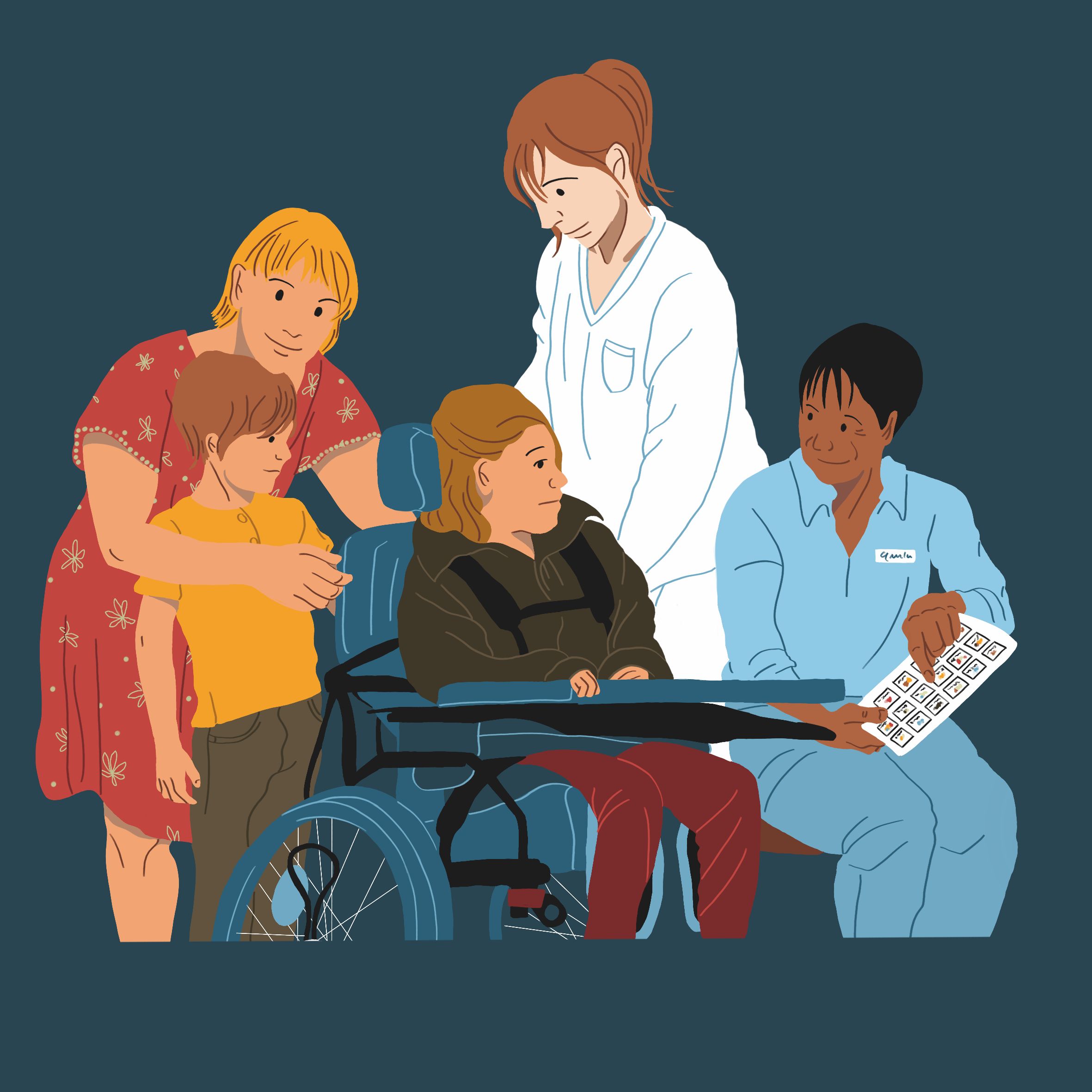
Menu

A promising new therapeutic approach for patients with arteriovenous malformations

malformations artérioveineuses
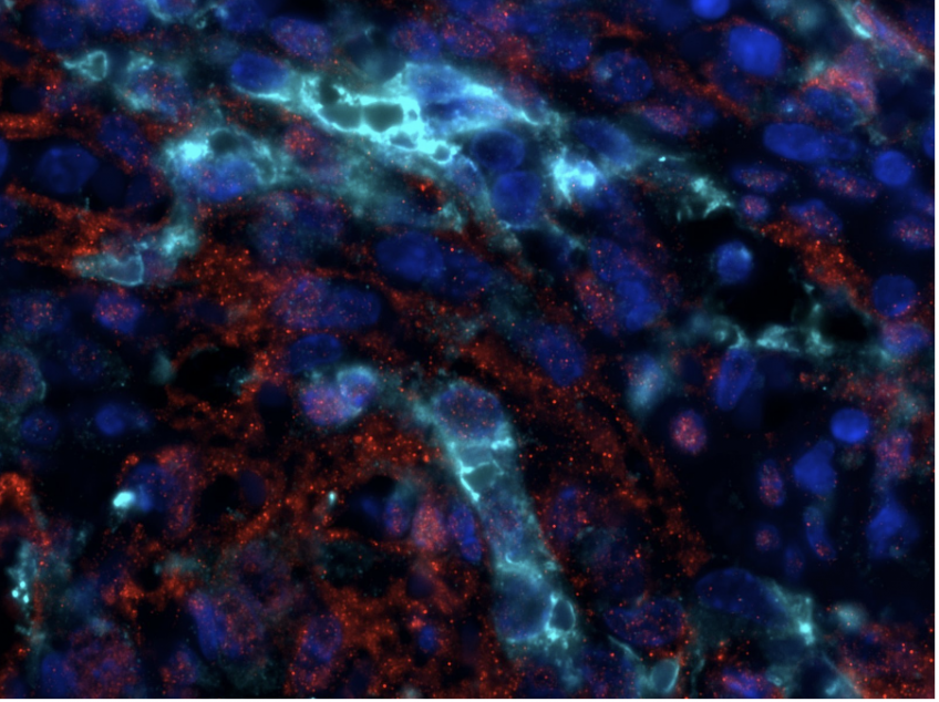
Phospho-ERK (red), Green Fluorescent Protein (cyan) and DAPI coimmunofluorescence on spleen sections from mice carrying a KRAS G12C endothelial mutation © Guillaume Canaud

The teams of the translational medicine and targeted therapies unit of the Necker-Enfants Malades AP-HP hospital, Inserm, Paris Cité University within the Necker–Enfants Malades Institute, coordinated by professors Guillaume Canaud (Université Paris Cité, AP-HP) and Laurent Guibaud (Hospices Civils de Lyon, Reference Center for Superficial Vascular Anomalies), conducted a study showing a promising effect of the anticancer drug sotorasib for arteriovenous malformations secondary to a mutation in the G12C type KRAS gene. The results were the subject of a publication published on July 17, 2024 in the New England Journal of Medicine.

An arteriovenous malformation (AVM) results from abnormal connections between arteries and veins. AVMs are frequently associated with symptoms such as pain, bleeding, heart failure, cosmetic deformity or organ compression. These malformations generally progress over time. AVMs are in most cases of genetic origin, either “germinal” and therefore familial, or sporadic due to a localized genetic mutation. In many of the latter cases, the gene responsible is the KRAS gene, a gene involved in cell growth, proliferation and survival. There are different types of KRAS mutation. At the moment, no drug treatment is approved for these pathologies.

Sotorasib, developed by Amgen, is an anticancer drug used to treat a type of lung cancer, advanced non-small cell lung cancer (NSCLC), with a mutation in the KRAS gene, the KRAS G12C mutation. The drug thus selectively targets the KRAS p.G12C protein.

The teams of Professors Guibaud and Canaud identified the presence of a KRAS G12C mutation in two adult patients with severe AVMs and without therapeutic resources. They then decided to create two mouse models developing vascular malformations secondary to a KRAS G12C mutation in order to better understand the pathophysiology of these malformations. Preclinical models have largely recapitulated the malformations observed in humans. Using these two models, they then tested and demonstrated the effectiveness of sotorasib in preventing the development of vascular malformations and significantly improving the survival of mice.

Based on these results, Professors Guibaud and Canaud obtained authorization for the use of sotorasib from the Amgen laboratory to administer it to these two patients as part of their therapeutic care.

In the weeks following the start of treatment, both patients noted a clear improvement in their symptoms (stopping of bleeding, healing of chronic skin ulcerations, disappearance of pain and recovery of deafness), a clinically visible reduction in malformation. This last result was then confirmed by MRI. After 2 years of follow-up, patients did not develop resistance to treatment.

This work demonstrates the interest in obtaining a molecular diagnosis for this type of rare disease and the possibility of repositioning highly targeted drugs developed for other indications, as the researchers had previously done for alpelisib in the syndrome of CLOVES and related syndromes.

These results will need to be confirmed by future studies on a larger number of patients. These drug repositionings open up new therapeutic fields, in particular for AVMs, for which these drugs could be combined with surgical treatment or in interventional radiology.

A valve made from human collagen opens up new avenues for the treatment of a paediatric heart disease

Image d'IRM montrant la valve pulmonaire reconstruite qui se ferme parfaitement après 7 jours d'implantation chez la brebis. © Fabien Kawecki/InsermMRI image showing the reconstructed pulmonary valve (circled in red) that is closing perfectly 7 days after implantation. © Fabien Kawecki/Inserm

Researchers from Inserm have developed a pulmonary valve using human collagen. A device that could ultimately be a game-changer in the treatment of paediatric heart diseases, such as tetralogy of Fallot. These findings form part of broader research conducted at BioTis (Inserm/Université de Bordeaux), a tissue bioengineering lab in Bordeaux which develops tissues made from biological material obtained from human cells. This study, published in Science Translational Medicine, opens up new therapeutic avenues in the longer term for young patients with tetralogy of Fallot, for whom the current treatment options continue to cause numerous complications.

Tetralogy of Fallot is a congenital heart malformation that affects one in 4 000 births. It involves pulmonary stenosis, which is when the outflow tract from the heart’s right ventricle to the pulmonary artery is narrowed. This prevents the normal flow of blood to the lungs, leading to reduced blood oxygen levels.

This abnormality can be corrected with surgery to widen the pulmonary pathway to restore normal blood flow. This involves removing the pulmonary valve, and then reconstructing it from either synthetic TeflonTM membranes or ‘biological’ leaflets made from chemically treated animal tissue.

Both solutions present major drawbacks – the first and foremost being a reaction from the immune system, which seeks to reject these foreign bodies. Together with a chronic inflammatory reaction, this phenomenon can also lead to other complications such as thrombosis and calcification[1]. What is more, the valves made from these materials are prone to the development of bacterial infections. Finally, they are not designed to adapt to the patient’s growth and changing morphology: this means that as the patient ages, other operations will be required to replace the initial valve.

A valve made from human cells

Therefore, the team led by Inserm researcher Fabien Kawecki wanted to find new solutions, and so has developed a ‘new-generation’ biological pulmonary valve, designed from collagen-rich leaflets produced by cells. Collagen is a structural protein highly abundant in the human body, which helps to support many tissues and organs. The researchers thus based themselves on the approach developed over the last decade at BioTis (Inserm/Université de Bordeaux), which consists of culturing human cells in the laboratory to obtain deposits of collagen-rich extracellular matrix.

These collagen deposits form leaflets that can be used to devise, as in this study, pulmonary valves. The major advantage is that since collagen does not vary from one person to another, these entirely biological and chemically undenatured leaflets are not considered by the body as foreign bodies to be rejected.

In the study, Kawecki and his team tested the use of their biological leaflets to reconstruct a pulmonary valve in an ‘organosynthetic’ heart model developed by their US collaborators at the Massachusetts Institute of Technology (MIT). It is a bioartificial heart that uses pneumatic muscles to reproduce the functioning of the human heart and to control heart beats. From this model it is possible to gather valuable data on the functionality of the valve. Then, working with cardiac surgeons at Bordeaux University Hospital, the scientists also implanted the valve for seven days in an animal model (sheep), performing the same surgical procedures and using the same tools as those used for this type of operation in humans.

‘Thanks to our two models, we have obtained proof of concept that the valve we have designed is functional and can easily be fitted following the same surgical procedures as in humans, which is promising if we are to move on to clinical studies in a few years’ time. Implanting our valve has restored the direction of blood flow through the pulmonary pathway without generating valve leakage. We also observed that after only 7 days of implantation, there was good integration of the valve with the animal’s native tissue. In addition, we have seen on our valve the presence of smooth muscle cells which will play an important role in its remodelling and growth,’ explains Kawecki.

Using the data collected in both models, the scientists were also able to develop a digital model that will allow the functionality and clinical utility of different biomaterials to be tested before being implanted in animals.

This digital model and that of the organosynthetic heart could be valuable tools for researchers and surgeons, enabling them in the future to test new biomaterials and medical devices, as well as to train in new surgical approaches before moving to animals and then humans,‘ explains Kawecki.

For the team, the next step is to implant the valve over longer periods of time (16 weeks, then one year) in animal models, to ensure that it functions correctly over the long term and adapts to the growth of the animal over time. Beyond that, if the results are conclusive, clinical trials could be envisaged.

The team has already filed a patent for the use of the lab-designed biomaterial as a pulmonary valve and hopes to test its utility in various cardiovascular diseases in adults and children in the future.


[1] Accumulation of calcium salts in organic tissues

A better understanding of Alzheimer’s disease: A study confirms the utility of caffeine as treatment avenue

Augmentation neuronale du récepteur A2A dans l’hippocampe de sourisIn red, the increased A2A receptors in the neurons of a mouse hippocampus. In blue, the cell nuclei (DAPI staining). © Émilie Faivre

In France, 900 000 people have Alzheimer’s disease or a related condition. The risk of developing Alzheimer’s depends on genetic and environmental factors. Among these factors, various epidemiological studies suggest that the regular consumption of moderate amounts of caffeine slows age-related cognitive decline and the risk of developing the disease. In a new study[1], researchers from Inserm, Lille University Hospital and Université de Lille at the Lille Neuroscience and Cognition research centre have taken a step further in understanding the mechanisms underlying its development. They have shown that the pathological increase in certain receptors in the neurons at the time of disease onset promotes a loss of synapses, and as such the early onset of memory impairments in an animal model of the disease. Their findings also confirm the utility of conducting clinical trials to measure the effects of caffeine on the brains of patients at an early stage of the disease. These findings have been published in Brain.

Alzheimer’s disease is characterised by disorders of memory, executive function and orientation in space and time. It results from the slow degeneration of the neurons, which begins in the hippocampus (essential for memory) and then spreads to the rest of the brain. Sufferers present two types of microscopic brain lesions: senile plaques (or amyloid plaques) and neurofibrillary degeneration (or tau pathology), which contribute to neuron dysfunction and disappearance[2].

Studies had already shown that the expression of certain receptors, known as A2A receptors, was found to be increased in the brains of patients affected by Alzheimer’s disease in the hippocampus. However, the impact of such dysregulation on the development of the disease and associated cognitive disorders had remained unknown until now. In a new study, a team led by Inserm researcher David Blum took a closer look at this question.

The scientists were able to reproduce an early increase[3] in the expression of the adenosine A2A receptors, as observed in the brains of patients, in a mouse model of Alzheimer’s disease that develops amyloid plaques. The objective was to assess the consequences of this increase on the disease and to describe the mechanisms involved.

The results of their research show that the increase in A2A receptors promotes the loss of synapses[4] in the hippocampus of ‘Alzheimer’s mice’. This causes the early onset of memory impairments in the animals. The scientists then showed that a dysfunction of certain brain cells – the microglial cells, partly responsible for the brain inflammation seen in the disease – could be involved in the loss of synapses, in response to an increase in the A2A receptors.

Similar mechanisms had already previously been described by the team, this time in another model of the disease developing tau lesions[5].

‘These findings suggest that the increased expression of the A2A receptors alters the relationship between neurons and microglial cells. This alteration could be the cause of an escalation of effects leading to the development of the memory impairments observed,’ explains Émilie Faivre, co-last author of the study and researcher at the Lille Neuroscience and Cognition centre (Inserm/Université de Lille/Lille University Hospital).


Caffeine: An interesting treatment avenue for the early prevention of cognitive decline?

Several studies had already suggested that regular and moderate caffeine consumption (equivalent to 2 to 4 cups of coffee per day) could slow the cognitive decline associated with ageing and the risk of developing Alzheimer’s disease.

In 2016, the same research team had described one of the mechanisms by which caffeine could have this beneficial effect in animals, reducing the cognitive disorders associated with Alzheimer’s disease. The scientists then showed that the effects of caffeine were linked to its ability to block the activity of the adenosine A2A receptors – the same receptors whose expression is abnormally increased in the brains of people with Alzheimer’s[6].

‘By describing in our new study the mechanism by which the pathological increase in A2A receptor expression causes a cascade of effects leading to a worsening of memory impairment, we confirm the relevance of therapeutic avenues that could act on this target. We therefore once again emphasise the utility of testing caffeine in a clinical trial on patients with early forms of the disease. Indeed, we can imagine that by blocking these A2A receptors, whose activity is increased in patients, this molecule could prevent the development of memory impairment or even other cognitive and behavioural symptoms,’ continues Blum, Inserm Research Director and co-last author of the study.

A phase 3 clinical trial[7], run by Lille University Hospital, is ongoing. Its objective is to evaluate the effect of caffeine on the cognitive functions of patients with early to moderate forms of Alzheimer’s disease.


[1]This research was supported by Fondation Alzheimer, FRM, ANR, CoEN (LICEND), Inserm, Université de Lille, Lille University Hospital and labEx Distalz (Development of Innovative Strategies for a Transdisciplinary Approach to Alzheimer’s Disease) as part of France’s Investments for the Future programme. 

[2] Read the Inserm Report on Alzheimer’s disease and consult its Graphic novel which explains the cellular and molecular mechanisms involved in its development. 

[3] At a stage during which the animals are not yet usually suffering from memory impairments.

[4] Zones that enable the transmission of information between neurons.

[5] Exacerbation of C1q dysregulation, synaptic loss and memory deficits in tau pathology linked to neuronal adenosine A2A receptor, Brain, Volume 142, Issue 11, November 2019, Pages 3636–3654, https://doi.org/10.1093/brain/awz288

[6] Read the press release

[7] The CAFCA phase 3 clinical trial is being conducted by neurologist Thibaud Lebouvier, in conjunction with the LilNCog laboratory and the Lille University Hospital Memory Centre. https://www.cafca-alzheimer.fr/

Immune cells that protect against post-stroke neurological damage

Visualisation, au sein de la barrière hémato-encéphalique, des macrophages associés au cerveau (CAM, en jaune), à l'interface entre un vaisseau sanguin (magenta) et des astrocytes (cyan), cellules de soutien des neurones en forme d'étoile.

Visualisation, au sein de la barrière hémato-encéphalique, des macrophages associés au système nerveux central (CAM, en jaune), à l’interface entre un vaisseau sanguin (magenta) et des astrocytes (cyan), cellules de soutien des neurones en forme d’étoile. © Dr Damien Levard

Ageing greatly increases the risk of ischaemic stroke. A team of researchers from Inserm, Caen-Normandy University Hospital and Université de Caen Normandie have looked at the role that immune cells known as central nervous system-associated macrophages (CAMs) could play in the neurological damage that occurs following a stroke. Their research shows that during the course of ageing these cells acquire a key role in regulating the immune response triggered in the wake of a stroke.  This research, to be published in Nature Neuroscience, highlights the importance of the presence of these cells at the interface between the blood and the brain in maintaining brain integrity.

The most common type of stroke is ischaemic stroke, which is caused by a blood clot obstructing an artery in the brain. Age is a major risk factor, with the risk of ischaemic stroke doubling every 10 years from the age of 55.

Ischaemic stroke is followed by inflammatory processes in the brain that may aggravate neurological lesions. Central nervous system-associated macrophages (CAMs) are immune cells located within the blood-brain barrier[1], at the interface between the blood circulation and the brain parenchyma[2]. Normally, the role of the CAMs is to monitor their environment, clean it of debris and other molecules from the brain parenchyma, as well as molecules from the blood that cross the blood-brain barrier, and alert other immune cells to the presence of pathogens. Little studied so far, they are nevertheless in an ideal anatomical situation to detect and react to external inflammatory signals and protect the brain parenchyma.

A research team from the Physiopathology and Imaging of Neurological Disorders unit (Inserm/Université de Caen Normandie) led by Marina Rubio, Inserm researcher, and Denis Vivien, professor and hospital practitioner at Université de Caen and Caen-Normandy University Hospital and head of the unit, has studied in mice and in human brain tissues how the role of CAMs evolves during ageing and their potential involvement in regulating the inflammatory response occurring in the brain after an ischaemic stroke.

First, the scientists sought to characterise how the role of CAMs and their biological environment change during the course of ageing. They observed that while the number of CAMs did not change with age, their functions did, with the appearance on their surface of the MHC II receptor – a specific molecule that plays a major role in the communication between immune cells (e.g. to coordinate the immune response to a pathogen). At the same time, the blood-brain barrier, which is intact in young brains, becomes more porous, allowing certain immune cells to pass from the blood to the parenchyma.

‘These observations suggest that the CAMs are capable of adapting their activity to the individual’s stage of life, state of health and the brain region in which they are located,’ specifies Rubio. Thus, in order to compensate for the age-related increase in blood-brain barrier porosity, they would strengthen their ability to communicate with other immune cells through further expression of the MHC II receptor. ‘Following an ischaemic stroke, this could help prevent an excessive immune response that would have more serious neurological consequences,’ adds the researcher.

The team then looked at the impact of these functional changes on the immune response in the brain parenchyma following an ischaemic stroke. To do this, it compared what happened after a stroke in a normal elderly mouse brain with what happened in the absence of CAMs or when their MHC II receptor was inhibited.

In these last two models, the researchers observed that during the acute phase of the ischaemic stroke and also in the days that followed, more immune cells from the blood crossed the blood-brain barrier, indicating its increased permeability coupled with an exacerbated immune response. This phenomenon was accompanied by a worsening of the neurological damage caused by the stroke.

These findings suggest that the CAMs acquire, during the course of ageing, a central role in orchestrating immune cell traffic after an ischaemic stroke, explains Vivien. And that, thanks to their capacity for adaptation, they ensure the ongoing close monitoring of the integrity of the blood-brain barrier and the intensity of the inflammatory response.’

The MHC II receptor located on the CAMs appears to be involved in this modulation as well as in the limitation of stroke-related neurological damage.

Further research for this team will aim to better understand the molecular mechanisms involved in the dialogue between the CAMs and the cells lining the internal wall of the brain’s blood vessels.

The objective will ultimately be to identify and develop new therapeutic targets that could enable the brain’s immune response to be modulated in a manner appropriate to each patient after a stroke,’ concludes Rubio.


[1]The blood-brain barrier separates the brain’s blood vessels from the brain parenchyma. It acts as a highly selective filter capable of allowing the passage of nutrients essential for the brain while protecting the parenchyma from pathogens, toxins and hormones circulating in the blood and which are likely to succeed in exiting the blood vessels. 

[2]The parenchyma is the functional tissue of the brain directly involved in neural activities and the transmission of nerve impulses. It is surrounded by the perivascular spaces and the meninges where the CAMs reside.

Toxoplasmosis: identification of a mechanism ensuring the immune surveillance of infection in the brain

Marquage de lymphocytes T cytotoxiques (CD8 en rouge et le marqueur de "résidence" CD103 en vert) logés dans le plexus choroïde d’un cerveau infectée par le parasite Toxoplasma gondii.Marking of cytotoxic T cells (CD8 in red and the ‘residence’ marker CD103 in green) lodged in the choroid plexus of a brain infected with the parasite Toxoplasma gondii. © Amel Aida

Toxoplasmosis is an infection caused by the parasite Toxoplasma gondii (T. gondii). In over one third of the human population, this parasite establishes a chronic infection of the brain which can have serious consequences in people with compromised immunity. Given the current lack of treatment to eliminate the persistent form of the parasite, a deeper insight into the immune mechanisms controlling this infection is essential if we are to hope to develop new therapeutic strategies. The study, conducted by Inserm researcher Nicolas Blanchard and his team at the Toulouse Institute for Infectious and Inflammatory Diseases (Infinity, Université Toulouse III Paul Sabatier, CNRS, Inserm), has shown that a category of immune cells known as resident CD8+ T cells plays a key role in detecting and neutralising the toxoplasmosis parasite in the brain. These findings, published in PNAS, make it possible to envisage new avenues for treatments to eliminate the persistent forms of toxoplasmosis.

Toxoplasmosis is a very common parasitic infection in humans. One in three people – or even one in two in some countries – has been exposed to this parasite in their lifetime. The parasite is transmitted by direct contact with the faeces of a feline carrier of T. gondii or by eating foods contaminated with it (poorly cooked meat, raw fruit and vegetables).

The consequences of this infection vary from one person to another. In healthy people, these are usually mild: while there may be fever and fatigue, the symptoms often go unnoticed. However, the parasite is not eliminated from the body. It can persist permanently in a so-called ‘latent’ form in the muscles, retina and brain. A growing body of data suggests that this chronic brain infection is associated with behavioural changes, or even an acceleration of neurodegenerative phenomena. What is more, in people with more fragile immunity, such as those with AIDS or using certain immunosuppressive treatments (e.g. in case of transplant), the consequences can be severe because the parasite can reactivate in the brain and cause potentially fatal inflammation (called brain toxoplasmosis or neurotoxoplasmosis).

At present, there is no treatment to eliminate the persistent form and permanently remove the parasite. A better understanding of the immune mechanisms that control the parasite, particularly in the brain, could suggest new therapeutic strategies aimed at stimulating natural immunity to the parasite in order to better contain or even eliminate it. 

The research team had previously shown that specific immune cells, called CD8+ T cells or ‘killer’ T cells, play a key role in controlling the parasite in the brain. However, we are dealing with a very diverse population of cells. For Inserm researcher Nicolas Blanchard and his team, it was crucial to identify which CD8+ T-cell subtype is involved, in order to elucidate the immune surveillance mechanisms of the parasite in the brain.

In 2009, a specific subtype of CD8+ T cells, referred to as ‘resident’ CD8+ T cells, was discovered.  These cells have the particularity of not patrolling the body but remaining stationary in the tissues, particularly the brain. The role of the brain-resident CD8+ T cell subpopulations in neutralising and eliminating the parasite had never previously been studied.

To study this role, the researchers used an animal model that mimics the latent T. gondii infection found in humans. Through selective elimination of the circulating or resident subpopulations, the team showed that the parasite is controlled in the brain by resident CD8+ T cells, as opposed to the other lymphocytes that patrol the lymphoid organs and tissues.

The researchers have also shown that the resident CD8+ T cells form thanks to signals issued by other immune cells, namely CD4+ T cells.

This is an interesting finding because it gives us a deeper insight into why people with HIV are potentially more vulnerable to brain toxoplasmosis. Indeed, HIV is known to reduce the number of CD4+ T cells, which could have a negative impact cascade on the formation of brain-resident CD8+ T cells, and therefore alter immunity to the toxoplasmosis parasite,’ explains Blanchard.

Based on these findings, the scientists will now be able to consider strategies in an attempt to improve the capacity of resident lymphocytes to fight brain infection.

‘Now that we have a better understanding of the mechanisms of surveillance of the toxoplasmosis parasite in the brain, we are conducting other research to understand the mechanisms deployed by the parasite to evade the control by CD8+ T cells and how we can try to neutralise these mechanisms,’ concludes Blanchard.

Avoiding kidney transplant rejection using liquid biopsy?

Human kidney cross section. 3d illustrationHuman kidney cross section on scientific background. 3d illustration © AdobeStock

The teams from the kidney transplantation department of Necker-Enfants Malades AP-HP hospital, Inserm and Paris Cité University, as part of the Paris Translational Research Center for Organ Transplantation (PARCC), coordinated by doctor Olivier Aubert and Professor Alexandre Loupy conducted a study on the benefit of liquid biopsy (cfDNA) as a technique for predicting kidney transplant rejection. This consists of detecting, in the blood of patients who have undergone a transplant, the DNA of their donor, with the aim of non-invasively predicting the rejection of the transplanted organ.

The results of this study were the subject of a publication published on June 2, 2024 in the journal Nature Medicine , accompanied by an editorial.

Allografts are the most commonly performed grafts, between two genetically different individuals of the same species. We speak of an allograft when the patient (or recipient) is grafted with cells from a healthy subject. Allograft rejection constitutes a major public health issue, which can have numerous consequences on the patient’s quality of life, even causing their death. Allograft rejections affect nearly 20% of patients in the following year.

The objective of this study is to show the usefulness of a liquid biopsy for kidney transplant patients. This technique consists of detecting, in the blood of patients who have undergone a transplant, the DNA of their donor, with the aim of predicting and non-invasively the rejection of the transplanted organ.

This study included nearly 3,000 kidney transplant patients from 14 transplant centers in Europe and the United States, all aged around 55 years, with a majority of men (61%). The cfDNA is integrated into a multimodal prediction algorithm¹ . Levels of cfDNA² have been shown to be strongly linked to different types of transplant rejection, including antibody-mediated rejection and T cell-mediated cellular rejection.

Thanks to this method, researchers will be able to determine, for each patient and in a non-invasive manner using a simple blood test, the probability of having a rejection of the transplanted organ. Additionally, analyzes revealed that adding cfDNA to existing monitoring models improves not only the detection of clinical rejections, but also subclinical rejections (not detectable with currently available tools), allowing for earlier therapeutic interventions. and more efficient.

Liquid biopsy, combining the usual transplant monitoring parameters with cfDNA, makes it possible to avoid unnecessary and invasive biopsies while detecting rejections more quickly with better precision. This approach can also reduce healthcare costs while considerably simplifying the care pathway for transplant patients. This non-invasive method offers a new way for monitoring transplant patients. Today, the liquid biopsy approach also extends to heart, lung and liver transplant recipients.


1. A type of artificial intelligence in which multiple data sources and numerous intelligent processing algorithms are combined to solve complex problems and achieve greater accuracy.

2. cfDNA levels indicate the intensity of inflammation and rejection of the transplanted organ.

Social inequalities widen after a breast cancer

cancer du seinA French-Swiss team has highlighted the long-term impact of socioeconomic inequalities on the quality of life of women who have had breast cancer. © Photo Angiola Harry / Unsplash

When it comes to health, inequalities can be seen at every level for women with breast cancer: prevention, screening, diagnosis, treatment, and survival. But what about their quality of life? A team from the University of Geneva (UNIGE), the University Hospitals of Geneva (HUG), Inserm, and Gustave Roussy has tracked nearly 6,000 women diagnosed with breast cancer over a 2-year period, showing that socioeconomic status has a major and lasting impact on their quality of life, despite identical medical treatment. These results from the UNICANCER-sponsored CANTO study, published in the Journal of Clinical Oncology, call for socioeconomic factors to be taken into greater account in support programmes for women with breast cancer.

Social and economic determinants (such as income and educational levels) impact how individuals cope with illness and are one of the main causes of inequalities in health. In cancer care, socioeconomic inequalities are present throughout the continuum of care, from prevention to diagnosis, treatment, and survival.

‘However, the extent of socioeconomic inequalities in the quality of life of women diagnosed with breast cancer and how these change during treatment was not known,’’ explains José Sandoval, an oncologist at the HUG Department of Oncology and a researcher in the Departments of Medicine and Community Health and Medicine at the UNIGE Faculty of Medicine, first author of this study. ‘‘We sought to quantify the inequalities in quality of life for these women, both at the time of diagnosis and in the following two years.’’

Nearly 6,000 women monitored over two years

The 5,900 women who took part in this study were treated in France for early breast cancer, a common form of cancer from which more than 80% of women recover.

‘‘Many of the women received heavy treatment in the first year following their diagnosis – including surgery followed by chemotherapy— followed by endocrine therapy in the second year. We followed them over two years to capture changes in quality of life over the medium term,’’ explains Gwenn Menvielle, research director at Inserm and at Gustave Roussy, who led this research.

The research team examined five areas of quality of life — general tiredness, psychological state, sexual health, and side effects — according to a number of socioeconomic indicators: level of education, household income, and perceived financial situation. Combining these elements produces a score where 0 indicates no inequalities.


Inequalities are increasing rapidly

At diagnosis, the inequalities in quality of life between the two socioeconomic extremes are notable, with a score of 6,7. The score increases to 11 during treatment, then remains at 10 two years after diagnosis, a higher score than at that time.

‘‘If we expected a certain degree of inequality at the start of the disease, the fact that these inequalities increase rapidly and persist for so long is a surprise,’’ mentions José Sandoval. ‘‘The impact on quality of life is much more pronounced for women with fewer resources, irrespective of the biological characteristics of their cancer, their age or the treatment they have received.’’

Why? The answers are to be found not in the treatment, which is similar for all women, but probably in all the elements of support around medical management.

‘‘Having the time, money, and access to information to take care of oneself, find support resources, and better manage the physical and psychological side-effects of the disease will probably be easier for women of high socioeconomic status than for, say, a single mother on a low income with no carer for her children,’’ points out José Sandoval. ‘‘These factors influence the disease and its consequences on patients’ physical and psychological health.’’

Taking better account of inequalities

Equal access to healthcare is not synonymous with the absence of inequality. The socioeconomic context can have a major impact on health status in the same way as biological characteristics.

‘‘When we talk about precision oncology, we need to consider the whole person, including their social dimension,’’ add the authors. ‘‘Our data concerns women treated in France, a country with equal healthcare access. In countries without a universal healthcare system, these inequalities are likely to be even more pronounced.’’

These results are part of the CANTO study: ‘‘étude des toxicités chroniques des traitements anticancéreux chez les malades porteurs de cancer localisé’’, supported by the French Government under the “Investment for the Future” program managed by the National Research Agency (ANR), grant n° ANR-10-COHO-0004.

Inserm publishes its Collective Expert Review on multiple disabilities

handicap© Julie Borgese

Inserm has published a new Collective Expert Review on the theme of multiple disabilities, commissioned by the French National Solidarity Fund for Autonomy (CNSA). During the 3 years it took to prepare it, a group of 12 experts reviewed over 3400 documents from the international scientific literature available as at the second half of 2023. The conclusions and recommendations of this Collective Expert Review provide useful new elements to improve care and help answer questions regarding the consideration, interactions and integration of people with multiple disabilities.

The term ‘multiple disabilities’ refers to the permanent consequences of a lesion (genetic or accidental) that occurs during the brain’s development and results in severe motor impairment and intellectual disability evaluated as severe to profound. Multiple disabilities are associated not just with extremely restricted communication, autonomy and mobility, but also with comorbidities, sensory impairment and behavioural disorders.

In France, the prevalence of multiple disabilities is currently estimated at around 0.3-0.5 people in every 1000.


Complex clinical care

The situations caused by multiple disabilities differ greatly from one person to another, with many disorders that are interlinked. Each must be taken into account when setting up care.

For example, epilepsy, which is very common, presents as a veritable secondary disability. As for respiratory disorders, they constitute the leading cause of mortality and emergency hospitalisation among people with multiple disabilities.

Other disorders that are commonly encountered include:

  • difficulty feeding oneself, digestive and nutritional disorders;
  • due to very limited mobility, bone fragility in children and osteoporosis in adults, excessively weak muscle tone, posture defects and orthopaedic deformities (scoliosis, hip dislocation, etc.);
  • frequent sleep disorders in children (significantly impacting the quality of life of those around them);
  • disruptions to puberty (late or early).

Pain, often multifactorial, is common, sometimes chronic from an early age, and rarely expressed through the usual modes of communication (such as verbal complaints). This makes it difficult to evaluate, leading to the risk of underestimating it. Generally dependent on a third party (healthcare professional or caregiver), such evaluation therefore raises ethical and methodological questions.

The expert group recommends:

  • for motor disorders: rehabilitation via adapted interventions aimed at promoting voluntary movements and motor learning; on a daily basis, prevention of the consequences of impaired motor activity by reducing passive activities (watching television for example) in favour of movement-based activities;
  • for intellectual disability: promotion of interaction-generating environments for people with multiple disabilities and their integration into everyday social spaces, with the appropriate conditions and trained personnel. An appropriate and soothing environment makes it possible to improve the common behavioural disorders (self-aggression, repetitive behaviours, etc.) that are largely linked to living environment;
  • for pain: systematically screen for its presence, evaluate its intensity, frequency and duration using specific validated tools and search for its cause(s) using a detailed examination;

more globally: generalise validated methods for evaluating quality of life, combining different objective approaches in a complementary manner (evaluations by medical staff, parents and other people close to the patient) and self-evaluation.

A French cohort of children and adults with multiple disabilities (Eval-PLH) is ongoing. Future data will make it possible to evaluate, for example, the mortality rate and causes of death of people with multiple disabilities.


Support and social integration of people with multiple disabilities

Alongside medical care, multiple disabilities involve lifelong comprehensive and individualised support, in order to offer people a life plan that is appropriate to their various needs and their personal development pathway. Evaluation of the skills, difficulties (medical, psychological, interpersonal) and methods of communication of people with multiple disabilities must be carried out regularly.

This support is crucial in terms of educational and social aspects, particularly for the core subject of being able to communicate, but also for learning, schooling, inclusion and social participation. People with multiple disabilities have the possibility to learn throughout their lives if the right arrangements are made. Some skills, if stimulated in early childhood, improve socialisation and communication over the long term. In addition, people with multiple disabilities can participate in various activities of daily and social life thanks to certain aids, methods and tools that make their environment more suitable.

The expert group recommends:

  • using the severity rating scale for multiple disabilities, validated in French to evaluate individual skills and difficulties;
  • enabling children with multiple disabilities to have access to education tailored to their needs and enabling them to develop their capacities to the fullest;
  • reflecting on the types of learning that benefit children with multiple disabilities in order to build a ‘tailored’ educational pathway within teaching units involving teams from both specialised institutions and ordinary schools;
  • implementing a combination of several modes of communication (voice, touch, gaze, gestures, etc.) and Augmentative and Alternative Communication (AAC) individually adapted to the person’s motor and cognitive abilities and enabling both communication and mutual understanding – this may be a succession of gestures (for example, inspired by sign language) or objects with a precise meaning, but also technological means.


The central roles of those close to people with multiple disabilities

Because of their dependence and extreme physical and psychological vulnerability, people with multiple disabilities need a high level of care and attention. The family, other people close to them and professionals are therefore highly impacted on the practical (care, day-to-day organisation) and emotional levels and play a prominent role in support. The evaluation of needs, their coordinated implementation and their adaptation to advancing age demand a multidisciplinary approach and complex coordination between carers.

While the French system (care for people with multiple disabilities, approval of reference and competence centres for multiple disabilities of rare causes) is likely to meet the various life-long needs of the people concerned, the coordination and continuity of the care pathway is not always optimal.

Thus the transition to adulthood, a continuous process that starts between 13 and 15 years of age, remains difficult with medical, social and legal implications for the individual and their family. The severity of multiple disabilities grows with age, consequently increasing the level of dependence. The end of life of people with multiple disabilities also raises many challenges relating to ethics and resources.

Intimacy and affectivity are essential for someone who is in a situation of total physical dependence and without a unified perception of their body. Affection and attention therefore play a decisive role in care and learning.

When it applies to someone whose mental life and psychological and emotional development develop in an atypical way, the question of sexuality finds itself confronted against communication problems and ethical questions.

Finally, in a context where the person is entirely dependent on the interpretations of their communication partners, the high level of vulnerability – physical, psychological and communicational – which characterises multiple disabilities reinforces the risks of abuse (voluntary or involuntary), which can accumulate.

The expert group recommends:

  • conducting early identification and diagnosis of multiple disabilities in children, involving families from the outset and providing adequate support;
  • offering early interventions while promoting care in inclusive early childhood environments in partnership with specialised services;
  • to prevent institutional abuse, the establishment of practice analysis groups, solid ongoing training, a culture of well-treatment and a monitoring unit in institutions and departments. However, the experts warn that these measures cannot replace sufficient human resources with the appropriate equipment;
  • to prevent parental abuse, take into account the psychological suffering of parents and encourage pair work and group- and multidisciplinary exchanges. Patient organisations and social media discussion groups are ways of limiting the effects of social exclusion, especially for parents forced to give up work;
  • to guard against forms of involuntary or passive abuse (laissez-faire, negligence, lack of knowledge, etc.) that may be linked to inappropriate care, interpersonal habits likely to intensify communicational vulnerability, or even an underestimation of the person’s cognitive abilities that may lead to a negation of their psychological life;
  • to recognise and take into account the person’s manifestations of sexuality, to question what the modes of this sexuality may be; to not neglect emotional life by clearly distinguishing it from the questions of sexuality.
For more information: the summary of the Multiple Disabilities Collective Expert Review will be published on 11 June on the dedicated page of the Inserm website. This will be followed by the full version of the Collective Expert Review on 12 June.

To consult them: inserm.fr/expertise-collective

The Inserm Collective Expert Reviews

Developed by Inserm since 1993, the Collective Expert Reviews constitute an approach to evaluating and summarising scientific knowledge on public health themes.

These Collective Expert Reviews respond to the requests from institutions wishing to have recent research data at their disposal. Their objective is to share knowledge and provide independent scientific insights into specific health questions, to aid public decision-making in the field of population health.

The scientific framework, bibliographic support, coordination and promotion of the Collective Expert Reviews are ensured by the Inserm Collective Expert Review Unit.

Cancer spread: targeting platelets to counter metastasis?

Scanning electron microscopy. Here we see how platelets (in blue/purple) attach to two tumour cells (in red) in a pre-clinical mouse model. © Maria Jesus Garcia Leon (unit 1109 Inserm/Université de Strasbourg)

What if our blood platelets[1], which play a major role in maintaining the integrity of our circulatory system, were not always on our side? Research teams from Inserm, Université de Strasbourg and the French Blood Establishment have studied their role in the process of metastasis formation. Their findings suggest that platelets, by binding specifically to circulating cancer cells, promote their survival not just in the bloodstream, but also within metastases. This research, published in Nature Communications, also shows that using treatments to target this binding could fight the formation of metastases without the same bleeding risk[2] as with conventional antiplatelets.

A metastasis is a ‘secondary’ tumour which is usually formed from cancer cells that have broken off from a ‘primary’ tumour before migrating through the blood or lymphatic vessels to settle elsewhere in the body. During their migration, these cancer cells encounter the blood platelets – which prove to be unexpected allies. By binding to the cancer cells, the platelets help them to survive the immune cells in the blood environment and exit the bloodstream to reach their metastasis site.

However, not all cancer cells receive the same protection because some bind to the platelets more easily than others. This dictates their capacity to survive in the blood circulation, target certain vascular regions and, as such, their ability to metastasise. Furthermore, in-depth analyses of lung metastases have shown the presence of large numbers of platelets which may play a role that differs from or complements the role they play in blood vessels.

Two teams led by Inserm researchers Jacky Goetz, from the Molecular Immuno-Rheumatology Unit (Inserm/Université de Strasbourg) and Pierre Mangin, from the Biology and Pharmacology of Blood Platelets: Haemostasis, Thrombosis, Transfusion Unit (Inserm/French Blood Establishment/Université de Strasbourg), studied the moments at which the platelets intervened during the migration of cancer cells to promote their survival and spread. They also looked at how to counter this alliance without using conventional antiplatelet drugs which, by altering bleeding cessation, present the risk of haemorrhage.

In a mouse model, the researchers artificially induced controlled falls in the number of platelets at different stages in the formation of lung metastases. They saw that by removing platelets early (while they were still circulating in the blood), they limited the exit from the blood vessel of the cancer cells with a high affinity for the platelets, and thereby inhibited the formation of metastases. Those cancer cells with low levels of platelet binding were also affected, but only when the platelet level was decreased later, when the lung metastases were already formed.

These observations suggest that as well as protecting the cancer cells in the bloodstream, the platelets could also protect them against the immune system later on – i.e. within the metastases themselves, helping them to proliferate there, explains Goetz. So the aim of our future research will be to understand how the platelets colonise growing metastases.’


But how can we circumvent the issue of bleeding risk associated with antiplatelet treatments?

One initial avenue could involve a specific protein found on the platelet surface: glycoprotein VI (GPVI). Previous research has suggested that it could modulate the pro-metastatic activity of platelets without causing bleeding. The expression of this protein could be inhibited by glenzocimab, a molecule currently being evaluated in patients for the treatment of stroke. When they used glenzocimab in their animal model, the scientists saw that it effectively reduced the development of already established lung metastases, without affecting the cessation of bleeding.

‘These observations reinforce the idea of the contribution of platelets to the formation of metastases after cancer cells leave the circulation, explains Mangin. In addition, our research highlights the possibilities of developing new therapeutic strategies which, unlike conventional antiplatelet treatments, would not disrupt bleeding cessation and could therefore be considered in oncology, particularly to reduce the progression of lung metastases. Our two teams are currently working to explore this potential,’ adds the researcher.

This study, carried out on experimental animal models, reconciles previous contradictory data on the role of platelets in the metastatic process.

Studies in humans, for example on cohorts of patients with long-term exposure to antiplatelet agents for cardiovascular indications, or to evaluate the efficacy of oncology treatments in patients on antiplatelet therapy, could be excellent indicators for verifying these findings,’ concludes Goetz.


[1]Platelets (thrombocytes) present in the blood are not cells per se, but fragments of large bone marrow cells: megakaryocytes. They play a major role in the rapid cessation of bleeding (haemostasis). As such, platelet counts that are too low can lead to clotting disorders and therefore a risk of bleeding (in the event of injury, for example).

[2] See footnote 1

Vulnerability of the placenta to air pollution: what effects on the unborn child’s development?

fœtus modélisé© AdobeStock

How does exposure to air pollution affect the proper course of pregnancy and the development of the unborn child? A research team from Inserm and Université Grenoble Alpes investigated the potential effects on placental DNA of exposure to three major airborne pollutants. When comparing the data obtained from around 1 500 pregnant women, it observed that exposure to these pollutants during pregnancy was associated with epigenetic changes[1] liable to alter the development of the foetus, particularly at the metabolic, immune and neurological levels. Its findings, to be published in The Lancet Planetary Health, also show that the periods of susceptibility to air pollutants differ according to the sex of the foetus, thereby impacting the development of girls and boys in different ways.

Exposure to outdoor air pollution presents a major risk for the proper course of pregnancy. It is particularly suspected of causing cardiometabolic, respiratory and neuropsychological pathologies in unborn children. However, while its physiological effects have been studied, the molecular mechanisms involved remain poorly understood.

The placenta is an organ that plays a key role in foetal development. Particularly vulnerable to many chemical compounds, it can be likened to an ‘archive’ that reflects the child’s prenatal environment: the epigenetic changes occurring in its cells partially reflect the mother’s environmental exposures during pregnancy. To study these changes, we usually measure the level of DNA methylation, one of the best known epigenetic mechanisms involved in gene control and expression.

A team led by Inserm researcher Johanna Lepeule at the Institute for Advanced Biosciences (Inserm/CNRS/Université Grenoble Alpes) investigated the impact of three air pollutants – nitrogen dioxide (NO2) and particulate matter (PM2.5 and PM10) – on placental DNA methylation. Thanks to data from three French mother-child cohorts[2], it was able to compare exposure to these pollutants and methylation levels in over 1 500 participants during their pregnancy.

Its findings show a significant impact of exposure to the three airborne pollutants on placental DNA methylation levels concerning genes involved in foetal development. One third of these changes were directly associated with indicators of child development (birthweight, birth length, head circumference, duration of pregnancy, etc.).

Other placental changes concerned genes involved in the development of the nervous system, immune system and the metabolism – including genes involved in the onset of neonatal diabetes or obesity. 

While these alterations in methylation are present in both sexes, the scientists also identified changes that have an additional impact and affect different genes depending on the sex of the unborn child. Two different gestation periods that are particularly vulnerable to epigenetic changes under the effect of the pollutants have emerged from this research: the start of pregnancy (1st trimester) in boys and the end of pregnancy (3rd trimester) in girls.

‘Our findings show that exposure to air pollution during pregnancy induces changes in placental DNA methylation specific to each of the two sexes, says Lepeule. This differentiated impact could contribute to alterations in the development and course of pregnancy that differ according to the sex of the unborn child.’

Thus in boys, significant alterations in methylation have been detected in genes that are critically involved in the development of the nervous system and intellect.

‘These observations support the growing number of studies linking exposure to air pollution during pregnancy with impaired neurodevelopment and/or reduced cognitive capacities, with greater vulnerability among male children,’ explains Lucile Broséus, Inserm researcher and first author of the publication.

In girls, the methylation affected genes involved in foetal development and the regulation of oxidative stress. It could therefore be associated with developmental defects liable to increase the risk of developing chronic metabolic diseases (hypertension, diabetes, obesity, etc.) later in life, but also with the occurrence of miscarriage or pre-eclampsia in the mother[3].

This research therefore provides new data on the epigenetic mechanisms involved in the deregulation of foetal growth under the effect of air pollution and which could cause long-term changes in the metabolism. 

Future studies may investigate whether placental epigenetic changes caused by exposure to air pollution during pregnancy persist after childbirth and how they could influence development during childhood, adds Lepeule. In addition, as this research has been carried out on French cohorts, its findings will have to be verified in populations from other geographical regions and with different genetic profiles,’ concludes the researcher.


[1] Epigenetic changes are materialised by biochemical marks present on DNA. Reversible, they do not lead to changes in DNA sequence but do induce changes in gene expression. They are induced by the wider environment: the cell receives signals informing it about its environment, and specialises itself accordingly, or adjusts its activity.

[2]EDEN, led by Inserm, Poitiers University Hospital and Nancy University Hospital; PELAGIE, led by Inserm; and SEPAGES, led by Inserm and Grenoble Alpes University Hospital.

[3]Pre-eclampsia is a complication of pregnancy characterised by elevated blood pressure and increased protein in the urine. It can occur in the middle of the second trimester or later, shortly before delivery or sometimes even after. Responsible for one third of the births of very preterm infants in France, this syndrome is a major cause of intrauterine growth retardation. If left untreated, it can lead to the death of the mother, child, or both.

Health of children born after medically assisted reproduction: no increased overall cancer risk, but a slightly increased leukaemia risk has not been ruled out

Embryon obtenu après une fécondation in vitroEmbryo obtained after in vitro fertilisation (five days after in vitro fertilisation and culture). © Prof. P. Fauque, Dijon University Hospital

Inserm scientists from the Epidemiology of Childhood and Adolescent Cancers team (EPICEA – Inserm JRU 1153)[1] and the EPI-PHARE scientific interest group (ANSM/Cnam), together with experts in medically assisted reproduction (MAR), have published in JAMA Network Open the findings of a large-scale study aimed at comparing the risk of cancer in children conceived by MAR with that of children conceived naturally. Involving over 8.5 million children born in France between 2010 and 2021, this is one of the largest studies conducted to date on the risk of cancer in children conceived by MAR.

Although the study does not show an increased overall risk of cancer among children born after MAR, it does suggest a very slight increase in their risk of leukaemia.

MAR concerns approximately 1 in 30 births in France (see box). The available data, which remain limited and heterogeneous, have suggested increased risks of certain health conditions, including cancers, among children conceived through it. Large-scale evaluation of the cancer risk is essential and constitutes a priority research objective, as a recent report from the French National Academy of Medicine shows[2].

Scientists from Inserm and EPI-PHARE, in conjunction with specialists in MAR, evaluated this cancer risk in one of the world’s largest cohorts of children born after MAR.

They used data from the French National Health Data System (SNDS)[3] to identify children conceived by MAR (artificial insemination, conventional in vitro fertilisation (IVF) or intracytoplasmic sperm injection (ICSI)) and to detect the onset of cancer in children conceived with or without the use of MAR.

All in all, the study concerned the 8 526 306 children born in France between 2010 and 2021, of whom 260 236 (3%) were conceived by MAR, and followed them up to a median age of 6.7 years.

During this follow-up, 9 256 children, including 292 conceived by MAR, developed cancer. The overall risk of cancer was no higher in the children conceived by MAR than in those conceived naturally.

However, a slight increase in leukaemia risk was observed in the children conceived by IVF or ICSI. This increase is very small – representing one additional case per 5 000 newborns conceived by IVF or ICSI having reached 10 years of age – and requires confirmation.

While the absence of an increased overall risk of cancer is reassuring, the epidemiological monitoring will be continued in order to better evaluate the cancer risk over the longer term. It is also necessary to continue research efforts in order to understand which mechanisms related to MAR techniques or parental fertility disorders could induce the increased risk of leukaemia, should this be confirmed.

Medically assisted reproduction (MAR) helps couples to conceive when doing so naturally would be difficult or impossible. The techniques most frequently used are artificial insemination,  conventional in vitro fertilisation (IVF) and intracytoplasmic sperm injection (ICSI) with the transfer of fresh or frozen embryos.

In this cohort, 60 106 of the children were born after artificial insemination, 133 965 after fresh embryo transfer and 66 165 after frozen embryo transfer following ICSI or conventional IVF.

For more information on these techniques, visit the websites of Inserm or the French Agency of Biomedicine.

This study received funding from the ANSM.


[1] The team is based at the Centre for Research in Epidemiology and Statistics – CRESS (Inserm/Université Paris Cité).

[2] Jouannet P, Claris O, Le Bouc Y. Rapport 23-07. Santé à moyen et à long terme des enfants conçus par fécondation in vitro (FIV). Bull Acad Ntle Med 2023; 207: 695-705.

[3] The SNDS, which groups France’s main existing public health databases, aims to improve knowledge of medical care and broaden the scope of research, studies and evaluations in the field of health.

fermer