Menu

MASH Discovery Redefines Subtypes with Distinct Risks : Shaping the Future of Fatty Liver Disease Treatment

La MASH est une pandémie croissante dans le monde entier, qui va de pair avec l'augmentation de l'obésité et du diabète. Il s'agit également d'un domaine où les besoins médicaux non satisfaits sont importants. Crédits : François PattouMASH is a growing pandemic worldwide, with obesity and diabetes on the rise. It is also an area of significant unmet medical need. © François Pattou

Metabolic dysfunction-associated steatotic liver disease (MASLD), formerly referred to as nonalcoholic fatty liver disease (NAFLD), impacts roughly 30% of the global adult population. The disease spans from benign fat accumulation in the liver (steatosis) to its more severe form, metabolic dysfunction-associated steatohepatitis (MASH, formerly nonalcoholic steatohepatitis or NASH). MASH represents a dangerous progression, with the potential to cause cirrhosis, liver cancer, type 2 diabetes, and cardiovascular disease.

Despite its prevalence, MASH remains highly heterogeneous. Not all individuals follow the same clinical trajectory, and conventional treatment approaches often fail to account for these differences. Recognizing this gap, a groundbreaking study led by Prof. François Pattou and Prof. Stefano Romeo has redefined MASH by identifying two distinct subtypes with distinct risks and outcomes.

This transformative research, conducted at Lille University Hospital as part of the RHU PreciNASH project coordinated by Inserm, was made possible through collaboration with leading scientific teams from Inria, CNRS, the University of Lille, Lille University Hospital, and the Pasteur Institute of Lille, alongside international partners from Sweden, Italy, Belgium, and Finland. Published in Nature Medicine, the study marks a pivotal shift in the understanding and treatment of MASH.

Two Subtypes of MASH, Two Distinct Risk Profiles

The study identified and validated two distinct types of MASH based on histology and liver imaging, using data from European cohorts and the UK Biobank:

  • Liver-Specific MASH: A genetically driven subtype with rapid progression of liver disease but a surprisingly low risk of cardiovascular complications.
  • Cardiometabolic MASH: A high-risk profile linked to type 2 diabetes and cardiovascular diseases, alongside comparable liver disease progression.

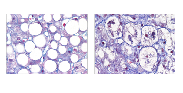
What makes this discovery groundbreaking is that both subtypes exhibit similar histological features under the microscope or on imaging, making them indistinguishable using traditional diagnostic methods. However, their markedly different clinical outcomes underscore the critical need for advanced diagnostic tools and personalized interventions.

Transforming Diagnosis and Treatment

This study empowers clinicians to move beyond one-size-fits-all approaches to treating MASH. By leveraging simple clinical markers—age, BMI, HbA1c, LDL cholesterol, triglycerides, and ALT—patients can be stratified into specific subtypes, enabling tailored treatments:

  • Liver-Specific MASH: Focus on therapies to halt liver damage and prevent progression to cirrhosis or liver cancer.
  • Cardiometabolic MASH: Emphasize aggressive management of metabolic and cardiovascular risks alongside liver disease treatment.

This research marks a turning point,” says Prof. François Pattou. “We now have a clear path to develop subtype-specific treatments that can improve patient outcomes.”

Why This Discovery Matters

MASH is the most severe manifestation of MASLD, with the potential for devastating health consequences. However, its heterogeneity has often been overlooked, leading to inconsistent treatment outcomes.

This manuscript offers a transformative perspective on MASH and its heterogeneous outcomes,” notes an anonymous reviewer. “Thoughtfully conducted on large, well-characterized cohorts, it opens new doors for precision medicine in this field.

The Science Behind the Subtypes

The study utilized data from the French ABOS cohort of 1,389 individuals with obesity and validated its findings across three European MASLD cohorts (Italy, Belgium, and Finland), comprising 1,099 participants, as well as imaging data (MRI) from over 6,000 UK Biobank participants. By integrating clinical traits with liver transcriptomics and plasma metabolomics, researchers uncovered distinct biological pathways driving each subtype.

This discovery sheds light on why current treatments often yield inconsistent results,” explains co-lead researcher Prof. Stefano Romeo. “It was a true ‘eureka’ moment for our team.”

A New Era for MASH Treatment

This breakthrough highlights the urgent need for subtype-specific care, paving the way for innovative treatments and personalized medicine. Future research will explore how these subtypes respond to lifestyle interventions, pharmacological therapies, and other treatments in diverse populations.

We’ve always known MASH was heterogeneous,” concludes Prof. Romeo. “Now, we finally have a roadmap to turn these insights into real-world solutions for patients.

Global Reach: Inserm Celebrates in Washington, DC, 60 Years of Research and Innovation

Affiche 60 ans de l'Inserm© Zebra /Inserm

In celebration of its 60th anniversary, Inserm is emphasizing its scientific collaboration with the United States and Canada. This privileged and fruitful relationship with North America will be highlighted during a day of scientific and institutional exchanges on October 29, 2024, at the French Embassy in Washington, DC. The event will be attended by Professor Didier Samuel, President and CEO of Inserm, along with representatives from numerous partner institutions. The agenda will include the initiation of new collaborations with leading American scientific institutions, the presentation of the 2024 Inserm International Award to a distinguished researcher, the strengthening of Inserm’s presence in Washington, DC, and the launch of the Inserm Abroad Network.

Since its founding in 1964, Inserm has made significant contributions to improving individual health. The Institute thus played a key role in several major medical advances, including cancer treatments, gene therapy, the discovery of HIV, and, more recently, the development of the Nutri-Score label. Inserm stands as a key actor in both national and international biomedical research, being the leading European health research organization and the second largest globally.


The United States: Inserm’s Primary International Partner

Among the 7,000 international collaborations that Inserm engages in annually, nearly one in five involves the United States. With a total of 3,000 joint scientific publications, the United States is Inserm’s primary partner.

Beyond publications, Inserm is involved in cooperative agreements focused on significant health issues for both countries, including:

  • Mental health and addiction, in collaboration with the National Institute on Alcohol Abuse and Alcoholism at the NIH;
  • Human genetics of infectious diseases, with the Rockefeller University;
  • Public health topics such as the relationship between health and the environment, aging, and chronic diseases, in partnership with the Mailman School of Public Health;
  • Issues related to blindness and vision, in collaboration with the University of Pittsburgh.

To further strengthen this cooperation, the two countries have established a virtual joint research unit, known as Joint Labs[1], in collaboration with the University of Pittsburgh (ophthalmology and vision). Such Joint Labs were also created with two Canadian institutions: Laval University (viral respiratory infections) and the University of Ottawa ( neuromuscular diseases).


The 2024 Inserm International Award

On October 29, Miriam Merad, a French-Algerian researcher and Chair of the Department of Immunology and Immunotherapy and of the Precision Immunology Institute at Mount Sinai School of Medicine (New York), will receive the 2024 Inserm International Award. Dr. Merad’s research focuses on cancer immunology and inflammatory diseases, examining the roles of innate and adaptive immune cells in these conditions. The 2024 Inserm International Award honors her dedication to Franco-American collaboration, particularly through her research partnerships with Inserm research teams.


Reinforcing Inserm’s Presence in the United States

The year 2024 also marks the arrival of a new director and team at Inserm’s Washington, DC, Representation Office, located within the French Embassy. This office represents a vital tool for enhancing Inserm’s influence among key institutional actors in the United States and Canada. It supports existing collaborations and promotes the development of new partnerships aligned with Inserm’s strategic research priorities.

“This event held in Washington, DC, reaffirms the significance of the Institute’s collaborations with the United States. By maintaining connections with local stakeholders and federal agencies, the Inserm office plays a crucial role in identifying new opportunities within the country,” states Fabien Agenes, the new Director of Inserm’s North American Representation Office.


Inserm Abroad: A New Network to Expand Inserm’s Global Reach

Simultaneously, October 29, 2024, will see the official launch of the Inserm Abroad Network, a new initiative that aligns with Inserm’s strategy to enhance its international attractiveness and influence. By uniting Inserm researchers and affiliates who work abroad within prestigious research institutions across various disciplines, the Inserm Abroad Network aims to address contemporary health research challenges and strengthen the Institute’s global partnerships.


For Professor Didier Samuel, President and CEO of Inserm, “All these new initiatives reflect the Institute’s commitment to scientific excellence, supported by a strong policy of international engagement. The United States is a historic partner of Inserm, and I am pleased that our 60th anniversary symbolizes our strengthened collaborations in key research areas essential for the health of our French and North American citizens.”


[1] “Joint Lab” is an Inserm label awarded to long-term collaborations of excellence between an Inserm laboratory and a foreign partner laboratory.

Predominance of zoonotic transmission of the mpox virus in the Democratic Republic of the Congo

mpoxColorized transmission electron micrograph of mpox virus particles (orange) found within an infected cell (brown), cultured in the laboratory. Image captured at the NIAID Integrated Research Facility (IRF) in Fort Detrick, Maryland. © NIAID

Central Africa, especially the Democratic Republic of the Congo (DRC), is highly affected by successive mpox outbreaks. Until now, the extent of genetic diversity of the virus had not been well characterised in this region of the world. For the first time, as part of the AFROSCREEN project[1] and the PANAFPOX project[2], teams from the Institut National de Recherche Biomédicale (INRB) in DRC, IRD and Inserm have provided important new information on the genetic diversity of mpox virus circulating in DRC and on the predominant route of transmission. Results of this work have just been published on the Cell website on 24 October 2024.  

Mpox is a viral zoonosis most likely transmitted from rodents to humans. The first case was reported in the Democratic Republic of the Congo (DRC) in 1970. This disease, which had been endemic mainly in rural and forested areas of West and Central Africa for several decades, spread around the world in 2022, including cases in European countries. For the first time, the disease has spread rapidly between individuals through sexual contact, a mode of transmission rarely observed before. This growing mpox outbreak has led to a declaration of a public health emergency of international concern.

The mpox virus can be divided into two major clades*. Clade I, the ‘historical’ strain of the virus, found in the Congo Basin and Central Africa, and clade II, present in West Africa, with clade IIb, found in Nigeria and responsible for the 2022 mpox outbreak.

The most affected country is DRC, where the number of cases has doubled in recent years, rising from around 3,000 in 2021 to 5,600 in 2022, and from over 14,000 in 2023 to over 20,000 by 1st September 2024. This increase is accompanied by an alarming expansion of the geographical spread, first in eastern DRC but also in urban areas including the capital city Kinshasa, and in neighbouring countries (Rwanda, Burundi, Kenya and Uganda) previously unaffected by mpox. On 14 August 2024, the substantial rise in mpox cases led the World Health Organisation to declare the mpox outbreak a public health emergency of international concern for the second time. These new infections have been attributed to clade Ib, a new variant of clade I with increased numbers of APOBEC3** mutations, which indicate that the virus has adapted to human hosts.

The study, conducted in DRC between February 2018 and March 2024, aimed to investigate whether the rising numbers of mpox infections in the country were due to zoonotic spillovers or viral evolution linked to human adaptation and sustained human-to-human transmission. A total of of 337 viral genomes from 14 out of 26 provinces were successfully sequenced. All new sequences from the South Kivu province, in eastern RDC, corresponded to the recently described clade Ib. This variant is associated with sexual contact and sustained human-to-human transmission, and the limited genetic diversity is compatible with its emergence in 2023. All other genomes from other provinces (i.e. 95% of cases) belonged to clade Ia, which is characterised by high genetic diversity and a low number of APOBEC3 mutations compared with clade Ib. The study results therefore suggest a predominance of zoonotic transmission of mpox in the human population. The co-circulation of genetically diverse viral lineages in small geographical areas even suggests multiple zoonotic introductions over a short period from one or more reservoir species.

For the first time, a large number of clade I mpox sequences have been analysed. This study has provided important new information about the genetic diversity of mpox viruses circulating in DRC, and shows that there are two modes of transmission: zoonotic transmission (clade Ia), which predominates, and human-to-human transmission, which is emerging (clade Ib) in South Kivu and is spreading rapidly to other regions in DRC and neighbouring countries. The presence of several clade I variants in urban areas, particularly Kinshasa, also highlights the need to continue monitoring the evolution and diversity of the virus in DRC as well as its modes of transmission. It is also urgent to better document the animal reservoirs involved in zoonotic transmission.


* a group of organisms,incliuding a particular organism and all of its descendants.

** APOBEC3 (Apolipoprotein B Editing Complex) are proteins that help protect against viral infections.


[1] Project coordinated by ANRS MIE in partnership with IRD and the Institut Pasteur, and financed by the French Development Agency (AFD).

[2] Multidisciplinary project with a “One Health” approach, funded by ANRS MIE.

Resistance mutations to nirsevimab are rare in respiratory syncytial virus (RSV)

vaccination d'un bébéNirsevimab is an antibody targeting the respiratory syncytial virus (RSV). Available in France since September 2023, it is indicated in neonates and infants for the prevention of bronchiolitis caused by RSV. © AdobeStock

Nirsevimab is an antibody targeting the respiratory syncytial virus (RSV). Available in France since September 2023, it is indicated in neonates and infants for the prevention of bronchiolitis caused by RSV. However, its widespread use raises the question of the emergence of resistance mutations. The POLYRES study, the largest prospective surveillance study of nirsevimab breakthrough infections to date, has just delivered its conclusions. This work, coordinated by Prof. Slim Fourati and Prof. Marie-Anne Rameix-Welti[1], was funded by the ANRS MIE with the support from the French Ministry of Higher Education and Research as part of the EMERGEN Consortium.[2] Scientists from AP-HP (including Henri Mondor University Hospitals), Inserm, Institut Pasteur and the Universities of Paris-Est-Créteil and Versailles-Saint-Quentin-en-Yvelines, members of the ANRS MIE virology network teams, have shown that nirsevimab resistance mutations in RSV are very rare. The results of this study have just been published in the Lancet Infectious Diseases on October 15, 2024.

Respiratory syncytial virus (RSV) is the main cause of bronchiolitis, a lower respiratory tract infection in infants. Two groups of RSV (RSV-A and RSV-B) circulate alternately or together. Every year, RSV is responsible for more than 33 million cases of bronchiolitis worldwide, leading to the deaths of 100,000 children, mainly in low-income countries. In France, the disease is responsible for around 480,000 cases a year. It is by far the most common cause of hospitalisation in children, leading to more than 26,000 paediatric hospitalisations every year. Nirsevimab, a new neutralising antibody* against the virus, became available in France in September 2023. This monoclonal antibody** targets a specific antigenic site (the epitope*** Ø) of the RSV surface F protein, which is involved in viral multiplication, and blocks the virus. Due to the genetically variable forms of RSV, there is a theoretical risk of the emergence of variants carrying mutations resistant to neutralisation by nirsevimab, even in the absence of selection pressure. This risk could increase with the widespread preventive use of nirsevimab. During the phase IIb/III clinical trials, only 48 RSVs infecting children treated with nirsevimab could be studied, and escape mutations# were found in two of them.  The aim of the POLYRES study was to assess the risk of viral escape from nirsevimab in a large cohort using a large, multicentre, real-life observational study conducted during the 2023-2024 winter season.

The study included 695 RSV infected infants, 349 of whom had received nirsevimab prophylaxis. RSV-A was the most dominant circulating virus this season and was found in 86.6% of infected children. The teams analysed the characteristics of RSV-A and RSV-B in nasopharyngeal swabs collected as part of the children’s routine care. Full-length sequencing of the viral genome was conducted to identify potential mutations in the Ø site, the nirsevimab binding site (genotypic analysis§ ). The ability of nirsevimab to inhibit viral multiplication in cell culture was also investigated (phenotypic analysis¥ ). Analysis of 472 RSV-A viruses (half from treated children) revealed no nirsevimab resistance mutation in the Ø site of the F protein. Of the 73 children infected with RSV-B, 24 had received nirsevimab prophylaxis. In these 24 children, two isolates of RSV-B had resistance mutations to the antibody. One mutation has been described before and the other is described here for the first time.


“This study is the largest surveillance study of nirsevimab virological failures to date. It was made possible thanks to collaborative synergy with the consortium of virologists at the ANRS MIE. It is a nationwide project that will help identify the resistance phenomenon associated with the widespread use of the drug. This type of study is essential for analysing the evolutionary dynamics of viruses, in the light of existing medical solutions” explains Prof Marie-Anne Rameix-Welti, head of the National Reference Centre for Respiratory Infection Viruses at the Institut Pasteur, and head of the M3P unit (Institut Pasteur, Inserm U1173).


“The low prevalence of nirsevimab resistance mutations in treated patients is reassuring. However, escape mutations have been observed in a few RSV-Bs from treated patients, prompting caution and highlighting the importance of active molecular surveillance in the context of future wider global use of nirsevimab. These results are essential in the fight against this disease and in anticipating any form of resistance“, adds Prof Slim Fourati, Head of the Virology Unit-Respiratory Viruses, CHU Henri Mondor, Inserm U955.

In conclusion, the results of the POLYRES study support the continued use of nirsevimab for RSV prophylaxis in all newborns worldwide.


* Neutralising antibodies are specific antibodies that prevent infection by blocking the virus from entering the target cells. They do this by forming an antigen-antibody complex which inhibits the biological activity of the antigen (a substance foreign to the body capable of triggering an immune response aimed at eliminating it).

** Monoclonal antibodies consist of a single type of antibody (polyclonal antibodies have several types). They are used in medicine.

*** Part of a molecule recognised by an antibody.

# Escape mutations enable the virus to thwart the action of antibodies in the human immune system.

  • Genotypic tests are based on the identification of mutations that confer resistance to the virus.

¥ Phenotyping, carried out using phenotypic tests, makes it possible to define the sensitive or resistant nature of the virus. This is done by culturing the virus in the presence of the antiviral being studied.

[1] Head of the National Reference Centre for Respiratory Infection Viruses at the Institut Pasteur and head of the M3P unit (Institut Pasteur, Inserm U1173)

[2] Coordinated by Santé publique France and ANRS MIE

Foods with low Nutri-Scores associated with an increased risk of cardiovascular diseases

photo d'un cœur avec logo NutriScoreL’alimentation serait responsable d’environ 30% des décès dus aux maladies cardiovasculaires. © Mathilde Touvier

Cardiovascular diseases are the leading cause of mortality in Western Europe, accounting for 1/3 of deaths in 2019. Diet is thought to be responsible for around 30% of such deaths. Nutrition-related prevention policies therefore constitute a major public health challenge for these diseases.

In an article to be published on 11 September 2024 in Lancet Regional Health – Europe, researchers from the Nutritional Epidemiology Research Team (CRESS-EREN), with members from Inserm, Inrae, Cnam, Université Sorbonne Paris Nord and Université Paris Cité, in collaboration with researchers from the International Agency for Research on Cancer (WHO-IARC), report an increased risk of cardiovascular diseases associated with the consumption of foods that rank less favourably on the Nutri-Score scale (new 2024 version) within the European cohort EPIC. A total of 345,533 participants from the cohort, spread across 7 European countries and followed for 12 years, were included in the analyses.

Officially adopted in France in 2017 (and in 6 other European countries since), the Nutri-Score aims to provide rapid information on the nutritional quality of foods and drinks to help and encourage consumers to compare them and choose those that offer a better nutritional quality. In parallel, it encourages manufacturers to improve the nutritional quality of their products.

The Nutri-Score has 5 categories, ranging from A (dark green – higher nutritional quality) to E (dark orange – lower nutritional quality). An algorithm ranks each product according to its levels – per 100 g – of energy, sugars, saturated fatty acids and salt (to limit) and proteins, fruits, vegetables and pulses (to favour).

A number of studies published in international scientific journals have shown the validity of Nutri-Score in characterising the nutritional quality of foods and its efficacy in guiding consumers towards more nutritious choices (over 140 publications). In particular, links between the consumption of foods with a less favourable Nutri-Score (lower nutritional quality) and an increased risk of cardiovascular diseases have so far been observed in French studies (SU.VI.MAX and NutriNet-Santé cohorts). Studies in France, UK, Spain and Italy have also seen similar associations with an increased risk of various chronic diseases as well as higher mortality.

In this new study, the researchers focused on the latest version of the Nutri-Score algorithm (updated in 2024, see box), linked to the risk of cardiovascular diseases, in a large population spread across 7 European countries, with the aim of providing new scientific evidence for validating the Nutri-Score on a European scale. It follows two studies published in 2018 and 2020 in the same population on cancer risk and mortality.

A total of 345 533 participants from the EPIC (European Prospective Investigation into Cancer and Nutrition) cohort were included in the analyses. During the follow-up (12 years, between 1992 and 2010), 16 214 participants developed a cardiovascular disease (6 565 of whom had myocardial infarction and 6 245 stroke). The findings show that the participants consuming on average more foods with less favourable Nutri-Score, reflecting lower nutritional quality, were at increased risk of cardiovascular diseases, particularly myocardial infarction and stroke. These associations were significant after a large number of sociodemographic and lifestyle factors were taken into account.

‘These findings confirm the relevance of Nutri-Score as a public health tool to guide consumers in their food choices with the goal of preventing chronic diseases’, emphasises Inserm researcher Mélanie Deschasaux-Tanguy.


‘They also provide key elements to support the adoption of Nutri-Score as a mandatory nutritional logo in Europe’, explains Mathilde Touvier, Inserm research director.


A new version of the Nutri-Score in 2024

Changes to the calculation of the Nutri-Score[2] were recently proposed by the international scientific committee responsible for its monitoring in order to improve its consistency with nutritional recommendations. This new version of the Nutri-Score is expected to come into force in 2024 with a gradual roll-out in the months to come. However, due to European labelling regulations, manufacturers are under no obligation to use Nutri-Score on their packaging.

While many companies and brands (over 1,400 in France) have so far committed to using Nutri-Score on their products, harmonisation at European level is needed to ensure the mandatory implementation of a single logo that is effective and useful for citizens. This harmonisation is envisaged as part of the European Commission’s Farm to Fork strategy.

[1] https://sante.gouv.fr/prevention-en-sante/preserver-sa-sante/nutrition/nutri-score/etudes-et-rapports-scientifiques/

[2] https://theconversation.com/en-2024-le-nutri-score-evolue-pourquoi-et-que-faut-il-en-retenir-221697

To find out more: watch the Inserm program “Nutri-Score, we tell you everything” (french only).

Ebola: a study in Guinea reveals persistent immunity five years after vaccination

In this work, scientists analyzed cellular immunity in 230 participants in Guinea © Aurélie Wiedemann

Epidemics of Ebola virus disease occur periodically in several sub-Saharan African countries. Two vaccines have already received WHO prequalification[1] against the Ebolavirus Zaire species. However, information on the long-term immune response to these vaccines is still insufficient. We need to consolidate our knowledge on this subject to continue developing the safest and most effective vaccination strategies possible, for both adults and children. In a new study conducted in Guinea, scientists from VRI, Inserm and Université Paris-Est Créteil (U955 Institut Mondor de recherche biomédicale)[2] have taken a further step in this direction. They have shown that the cellular immune response induced by three different vaccine strategies is maintained for up to five years after vaccination. These results, which support current vaccine strategies against Ebola, are published in Nature Communications.

The Ebola virus causes high fevers and hemorrhages, often resulting in death. Many countries in sub-Saharan Africa regularly face epidemic outbreaks.  the Ebola virus caused the largest epidemic known to date in West Africa in 2014. It has since re-emerged several times in the DRC, but also in Guinea. Vaccination is today one of the most effective tools for combating the disease, and one of the major challenges for research and public health strategy is to continue improving knowledge of the immune response induced in the long term by currently available vaccines.

Since 2019, two vaccines have obtained WHO prequalification against the Ebolavirus Zaire strain: the rVSVΔG-ZEBOV-GP vaccine (Ervebo®), developed by Merck, Sharpe & Dohme, Corp. and the vaccine regimen comprising Johnson & Johnson’s Ad26.ZEBOV (Zabdeno®) vaccine and Bavarian Nordic’s MVA-BN-Filo (Mvabea®) vaccine.

In 2022, the international PREVAC consortium (see box at the end), comprising Inserm, NIH and the London School of Hygiene and Tropical Medicine (LSHTM), published a study in the New England Journal of Medicine examining the safety and efficacy of three vaccine regimens:

  • The first vaccine regimen tested consisted of one dose of Ad26.ZEBOV vaccine, followed 56 days later by one dose of MVA-BN-Filo;
  • The second regimen involved a dose of rVSVΔG-ZEBOV-GP;
  • Finally, the third schedule began with a dose of rVSVΔG-ZEBOV-GP, followed 56 days later by a booster dose of the same vaccine.

Published results showed a high serum antibody response  12 months after vaccination. However, it was essential to obtain information on the long-term maintenance of the response, and in particular regarding the cellular response (see box below).

Humoral response and cellular response

Adaptive immune responses fall into two broad categories: the humoral response, based on the production of antibodies, by B lymphocytes, that recognize and neutralize the virus before it infects cells, and the cellular response, where CD8+ T lymphocytes identify and destroy already-infected cells to limit the spread of the virus. CD4+ T lymphocytes play a crucial role in helping B lymphocytes produce antibodies, boosting the effectiveness of the immune response.

In this study, scientists looked specifically at the short-, medium- and long-term (5 years) cellular response in participants following vaccination with three different vaccine regimens.

In December 2023, the 5-year follow-up of participants in the PREVAC clinical trial was completed. The results are being analyzed and will enable assessment of long-term immunity. sIn an ancillary study, scientists analyzed cellular immunity in 230 participants in Guinea, just after vaccination, at one year and five years post-vaccination.

This is the first study from the PREVAC consortium to look specifically at participants’ cellular immune response. It completes the knowledge already acquired on the humoral response at one year and offers the first follow-up results at 5 years”, emphasizes Aurélie Wiedemann, immunologist at VRI and the Institut Mondor de recherche biomédicale (Inserm/Université Paris Est Créteil) and first author of the study.

Using blood samples taken in Conakry, the scientists were able to analyze the response of CD4+ and CD8+ T lymphocytes to vaccination. They showed the presence of anti-Ebola CD4+ T cells five years after vaccination, regardless of the vaccination regimen. The persistence of these responses is important for the maintenance of humoral immune memory in the event of exposure to the Ebola virus. In a subgroup of volunteers, the authors show a correlation between CD4+ T cell response and the quantity of specific antibodies in the long term.

While CD4+ T response is important for maintaining an antibody response, the presence of cytotoxic CD8+ T cells is also crucial for effective antiviral protection. A specific CD8+ T cell response was demonstrated in individuals vaccinated with two of the three vaccine regimens.

These results will shortly be supplemented by humoral response data – on antibody production – from all PREVAC consortium countries, on a larger number of participants. Nevertheless, these results are promising and suggest that vaccination against the Ebola virus can induce long lasting immunity. They also pave the way for adjusting current vaccination strategies, by making it possible to assess, for example, the need for a long-term booster vaccination”, explains Prof. Yves Lévy, Director of the VRI and final author of the study.

In 2020, the team also published a study in Nature Communications on the immunity of Ebola survivors two years after discharge from hospital. One of the next avenues of research could be to compare the long-term immune response of these survivors with that induced by vaccination, in order to identify possible correlates of protection [1] against infection, as these are currently undetermined.

Thus, this new study could help identify vaccine responses that would be effective against the infection, improve current vaccine strategies, and define long-term booster vaccine strategies to maintain protection for particularly at-risk individuals such as healthcare workers in Africa.

About PREVAC

PREVAC (Partnership for Research on Ebola Vaccinations; NCT02876328) is an international consortium conducting researches in West Africa to evaluate the safety and efficacy of Ebola vaccination.

The project is co-funded by Inserm, NIAID, the London School of Hygiene & Tropical Medicine (LSHTM) and the College of Medicine and Allied Health Sciences (Comahs), with support from Guinea, Liberia, Mali and Sierra Leone. On-the-ground support from the NGO Alima was also crucial in encouraging the population to take part in the research and in monitoring the volunteers. Manufacturers Merck and Janssen supplied the vaccines used in the trial.

The project also benefited from additional funding to continue long-term follow-up of volunteers (PREVAC-UP project coordinated by Inserm) via the European and Developing Countries Clinical Trials Partnership (EDCTP2) program supported by the European Union.


[1] Prequalification means that a vaccine meets WHO standards of quality, safety and efficacy. On the basis of this recommendation, UN agencies and the Gavi Alliance can purchase the vaccine for at-risk countries.

[2] This analysis was carried out in collaboration with the SISTM team at the Bordeaux Population Health Research Center (UMR 1219 Université de Bordeaux/Inserm).

[3]  These are immunological markers associated with protection against infection: for example, post-vaccination antibody levels against hepatitis B are a good correlate of protection. In other words, in the context of vaccination, they designate the parameters that scientists monitor to find out whether the vaccine works and protects effectively against infection.

A Parkinson’s treatment could delay progression of one of the forms of AMD

DMLAAge-related macular degeneration (AMD) is the leading cause of visual disability in people over 50 years of age. © Adobe Stock

Age-related macular degeneration (AMD) is the leading cause of visual disability in people over 50 years of age. Improving the treatment offering for patients is a major challenge for research. In a new study, a team of researchers from Inserm, CNRS and Sorbonne Université at the Vision Institute[1] in Paris describes the efficacy of dopaminergic drugs in slowing the progression of one of the forms of the disease, namely the neovascular or ‘wet’ form which is characterised by the proliferation of dysfunctional blood vessels under the retina. These specific drugs are already used in the treatment of Parkinson’s disease. These findings have been published in The Journal of Clinical Investigation.

AMD  is a multifactorial disease of the retina that affects people over 50 years of age. It is when part of the retina – the macula – degenerates, with the potential loss of central vision. Although highly incapacitating, it never causes complete blindness since the peripheral part of the retina remains intact.

There are two forms of the disease whose prevalence is roughly equivalent: the neovascular form – known as the ‘exudative’ or ‘wet’ form and the atrophic – ‘advanced dry’ form (see box).

While there is currently no curative treatment for the dry form of the disease, the neovascular form can be slowed down by regular injections administered directly into the eye (intravitreal injections). Although necessary, these injections can represent a major therapeutic burden due to their frequency, which is monthly or bimonthly depending on the course of the disease. It is therefore useful to continue to identify new alternatives for patients.


Medicines for Parkinson’s

Previous epidemiological studies have already shown a possible association between Parkinson’s disease and a reduced risk of neovascular AMD[2]. In this new study, researchers from Inserm, CNRS and Sorbonne Université at the Vision Institute explored the underlying mechanisms that would explain this potential protection.

In cell and animal models, the scientists have shown that L-Dopa, a drug in the dopaminergic family[3] used to treat Parkinson’s disease, activates a specific receptor in the brain known as DRD2. This activation blocks the formation of new blood vessels in the eye, which is a key process in the development of neovascular AMD.

To go further, the team then analysed the health data of over 200 000 patients with neovascular AMD in France[4]. They showed that those patients who took L-Dopa or other drugs that inhibit the DRD2 receptor (DRD2 agonists) to treat their Parkinson’s disease developed neovascular AMD later in life and required fewer intravitreal injections. Indeed, such patients developed the disease at 83 years of age instead of 79 years of age for the other patients.

‘These findings open up new perspectives for patients with wet AMD. We now have a serious avenue for delaying the progression of this disease and reducing the burden of current treatments’, explains Florian Sennlaub, Inserm Research Director at the Vision Institute (CNRS/Sorbonne Université/Inserm).

Thibaud Mathis, university professor and hospital practitioner in the ophthalmology department of Croix-Rousse Hospital-Hospices civils de Lyon, and researcher at Université Lyon 1, as well as at the Vision Institute, agrees: ‘These findings suggest that dopaminergic drugs, beyond their role in Parkinson’s disease, could have a beneficial effect in the prevention and treatment of neovascular AMD.’

While more in-depth clinical studies will be needed to confirm these findings and evaluate the efficacy and safety of these drugs in the treatment of AMD, this discovery opens up encouraging new perspectives for the fight against the neovascular form, offering hope of a more effective and less burdensome treatment for patients.


Two forms of AMD

Wet AMD is characterised by the proliferation of new dysfunctional vessels under the retina. Blood can leak through their walls and lead to the formation of macular oedema from which blood sometimes escapes, leading to retinal haemorrhages.

The wet form of AMD progresses rapidly if not managed. Previously, loss of central vision could occur within weeks or even days. This process can now be stopped thanks to drugs (anti-VEGF) injected into the eye, which inhibit the growth of new vessels. However, after several years of treatment, the disease can progress to an atrophic form.

In atrophic or ‘advanced dry’ AMD, the photoreceptors in the macula gradually disappear, followed by the retinal pigment epithelial cells. This process generates holes of increasing size in the macula, visible by simply observing the retina (dilated eye exam). This process is slow and usually takes between five and ten years before the patient loses their central vision. Currently, no treatment for this form of AMD is approved in Europe.

Mixed forms of the disease may be observed, and each of these two forms may precede the appearance of the second.


[1]This research is the result of collaboration with teams from Université de Lyon, Lyon University Hospital, Université de Bourgogne and the Brain Institute in Paris.

[2]Levodopa Is Associated with Reduced Development of Neovascular Age-Related Macular Degeneration, Max J Hyman et al. Ophthalmology retina 2023

[3]Dopaminergic drugs provide the dopamine needed for the brain to function. In Parkinson’s disease, dopamine reduces the intensity of tremor, rigidity and akinesia.

[4] Data from the French national health information database (Système National des Données de Santé [SNDS]).

Hepcidin, iron hormone in the skin: a new target in the treatment of psoriasis?

Présence d’hepcidine (visualisée en marron) dans l’épiderme d’un patient souffrant de psoriasis pustuleuxPresence of hepcidin (in brown) in the epidermis of a patient with pustular psoriasis. © Élise Abboud

Psoriasis is a chronic inflammatory disease characterised by the rapid and excessive multiplication of skin cells. Although research is progressing and certain treatments are already able to improve the daily lives of patients, this disease remains incurable. The team led by Carole Peyssonnaux, Inserm Research Director at Institut Cochin (Inserm/CNRS/Université Paris Cité) has shown that a hormone that regulates iron in the body, called hepcidin, is produced by the patients’ skin and is essential for triggering psoriasis. This discovery opens up new avenues for treatment. Drugs that block the action of hepcidin could be a therapeutic alternative in psoriasis. These findings have been published in Nature Communications.

Psoriasis is a chronic inflammatory disease that primarily affects the skin. It is common and affects 2 to 3% of the world’s population. Despite many treatment options available to improve patient care, psoriasis remains a chronic condition with no definitive cure.

Characterised by red patches covered with scales, the disease manifests as an excessive proliferation of epidermal cells and an excess of immune cells in the skin, accompanied by a local inflammatory reaction.

Over the last few decades, much progress has been made in understanding the disease, such as the identification of certain genetic factors. Several studies, whose findings are still little known by the scientific community, have also shown that there is an accumulation of iron in the skin of psoriasis patients. We know that the regulation of iron levels in the body is controlled by a hormone called hepcidin. While hepcidin is primarily synthesised by the liver, it can be produced by other organs or tissues under disease conditions.

For years, the Iron and Immunity team at Institut Cochin, led by Inserm Research Director Carole Peyssonnaux, has studied hepcidin closely. Despite the proven presence of iron in the epidermis of psoriasis patients, the production by the skin and the potential role of this ‘iron hormone’ in psoriasis had never been investigated. The researchers therefore decided to study this avenue more closely.

The team[1] started by showing that hepcidin was expressed in the skin of patients with psoriasis, particularly in severe forms such as pustular psoriasis, which is characterised by the accumulation of a type of white blood cell – neutrophils – within the epidermis.

To further study the role of hepcidin in psoriasis, the team then developed new mouse models in which the hepcidin gene was specifically inactivated or overexpressed in the epidermis. The scientists then showed that when this gene was activated, certain characteristics of psoriasis were induced, including skin lesions and the recruitment of neutrophils in the epidermis. Conversely, when the gene was inactivated, the psoriasis markers disappeared.

‘Hepcidin plays a key role in the development of psoriasis. Based on our findings, we show that when psoriasis is triggered, the hepcidin produced by the epidermis plays a crucial role in retaining iron in the skin cells. With iron being an essential metal for cell proliferation, this retention promotes cell division in the epidermis of “psoriatic” skin. What is more, hepcidin-mediated iron retention also contributes to the recruitment of neutrophils, another characteristic of psoriatic skin lesions, particularly pustular’, explains Peyssonnaux.

The next step would be to explore these findings in greater depth, with the goal of developing drugs to block the action of hepcidin and therefore potentially benefit patients with psoriasis, particularly those suffering from an acute and resistant form. With this in mind, the team is developing, with the support of Inserm Transfert[2], new drugs capable of neutralising hepcidin, in order to test them in animal models of psoriasis.

‘In the future, if our findings prove conclusive, such drugs could be used as maintenance therapy following a flare-up, during phases of remission, to prevent recurrence of the disease. Additional studies will determine whether hepcidin also plays a role in other inflammatory skin diseases’, concludes Peyssonnaux.


Read our report (only available in French) : Psoriasis, des traitements le plus souvent efficaces

[1]In collaboration with the team of Selim Aractingi (Cochin Hospital) and Hervé Bachelez (Saint-Louis Hospital)

[2] Patent WO2016/146587 / EP3268027B1 and US11203753B2

Scientists identify immune cells responsible for cancer

Illustration médicale en 3D de l'anatomie masculine - Cancer du côlon ; côlon descendant.Around 30% of cancers develop in the wake of chronic localised inflammation. This is particularly the case of certain colorectal cancers, or cancers of the small intestine, liver or pancreas. © Adobe Stock

Nearly one in three cancers develops following chronic inflammation, whose origin remains unclear. In a new study, researchers from Inserm, CNRS, Université Claude-Bernard Lyon 1 and the Léon Bérard Centre at the Cancer Research Center of Lyon[1]identified lymphocytes involved in the inflammatory processes and that are thought to be implicated in the generation of these cancers. This research opens up new avenues in terms of prevention and treatment, and its findings have been published in Nature Immunology.

Around 30% of cancers develop in the wake of chronic localised inflammation. This is particularly the case of certain colorectal cancers, or cancers of the small intestine, liver or pancreas. However, many questions remained unanswered regarding their development. Is one specific immune cell responsible for the inflammatory process that leads to cancers, or more than one? And if so, which?

Answering these questions is one of the objectives of Inserm Research Director Julien Marie[2] and his team at the Cancer Research Center of Lyon (Inserm/CNRS/Université Claude-Bernard Lyon 1/Léon Bérard Centre) in order to better understand how the disease is initiated.

The researchers were particularly interested in TH17 lymphocytes – a population of immune cells which are already known to be involved in many inflammatory diseases, such as multiple sclerosis and Crohn’s disease.


Cells that cause cancer

The hypothesis was that TH17 cells are not a homogeneous population, but can actually be divided into several subgroups. Using single-cell RNA sequencing approaches, the scientists demonstrated this heterogeneity of TH17 cells within the gut.

‘More specifically, this study shows for the first time that there are actually eight TH17 subtypes with distinct roles. One of them has a tumorigenic role, which means that when certain activation barriers are removed, it will contribute to the development of cancers. On contact with these TH17 cells, the previously healthy gut cells become cancerous’, explains Marie.

The scientists then showed this tumorigenic population to be increased in patients at high risk of cancer. Finally, they also identified that a protein – cytokine TGFβ – is capable of inhibiting the formation of tumorigenic TH17 cells.

‘This study may make clinicians stop and think about the long-term use of immunotherapies in cancer patients, whose aim is to stimulate lymphocytes’, emphasises Marie.

While these therapies have transformed cancer care, they are also known to cause chronic gut inflammation. Therefore it is important to consider, for a given patient, the risks of immunotherapy being accompanied by the emergence of tumorigenic TH17 lymphocytes, which could eventually lead to the development of another cancer. Furthermore, this study lays the foundations for the development of new cancer preventive therapies by blocking the appearance of the TH17 subtype implicated by the scientists in this research.


[1]Scientists from the Institute of Molecular Genetics of Montpellier (CNRS/Université de Montpellier) also participated in this research.

[2]Julien Marie is the winner of the Bettencourt Coups d’Élan Prize for French Research.
Created by the Bettencourt-Schueller Foundation in 2000, this prize had been awarded to 78 French laboratories and over 900 researchers until 2021.

Rapid rollout of smallpox vaccination reduces the risk of mpox

VaccinationVaccination © Inserm/Depardieu, Michel

The ANRS DOXYVAC trial, promoted and funded by ANRS Emerging Infectious Diseases, and conducted by research teams from Inserm, AP-HP, Université Paris Cité and Sorbonne Université (France), shows that a rapid rollout of smallpox vaccination with MVA-BN (Modified vaccine Ankara) among HIV-positive men who have sex with men significantly reduces the risk of mpox by 99%. The results of this study were published in The Lancet Regional Health-Europe on 31 July 2024.

In May 2022, cases of mpox, formerly known as monkeypox, were reported in more than 100 countries where the disease was not endemic. In France, the first case was reported on 19 May, with a rapid increase in infections among men who have sex with men (MSM).

The ANRS DOXYVAC trial, which began in 2021, was designed to study how to improve protection against sexually transmitted infections (STIs) in HIV-positive men who have sex with men.* So, when it became apparent during the course of the study that mpox cases could occur in the trial population, a smallpox vaccination was conducted, as soon as preventive  vaccination in multi-partner MSM was recommended  by French Health Authorities (HAS) on 11 July 2022.

To date, there is no specific vaccine against mpox. However, the virus is closely related to that of human smallpox, and the smallpox vaccine has been shown to be more than 95% effective in protecting against mpox. The vaccine used in the trial, Imvanex® (MVA-BN: Modified vaccine Ankara) from Bavarian Nordic, is a third-generation vaccine. This type of vaccine is produced from smallpox virus with the same antigens as historical smallpox. More specifically, it contains a highly attenuated form of the vaccinia virus known as “modified vaccinia virus Ankara”, a virus that does not cause disease in humans and cannot reproduce in the cells of people who are vaccinated. This means that the vaccine cannot cause local or systemic infections, particularly in immunocompromised people (such as those infected with HIV).

The aim of the analysis among participants of the ANRS DOXYVAC trial was to assess the incidence of mpox infection in participants before (9 May-10 July 2022) and after the launch of the MVA-BN vaccination campaign (from 11 July 2022), and to study the respective effects of vaccination and sexual behaviour adopted during the epidemic period on changes in incidence.

Of the 472 participants included in the analysis, 20% had been vaccinated against smallpox in childhood. The incidence rate** of mpox among trial participants, all of whom were on HIV pre-exposure prophylaxis (PrEP) and had a history of bacterial STIs, was high (49.3 per 1,000 participant-months between 9 May and 20 September 2022).

Vaccination implementation was rapid: 86% (341/398) of eligible participants had received at least one dose of MVA-BN vaccine by 20 September 2022. People were also particularly receptive to the prevention messages and recommendations, especially those with more than ten sexual partners in the last three months who are most at risk. Their sexual behaviour changed significantly before and after 9 May, leading to a reduction in the proportion of people with more than 10 partners in the last 3 months (45% compared with 38%). A significant reduction in the incidence rate of mpox was observed between the period prior to vaccination (67.4 per 1,000 months between May 9 and July 10, 2022) and the period following the launch of the vaccination campaign (24.4 per 1,000 months between July 11 and September 20, 2022).

This trial demonstrated that the rapid implementation of smallpox vaccination with MVA-BN in MSM undergoing PreP significantly reduced the risk of mpox, with an estimated 99% reduction in incidence between the two periods. The reduction in risky sexual behaviour among those most at risk also probably contributed to the reduction in incidence, but to a lesser extent than the vaccination programme.

This study emphasises that identifying and prioritising at-risk populations, delivering targeted prevention messages and awareness campaigns, ensuring the availability of a smallpox vaccine and, above all, the rapid rollout of vaccination to people at risk should enable health authorities to control a future mpox epidemic like the one that occurred in 2022.


* DOXYVAC has demonstrated the efficacy of post-exposure doxycycline in reducing the occurrence of chlamydial infections, syphilis and, to a lesser extent, gonococcal infections.

** Incidence is the number of new cases of a disease in a year in a given population (not to be confused with prevalence, which refers to the number of sick people at a given time). The incidence rate of a disease corresponds to the number of individuals having contracted a disease per 1,000 people exposed to the risk of this disease (in the DOXYVAC trial, it is calculated for one month) (https://www.ined.fr/en/glossary/incidence-of-a-disease/).

Fighting leukaemia by targeting its stem cells

cancer cell made in 3d softwareCancer cell made in 3d software © Fotalia

Acute myeloid leukaemia is one of the deadliest cancers. Leukaemic stem cells responsible for the disease are highly resistant to treatment. A team from the University of Geneva (UNIGE), University Hospital of Geneva (HUG), and Inserm has made a breakthrough by identifying some of the genetic and energetic characteristics of these stem cells. Notably, a specific iron utilisation process. This process could be blocked, leading to the death or weakening of these stem cells without affecting healthy cells. These results, published in Science Translational Medicine, pave the way for new therapeutic strategies.

Acute myeloid leukaemia (AML) is the most common blood and bone marrow cancer in adults. Caused by an increase in immature cells that rapidly destroy and replace healthy blood cells (red and white blood cells and platelets), it is lethal in half of those affected under the age of 60, and in 85% of those over that age.

This unfavorable prognosis may be due to the presence of so-called ”dormant” or ”quiescent” leukaemic stem cells (LSCs), which evade chemotherapy. Often invisible, these cells can ”wake up” and reactivate the disease after an apparently successful course of treatment. Developing therapies that target these cells directly is therefore a major research challenge. However, the mechanisms controlling them are poorly understood.

By identifying genetic and metabolic characteristics specific to LSCs, a team from the UNIGE, HUG, and Inserm is providing new insights, as well as ways of combating the disease. These results, published in Science Translational Medicine, pave the way for a new therapeutic target and its clinical application.


A distinctive genetic signature

Using advanced bioinformatics techniques, and in collaboration with the team of Dr Petros Tsantoulis from the Department of Oncology and Precision Oncology at the HUG, we first established that these quiescent cells contain a unique genetic signature consisting of 35 genes. When we used this signature in large clinical databases of patients with AML, we were able to show that this signature was strongly linked to the prognosis of the disease,” explains Jérôme Tamburini, associate professor in the Department of Medicine and the Centre for Translational Research in Onco-haematology (CRTOH) in the UNIGE Faculty of Medicine and at the Swiss Cancer Center Léman (SCCL), staff physician in the Division of Oncology at HUG, who led this research.


Locking a specific nutrient

The study also highlights a metabolic difference between dormant and active leukaemic stem cells. In general, to survive, cells trigger chemical reactions that enable them to break down nutrients and thus produce energy. This also involves ”autophagy”, a process that allows cells to recycle cellular components to generate new ones and to provide energy in case of a lack of external nutrients. Scientists have discovered that dormant leukaemic stem cells depend on ”ferritinophagy”, a specific form of autophagy targeting ferritin, the main iron storage molecule.

‘This process is mediated by a protein called NCOA4. It controls the availability of iron in cells. By inhibiting it, either genetically or chemically, we observed that leukaemia cells, especially dormant stem cells, are more likely to die, whereas healthy blood stem cells remain intact,” reveals Inserm researcher Clément Larrue, a former post-doctoral researcher in Jérôme Tamburini’s group, currently a post-doctoral researcher at the Toulouse Cancer Research Center, and first author of the study.


Towards clinical trials

Experiments conducted with mouse models have confirmed that blocking the NCOA4 protein reduces tumour growth, viability and self-renewal of leukaemic stem cells. Targeting ferritinophagy through this inhibition pathway could therefore be a promising therapeutic strategy. The compound used to block NCOA4 is in the early stages of development for future clinical trials, under the direction of one of the study’s co-authors, Jun Xu, a professor at Sun Yat-Sen University in China.

The next step for the UNIGE team will be to further explore further the mechanisms of ferritinophagy and its association with mitophagy, another key mechanism in the regulation of LSCs. This new stage of research is supported by the Swiss Cancer League.

fermer