Menu

A Toxin Causing DNA Damage Found in Patients with Urinary Tract Infections

Bactérie E Coli

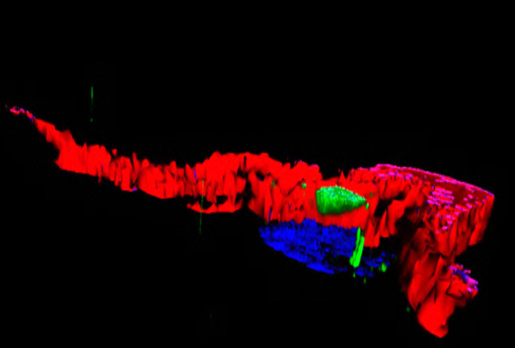
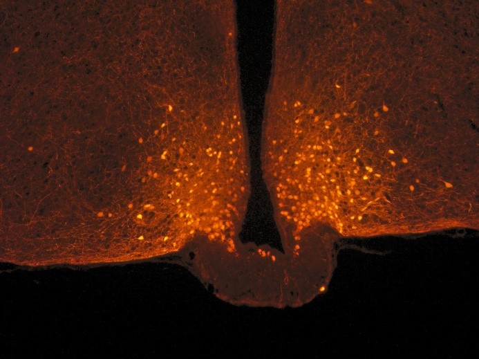
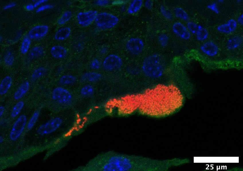
During a urinary tract infection, uropathogenic Escherichia coli bacteria (in red, their DNA) form bacterial communities within superficial cells of the bladder (in blue, their nuclei; in green, their cellular sugar content), here 6 hours following an infection reproduced in mice. © Inserm/Motta, Jean-Paul/Chagneau, Camille

Urinary tract infections affect over 50% of women, in some cases recurrently. E. coli bacteria are very often implicated in their development. For the first time, researchers from Inserm, Toulouse University Hospital, INRAE, Université Toulouse III – Paul Sabatier and École nationale vétérinaire de Toulouse have identified the presence of a toxin produced by these bacteria in the urine of patients, which is thought to damage bladder cell DNA. These findings pave the way for new reflection on how to refine the treatment of patients prone to recurrent urinary tract infections. The study was published in Plos Pathogens on February 25, 2021.

Every year, 150 million people are affected by urinary tract infections. These are more common in women, with more than one in two experiencing them at some point in their lives. This represents a major public health problem, especially since the frequent need for antibiotic treatment is promoting the emergence of antibiotic resistance.

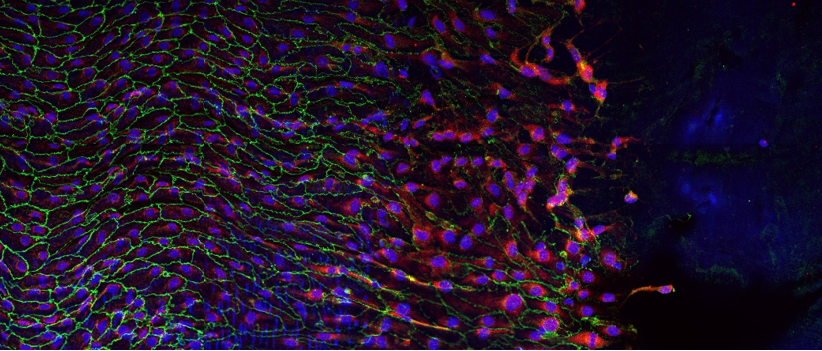
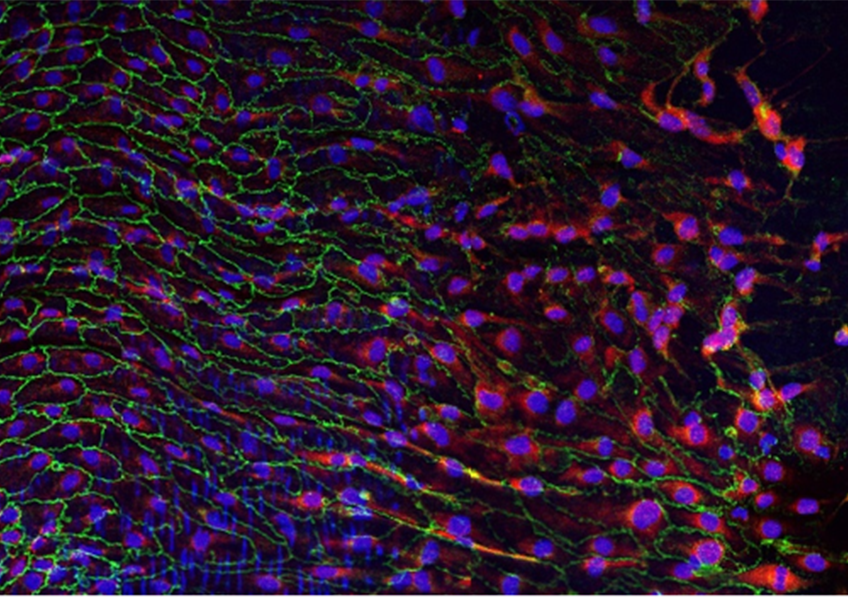
Urinary tract infections develop when the urogenital area is contaminated with bacteria from the gut microbiota. Escherichia coli (E. coli) bacteria are implicated in 80% of these infections[1] and have been the subject of several years of study by Eric Oswald and his team at the Digestive Health Research Institute (Inserm/INRAE/Université de Toulouse III Paul Sabatier/École nationale vétérinaire de Toulouse) in collaboration with various research teams in Toulouse.[2]

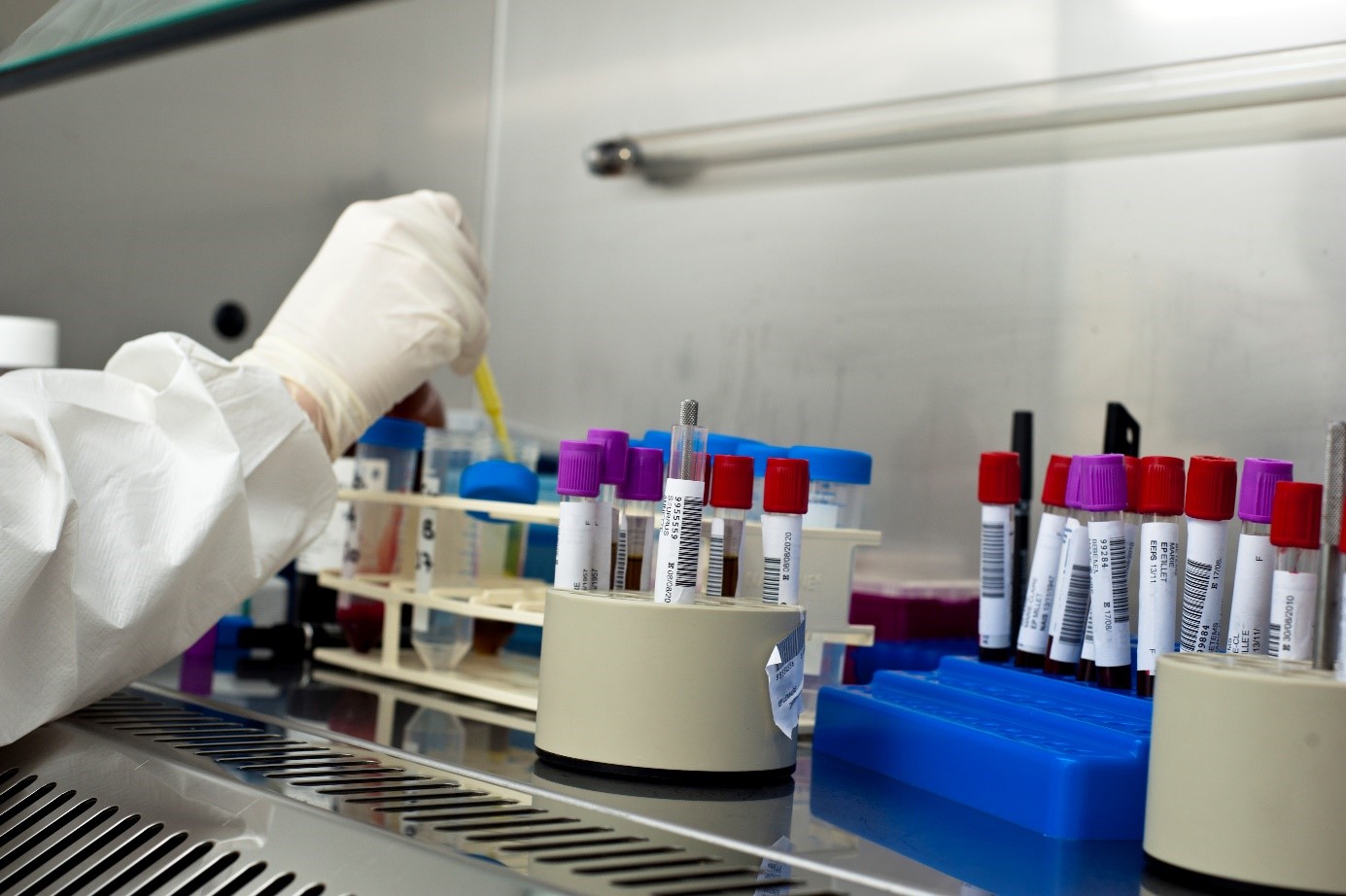
The scientists are particularly interested in the virulence factors of these bacteria, namely their ability to infect or damage the tissues of the host. They had already shown that under certain conditions E. coli in the intestinal tract can produce a toxin, colibactin, which is associated with an increased risk of colorectal cancer. In this new study, the team analyzed the urine samples of 223 adults with a urinary tract infection linked to the presence of E. coli and who were being treated in the emergency room of Toulouse University Hospital.

They identified a biomarker reflecting the presence of colibactin produced by the E. coli in at least 25% of the urine samples collected. This is the first time that this toxin has been identified in the context of a urinary tract infection and that researchers have provided direct evidence of its production during infection in humans.

DNA damage in mice 

In an attempt to better understand and characterize the effects of colibactin in the context of urinary tract infections, the researchers turned to animal models, showing in mice that the toxin is produced during urinary tract infection with E. coli and that it induces damage to the DNA in the bladder mucosa cells.

“These experiments allow us to step outside of a highly theoretical framework and show that, during a urinary tract infection, colibactin can have a genotoxic effect: the damage to the DNA does not completely repair itself and genetic mutations can occur. While we can only speculate on the impact of these mutations at this time, it is likely that they are linked to an increased risk of bladder cancer,” clarifies Oswald.

While these findings in animal models cannot in their current state be applied to humans, the researcher and his team believe that they could nevertheless lead to more extensive and targeted monitoring of people who are prone to recurrent urinary tract infections.

In addition, a better understanding of the links between gut microbiota and recurrent urinary tract infections is considered a priority. “We could consider implementing more specific management of patients suffering from regular urinary tract infections, with systematic screening for colibactin markers in their urine.And, more proactively, propose therapeutic approaches aimed at modulating the composition of their gut microbiota, which represents the main reservoir for the E. coli implicated in these infections,” emphasizes Oswald.

More particularly, the team is working on several research projects surrounding probiotics and the intestinal reservoir to limit harmful populations of E. coli in the microbiota and promote the emergence of “good bacteria”. A domain in which they have patented with Inserm-Transfert a non-pathogenic strain of E. coli that is capable of waging “biological warfare” on uropathogenic strains.


[1] This is referred to as uropathogenic Escherichia coli (UPEC).

[2] In particular, the coordination of a French National Research Agency (ANR) project in partnership with the company VibioSphen and an Inserm team working on iron metabolism.

Discovery of a Mechanism Explaining the Beneficial Action of Cancer Drug Tamoxifen on the Cardiovascular System

Image showing the endothelial healing process in mice 3 days following carotid artery injury. © Coralie Fontaine.

A commonly used treatment in some forms of breast cancer, tamoxifen acts on the cancer cells by blocking the estrogen receptor (ER)a and thereby inhibiting their proliferation. However, the action of this drug appears to be more complex than that, with the addition of protective effects on the arteries that could reduce the risk of developing cardiovascular disease. Researchers from Inserm and Université Toulouse III – Paul Sabatier at the Institute of Cardiovascular and Metabolic Diseases have studied the effects of tamoxifen on the arteries in animal models in order to better understand its mechanism of action and refine its clinical use. Their findings have been published in the journal Circulation Research.

Following breast cancer, women are at increased risk of developing cardiovascular disease. Several studies have confirmed this association, highlighting risk factors common to both disease types as well as the toxicity of certain cancer treatments, such as chemotherapies, to the cardiovascular system. However, experimental and clinical data suggest that tamoxifen – a hormone therapy that reduces the risk of recurrence of certain forms of breast cancer[1] – also has protective effects against cardiovascular disease.

In the cancer cells, tamoxifen acts as an anti-estrogen: without eradicating the production of this hormone, it takes its place in its receptors (the ERa receptors), thereby blocking the proliferation of these cells.

However, the action of this drug is not limited to blocking the estrogen receptors of the cancer cells. In other cell types it could have the opposite effect of activating these same receptors, as estrogens do.

Different mechanisms of action

In their study, the researchers have shown that tamoxifen accelerates arterial healing by promoting the renewal of the endothelial cell layer that protects the arteries, thereby revealing a novel beneficial effect of this drug in terms of cardiovascular risk.

In order to explain this novel beneficial action of tamoxifen, the team shows that contrary to its inhibiting effect on the cancer cells, the drug mimics the action of the estrogens in the arteries, bringing about their healing.

However, the cellular mechanisms involved are thought to be different.

Whereas estradiol (the main estrogen) induces this effect by directly activating the estrogen receptors in the arterial endothelial cells, the researchers show that tamoxifen produces this same effect on the arteries by also activating the estrogen receptors, but in another cell type (the underlying smooth muscle cells).

This research therefore shines a new light on the action of tamoxifen, showing that this molecule can mimic the action of estrogens by targeting the different functions of their receptors in different cell types.

These findings could have various clinical implications, particularly because they enable a deeper understanding of the spectrum of action of this drug that is prescribed to thousands of patients in oncology.

They are also expected to generate further research on this molecule that has been in use for 40 years.

“The vision we have of tamoxifen at present is that of a hormone therapy that blocks the receptors present on the cancer cells, but this only partially explains its action. Our study emphasizes that this drug mimics estrogens by targeting pathways that are not always the ones we expect. We have revealed a protective effect on the arteries through an indirect action on the endothelial cells, but this action could also affect the immune system cells, which play a key role in the immune surveillance of tumors”, emphasize Jean-François Arnal, professor at Université Toulouse III – Paul Sabatier, and Coralie Fontaine, Inserm researcher, who have coordinated this research.


[1]So-called “hormone-dependent” cancers, for which the cancer cells express the estrogen receptor

A Disruption of the Microbiota Is Linked to the Formation of a Molecule Promoting Type 2 Diabetes

Diet plays a major role in the composition of our gut microbiota. From what we consume, the gut bacteria produce organic compounds known as metabolites, which can affect our health. © Adobe Stock

An imbalanced diet has been linked to a disruption of the gut microbiota, which promotes metabolic diseases such as diabetes. Researchers from Inserm, Sorbonne Université, Paris hospitals group AP-HP and the French National Research Institute for Agriculture, Food and Environment (INRAE) in collaboration with a Swedish team have shown, in a large European cohort, that changes in the composition of the gut microbiota lead to increased blood levels of the molecule imidazole propionate. A molecule known to render the body’s cells resistant to insulin, thereby increasing the risk of developing type 2 diabetes. Their findings have been published in Nature Communications.

Diet plays a major role in the composition of our gut microbiota. From what we consume, the gut bacteria produce organic compounds known as metabolites, which can affect our health if they are present in too large or too small a quantity in our body.

Previous studies have shown that changes in the makeup of the gut microbiota and the production of certain metabolites can directly influence the development of type 2 diabetes.

For example, they have revealed that lower levels of the bacteria that produce butyrate, a fatty acid known to improve insulin sensitivity, are linked to an increased risk of diabetes.

Other recent research suggests that an alteration of the gut microbiota disrupts the metabolism of histidine, an amino acid found in many foods, leading to increased levels of the metabolite imidazole propionate. This molecule blocks the action of insulin, preventing it from lowering blood glucose levels.

The present study published in Nature Communications confirms these initial findings in a large European cohort of 1990 participants from France, Germany and Denmark, called METACARDIS. Coordinated by Inserm, the objective of this cohort is to study the impact of changes in the gut microbiota on the onset and progression of cardiometabolic diseases and associated pathologies. “METACARDIS is a unique and valuable database in that it allows us to access very detailed characteristics of each person enrolled in the cohort with large amounts of phenotypic, metabolic, and bacterial genetic data,” emphasizes the project’s coordinator, physician Karine Clément, teacher-researcher in nutrition at Sorbonne Université.

She and her colleagues show that in the cohort, subjects with prediabetes[1] or type 2 diabetes do indeed have higher levels of imidazole propionate in their blood. The gut microbiota of these subjects is also characterized by a significant depletion of bacteria.

The researchers suggest that these alterations in the bacterial composition of the microbiota are linked to an imbalanced diet. They cause a disruption in the metabolism of histidine, which in turn leads to an increase in imidazole propionate and problems regulating blood glucose. The risk of developing type 2 diabetes then becomes higher.

“Our study suggests that people with poor diets have increased levels of imidazole propionate and that there is a clear link between the depleted composition of the microbiota, diet and type 2 diabetes. Its aim is to convey a message of prevention, emphasizing that a more varied diet can enrich the microbiota. This study also has therapeutic implications since we could envisage the future development of drugs to modify the synthesis of certain metabolites such as imidazole propionate,” explains Clément.

A number of questions continue to be raised and are expected to be elucidated in future research based on METACARDIS. In particular, the researchers want to understand how the elevation of one or more metabolites can predict, in people with diabetes, the risk of developing other complications, such as those affecting the cardiovascular system. They also want to study how increased imidazole propionate levels in people with prediabetes could increase their risk of developing type 2 diabetes earlier on in their clinical course.


This large-scale research project, based on close collaboration between several European scientific teams, has received support from the European Community (7th Framework Programme FP7-Metacardis), as well as from the Leducq Foundation.

[1] Prediabetes is a blood glucose disorder at a less advanced stage than diabetes itself. It is characterized by fasting blood glucose levels of between 1.10 g/L and 1.25 g/L (normal fasting blood glucose is below 1.10 g/L). The risk of going on to develop type 2 diabetes is increased.

Links between nutrition and the brain: how a maternal omega-3 deficiency can influence the behavioural development of offspring in animals

Animal and vegetable sources of omega-3 such as salmon, avocado, flax seeds, eggs, butter, nuts, almonds, pumpkin seeds, parsley leaves and colzal oil. © Fotolia


Omega-3 fatty acids* are essential, necessarily supplied by the diet and indispensable to brain development. Scientists from INRAE and University of Bordeaux, working in collaboration with INSERM, Laval and Toronto Universities in Canada and other partners (Harvard, Fondation Basque, etc.) have focused in particular on the impact of the maternal diet during gestation and lactation on the brain development of their offspring. They have thus shown for the first time in mice how an insufficient intake of omega-3 in the mother can alter the development of neuronal networks in the offspring, causing memory deficits. They have also deciphered the molecular mechanisms underpinning these effects. This unprecedented work, which is the result of several years of research, is published on 30 November 2020 in Nature Communications.

Essential fatty acids (omega 3 and 6) are massively incorporated in the brain of offspring via the maternal diet during gestation and lactation. Patchy scientific findings indicated that an insufficient consumption of these fatty acids by the mother during the perinatal period constitutes a risk factor for cognitive deficits in children (language, memory, learning, etc.). But what is the causal mechanism?

INRAE scientists from the Nouvelle-Aquitaine Research Centre and University of Bordeaux, and their colleagues, focused on a particular cell type in the brain: the microglial cells (or microglia) that participate in the shaping of the neuronal networks sustaining memory abilities. These brain macrophages lie at the interface between the environment and neurons.

During brain development, the microglia “sculpt” neuronal networks by “engulfing” useless synapses – the connectors between neurons – and only retaining those that are essential for satisfactory brain functioning.


The scientists focused their studies on a mouse model to determine whether maternal omega 3 status – and hence that of offspring – could exert an effect on microglia activity.

Omega 3 deficiency impacts the activity of a particular cell type in the mouse brain

The results showed for the first time that an insufficient intake of omega 3 via the maternal diet affects the activity of microglia in the developing brain; these cells adopt abnormal functioning and become hyperphagic; i.e. they lose the ability to recognise the synapses that needed to be deleted, hence “engulfing” too many of them. The neuronal network is thus poorly formed, causing deficits in the offspring memory capacities. The scientists were also able to decipher the molecular mechanisms responsible for this abnormal microglial activity.

To study this link between omega 3 intake and brain development, the scientists also developed several innovative technologies to evaluate the modification of microglial behaviour towards synapses, to analyse their lipid content, and to test different molecules in order to identify those responsible for this dysfunction and determine how it could be restored.

This work offers new perspectives for research, and studies will continue in humans in order to better understand the links between omega 3 and brain development.

In the general population, many pregnant women are deficient in omega 3, and the early identification of individuals at risk could enable preventive measures in order to counterbalance this deficiency.

* Omega 3 fatty acids are a family of essential fatty acids. This contains the fatty acids that are essential to developing satisfactory functioning of the body, but they can only be supplied by diet. They are found in numerous vegetable oils (walnut, rapeseed, linseed, etc.) and in the flesh of fatty fish.

An Immune “Signature” to Identify Diabetic Patients at Risk of Developing Severe Covid-19

Researchers have identified biomarkers in blood samples taken from diabetic patients. © Inserm/Latron, Patrice

Type 2 diabetes is a risk factor for the development of a severe form of Covid-19. Identifying the immune- and inflammatory markers associated with these severe forms of the disease in this patient population would enable earlier and more appropriate care. Researchers from Inserm, the Paris hospitals group AP-HP and Université de Paris have identified an immune signature in hospitalized diabetic patients that would make it possible to predict the risk of admission to intensive care. Their findings have been published in EMBO Molecular Medicine and supplement those of other studies published in recent months on the identification of biomarkers predictive of severe forms of Covid-19.

In the early months of the Covid-19 pandemic, type 2 diabetes was identified as a risk factor for developing a severe form of the disease and has been linked to higher mortality. Therefore, understanding why this is and identifying biomarkers to predict which diabetic patients will progress to a severe form of Covid-19 requiring intensive care constitutes a research priority in order to improve their care and increase their chances of survival.

Type 2 diabetes is characterized by chronic inflammation, related to the disruption of adipose tissue which produces lipids recognized as “danger signals” by certain cells of the immune system. The immune response is then deregulated, leading to local and then systemic inflammation.

As part of the team led by Inserm Research Director Nicolas Venteclef at Cordeliers Research Center (Inserm/Université de Paris/Sorbonne Université), researchers Fawaz Alzaid and Jean-Baptiste Julla prepared an observational study in a hospital setting. It was conducted at the University Center for the Study of Diabetes and its Complications led by Jean-François Gautier, a diabetologist researcher at Lariboisière Hospital AP-HP. The objective was to better understand the link between pre-existing inflammation in diabetes and the risk of developing a severe form of Covid-19. The scientists sought to characterize the immune and inflammatory “signatures” of diabetic patients hospitalized following infection with SARS-CoV-2 and who presented severe symptoms of the disease.

They looked at the immune response of 45 patients hospitalized with Covid-19, thirty of whom had type 2 diabetes. Among the study participants, 35% of the diabetic patients developed a severe form of the disease requiring a stay in intensive care, compared to 25% of the non-diabetic hospitalized patients.

The researchers analyzed blood samples from all of the patients. They found that those most severely affected had fewer lymphocytes (a type of white blood cell) than those who had not been in intensive care. The team observed particularly low levels of cytotoxic CD8+ lymphocytes, immune cells particularly involved in the antiviral response with important functions of recognizing and eliminating infected cells. This was observed in all of the intensive care patients, regardless of diabetic status.

However, the diabetic patients having required intensive care differed from non-diabetic patients in the same case because they also had fewer monocytes (another type of white blood cell) in their blood. Changes in the morphology of these monocytes were also observed, as these immune cells in patients with type 2 diabetes had a larger average size than those found in blood samples from non-diabetic patients.

Finally, the researchers noted an increased presence of inflammatory markers associated with the type 1 interferon pathway, powerful antiviral molecules.

“These findings have major clinical implications as they suggest that there is an immune- and inflammatory signature specific to diabetic patients at risk of developing severe Covid-19. If physicians notice a decrease in monocyte frequency and a change in their morphology, they have the possibility to identify patients who will require further follow-up and potentially a place in intensive care. This will make it possible to refine and improve care,” explains Inserm researcher Fawaz Alzaid.

This research also provides data to support ongoing clinical studies that suggest the importance of a disruption of the type 1 interferon pathway in the development of severe forms of the disease, and the potential therapeutic value of anti-interferon drugs, already highlighted in recent research involving Inserm, published in Science.

A New Therapeutic Target for Type 2 Diabetes Discovered Thanks to a Rare Disease

An image representing a 3D photo of a human adipocyte: in green the ALMS1 protein reservoir, in red a part of the cytoskeleton, and in blue the cell nucleus. © Vincent Marion

A new therapeutic target for type 2 diabetes has recently been identified by researchers from Inserm and Université de Strasbourg, in collaboration with several European hospitals. The target in question is ALMS1, a protein whose function is still poorly understood. It has come to light thanks to the study of a rare disease, Alström syndrome, which affects different organs and associates childhood obesity and type 2 diabetes. This research paves the way for the development of a new drug and has been published in Diabetes.

Obesity and type 2 diabetes are strongly intertwined. Around 80% of obese subjects develop the disease, although the reasons for this association have not yet been clearly established. In order to study the links between them, the team of Inserm researcher Vincent Marion at the Laboratory of Medical Genetics (Inserm/Université de Strasbourg) focused on Alström syndrome, an extremely rare monogenic[1] disease that affects multiple organs, and leads to both obesity and type 2 diabetes.

This disease is caused by mutations in the ALMS1 gene coding for a protein whose function is still poorly understood. “The fact that it is a monogenic disease provided a starting point for studying the complex mechanisms of type 2 diabetes,” emphasizes Marion. The team found that abnormalities in adipose tissue caused by loss of ALMS1 function led to type 2 diabetes in people with Alström syndrome. What is more, in animals, restoring the function of this protein restored blood glucose balance. The researchers have therefore identified a new therapeutic target for type 2 diabetes: the ALMS1 protein.

These findings are the result of several years of research based on different clinical and experimental approaches, carried out in vivo in subjects with Alström syndrome and in a mouse model for the disease, as well as on in vitro observations. The researchers have identified abnormalities in the structure and function of adipose tissue in people with Alström syndrome that are far more significant than those found in obese individuals with the same body mass but without the disease. In mice, these abnormalities were associated with the inability of adipocytes, which make up the adipose tissue, to absorb glucose. “By preventing adipocytes from absorbing glucose, the loss of ALMS1 function is directly responsible for type 2 diabetes, making it a very interesting therapeutic target,” explains Marion.

ALMS1 therapeutic target in diabetes

In the study published in Diabetes, the researchers wanted to evaluate the therapeutic relevance of this protein by restoring the expression of the ALMS1 gene in their mouse model. Doing so restored blood glucose balance in these animals by increasing their glucose absorption.

The researchers also worked in vitro with adipocytes from people with Alström syndrome in order to understand the underlying molecular mechanisms that explain why this protein helps restore blood glucose balance. They found that in these adipose tissue cells, the ALMS1 protein acts far downstream of an insulin-controlled molecular signal chain.

“Thanks to this research using a rare disease model, we have discovered a molecule that by itself is capable of increasing glucose absorption by the adipocytes and maintaining good blood glucose balance. This makes it a very good therapeutic target for type 2 diabetes in general, whether or not it is associated with obesity,” explains Marion.

By identifying and using a molecule capable of targeting this ALMS1 protein in subjects with type 2 diabetes, the hope is to improve diabetes control, regardless of the level of circulating insulin in these individuals. A peptide is already under development.

Preclinical animal testing is being finalized and clinical trials are expected to begin in 2021 in subjects with type 2 diabetes, whether or not they are obese. Ultimately, if this drug candidate proves to be safe and effective, it could be prescribed alone or in combination with other diabetes drugs that target other molecular mechanisms.

On the strength of these results, the researcher has founded the company ALMS Therapeutics, in order to capitalize on this discovery.

[1] Genetic disease resulting from the mutation of a single gene

For diabetic patients confined during the Covid 19-epidemic, the AP-HP, in partnership with Inserm and University of Paris, launches CoviDIAB, a national application for information, prevention and support online

©Dimitri Karastelev on Unsplash

The AP-HP, the Federation of Diabetological Hospital Services (Bichat Hospital, Professor Roussel, Cochin Hospital, Prof. Larger, Lariboisière Hospital, Professor Gautier) and the AP-HP Connected Health Responsibility Centre (Dr Boris Hansel Pr Patrick Nataf) offer this week to diabetic patients confined at home during this period of Covid-19 outbreak a national programme of information, prevention and support for their health.

“Diabetes is one of the situations at risk of severe COVID-19 infection,” recalls Professor Ronan Roussel, diabetologist at the Bichat Hospital. “According to preliminary Chinese studies, it multiplies by 2 to 4 the risk of death due to this infection and patients with diabetes are likely to question their personal risk.” To meet this expectation, the Federation of diabetology proposes, at national level, concrete assistance based on e-health.

“We have identified three immediate needs: informing in real time, answering practical questions and directing diabetic patients to appropriate care in case of symptoms” said Dr. Boris Hansel, co-director of the Connected Health Responsibility Centre at Bichat hospital.

A collaborative initiative of teams of the AP-HP, Inserm and University of Paris.

The application is based on the IRIADE platform. Diabetic patients can register themselves or through a doctor on the website www.covidiab.fr

Therefore, the diabetic patient accesses a media library updated daily. Notifications are sent every time an information useful for diabetic patients is published. In addition, through questionnaires, personalized advice are provided. Finally, questions on health status are regularly asked to refer the patient to specific treatments according to updated recommendations (GP, EMS, ER) if necessary by promoting teleconsultation. The system provides for physicians who wish it the supervision of individual alerts allowing them to work directly with their patients.

LIVE video sessions are offered to Covidiab registrants with diabetologists, hygienists and infectiologists.

The application is intended for all diabetic patients residing in France (mainland and overseas).

“The project involves teams  INSERM U1148 (LVTS connected health director Didier Letourneur)  and U1138 (IMMEDIAB, Director: Nicolas Venteclef) , AP-HP Research Foundation  and University of Paris. 

This project has also received the support of the Youtube channel PuMS: https://www.youtube.com/pumsuniv 

The origin of satiety: brain cells that change shape after a meal

POMC neurons (orange dots) in the hypothalamus of a mouse, located at the base of the brain. Photo taken from a mouse using a confocal microscope.© Danaé Nuzzaci / CNRS / CSGA

You just finished a good meal and are feeling full? Researchers from the CNRS, Inrae, University of Burgundy, Université de Paris, Inserm, and University of Luxembourg1 have just revealed the mechanisms in our brains that lead to this state. They involve a series of reactions triggered by a rise in blood glucose levels. This study, which was conducted on mice, is published in Cell Reports on 3 March 2020.

The neuronal circuits in our brain governing feelings of hunger and satiety can modify their connections, thereby adjusting feeding behaviour to living conditions and maintaining a balance between food intake and calorie expenditure. Scientists suspect that this plasticity could be altered for obese subjects.

In a new study conducted on mice, a team led by Alexandre Benani, a CNRS researcher at the Centre for Taste and Feeding Behavior (CNRS/Inrae/University of Burgundy/AgroSup Dijon), has shown that these circuits are activated on the time scale of a meal, subsequently regulating feeding behaviour. However, this activation does not occur through a change in the circuit’s “connections.”

Remodelling of the satiety circuit of POMC neurons after a balanced meal. Red box: area corresponding to the photo at right.© Alexandre Benani / CNRS / CSGA

Scientists focused on POMC neurons in the hypothalamus, located at the base of the brain, which are known for limiting food intake. They are connected to a large number of neurons from other parts of the brain, with the connections of this circuit being malleable: they can be made and unmade very quickly based on hormonal fluctuations. Researchers observed that this neuronal circuit is not modified after a balanced meal, but that other nerve cells associated with POMC neurons, known as astrocytes, actually change form.

Astrocytes are star-shaped nerve cells that were first studied for their supporting role with respect to neurons. Under usual conditions, they sheathe POMC neurons and act somewhat like brake pads by limiting their activity. After a meal, blood glucose levels (glycaemia) temporarily increase, with astrocytes detecting this signal and retracting in less than one hour: once this “brake” is released, POMC neurons are activated, ultimately promoting the feeling of satiety.

Surprisingly, a meal that is high in fats does not lead to this remodelling. Does this mean that lipids are less effective in satisfying hunger? The scientists are trying to determine whether they trigger satiety through another circuit. It also remains to be seen whether sweeteners have the same effects, or whether they lure the brain by providing an addictive sensation of sweetness without satisfying hunger.


1 The study was led by the Centre for Taste and Feeding Behavior (CNRS/Inrae/University of Burgundy/Agrosup Dijon), in close collaboration with colleagues from the Institute of Molecular and Cellular Pharmacology (CNRS/Université Côte d’Azur), Institute for Functional Genomics (CNRS/Inserm/ University of Montpellier), and University of Luxembourg, along with contributions from l’Unité de biologie fonctionnelle et adaptative (CNRS/Université de Paris) and Institute of Psychiatry and Neuroscience of Paris (Inserm/Université de Paris).


A Vaccine Against Chronic Inflammatory Diseases

The colon viewed using a confocal microscope, showing the microbiota bacteria (in red), the intestinal mucous (green), the intestinal cells (purple) and their DNA (blue). Credits: Benoît Chassaing

In animals, a vaccine modifying the composition and function of the gut microbiota provides protection against the onset of chronic inflammatory bowel diseases and certain metabolic disorders, such as diabetes and obesity. This research was conducted by the team of Benoît Chassaing, Inserm researcher at Institut Cochin (Inserm/CNRS/Université de Paris), whose initial findings have been published in Nature Communications.

Chronic inflammatory bowel diseases, such as Crohn’s and ulcerative colitis, are linked to abnormalities of the gut microbiota in humans and in animals. The subjects concerned generally present reduced bacterial diversity in their intestinal flora along with excessive levels of bacteria that express a protein called flagellin, which favors their mobility. This enables them to penetrate the layer of mucous that covers the intestinal wall and is usually sterile. The purpose of this layer is to form a bacteria-resistant wall between the internal digestive tract and the rest of the body, thereby protecting it from the risk of inflammation linked to the presence of the billions of bacteria of the intestinal flora.

Previous research had already shown that antibodies are naturally found within this mucous layer, some of which are directed against flagellin. This means that the body spontaneously develops immune protection against flagellin, making it possible to control the presence of the bacteria that express it. With the aim of reducing the risk of chronic inflammation, Inserm researcher Benoit Chassaing and his colleagues had the idea of stimulating this anti-flagellin antibody production in order to reduce the presence of the bacteria that express flagellin in the gut microbiota.

As described in their study published in Nature Communications, the researchers administered flagellin to mice intraperitoneally, thereby inducing a marked increase in the anti-flagellin antibodies, particularly in the intestinal mucosa. The researchers then applied a protocol in order to induce chronic intestinal inflammation, whereupon they observed that immunizing against flagellin gave the animals significant protection from intestinal inflammation. In addition, detailed analysis of their microbiota and intestines revealed not just a reduction in the levels of bacteria that strongly express flagellin but also their absence in the intestinal mucosa, as opposed to the unvaccinated group.

Given that excess flagellin in the gut microbiota has also been linked to metabolic disorders such as diabetes and obesity, the researchers tested their vaccine strategy in mice exposed to a high-fat diet. Whereas the unvaccinated animals developed obesity, the vaccinated animals were protected.

This vaccine strategy can be envisaged in humans, because such abnormalities of the microbiota have been observed in patients with inflammatory and metabolic diseases. With this in mind, we are currently working on a means of locally administering flagellin to the intestinal mucosa“, explains Chassaing. The researchers are considering, for example, the possibility of developing ingestible flagellin-filled nanoparticles. Finally, in addition to the preventive aspect, they now wish to test this vaccination in curative mode, in animals already presenting chronic inflammatory disease or metabolic disorders.

Metastasis: When Cancer Cells Hitch a Ride Through the Bloodstream

Imagerie de billes fluorescentes filmées à très haute vitesse

Des billes fluorescentes sont filmées à très haute vitesse, et analysées pour suivre leur trajectoire, pour comprendre l’influence des forces fluidiques sur l’arrêt d’une cellule tumorale sur un tapis de cellules endothéliales. ©Harlepp S. /Goetz J

Circulating tumor cells use the blood and lymphatic networks to disseminate within the body, forming metastases distant from the primary tumor. Inserm researcher Jacky Goetz and his Tumor Biomechanics team at the Molecular ImmunoRheumatology laboratory (Inserm/Université de Strasbourg) have helped to show that the flow properties of these biological fluids have a huge influence on the risk of developing metastases. They observed that the slowdown of blood flow where the arteries branch out gives cancer cells the opportunity to attach to and pass through the vessel wall, and then colonize the tissues. The team describes its work in a review article published in Nature Reviews Cancer.

Cancer cells largely exploit the biological fluids in order to disseminate within the body and form metastases in locations remote from the primary tumor. While some access the bloodstream directly, others do so first by escaping from the tumor via the interstitial fluid and lymphatic network to then colonize the lymph nodes before joining the bloodstream.

Jacky Goetz, Inserm Research Director of the Tumor Biomechanics team at the Molecular ImmunoRheumatology laboratory (Inserm/Université de Strasbourg), has been studying these mechanisms for a number of years. His research has been showcased in Nature Reviews Cancer.

Goetz and his team developed a zebrafish embryo model to study the links between blood flow properties, the formation of circumstances favorable to metastases, and their development in the body. The transparency of this animal made it possible to observe in vivo and in real time using a microscope the displacement of cells or tumor extracellular vesicles that had been rendered fluorescent. This enabled the scientists to calculate with precision the flow characteristics — the speed and pressure exerted on the cells — and to correlate these data with the development of metastases.

The researchers were able to observe that the cancer cells circulate rapidly in the large arteries without the ability to stop. However, when the arterial diameter narrows and the blood network branches out, the blood flow slows markedly, giving the tumor cells the opportunity to attach to the vessel wall, pass through it — a phenomenon known as extravasation —, and then colonize the tissues. The team identified a certain number of “hotspots” for this extravasation, corresponding exactly to the most common metastatic sites in humans, there where the blood network is made up of many small capillaries: the brain, lungs and liver. This research was confirmed in collaboration with teams from Germany, in a cohort of 100 patients with brain metastases. Goetz’ team also discovered that it is possible in the zebrafish to change the location of the hotspots, and therefore that of the metastases, by changing the speed of the blood flow.

Also with the help of the zebrafish, they observed that metastatic formation was preceded by the release of tumor‑derived extracellular vesicles. These contain proteins, DNA, RNA, and appear to act as veritable scouts for the tumor cells, possibly to prepare their implantation. The research team showed that the behavior of these vesicles in the blood is similar to that of the tumor cells, and also depends on the force of the blood flow.

Finally, the researchers correlated the force of the flow with the action of two proteins located on the tumor cell surface, which can only act when the blood flow is slowed down. The first, CD44, acts like a brake by attaching to the vessel wall. The second, α5ß1 integrin, enables the cell to stop and to pass through the vessel wall and out of the blood circulation. In zebrafish and mice, the absence of α5ß1 integrin strongly slows metastatic growth.

“Overall, this research shows that if we are to prevent the development of metastases, we cannot just focus on the inherent properties of the tumor, its microenvironment or that of the metastases, we must also consider the role played by the biological fluids. Preventing the circulating cells from stopping or from attaching to the vessel wall could, for example, reduce this risk”, concludes Goetz.

Schéma montrant la circulation, adhérence et extraction des cellules et vésicules tumorales pour former une métastase en fonction de la variation de la vitesse du flux sanguin

Circulation, adherence and extravasation of tumor cells and vesicles to form a metastasis according to changes in blood flow speed. ©Jacky Goetz / Nature Reviews Cancer, 2019


French

English

Cellule cancéreuse

Cancer cell

Globule rouge

Red blood cell

Vésicule extracellulaire issue de la tumeur

Tumor‑derived extracellular vesicle

Remodelage de la paroi endothéliale

Endothelial wall remodeling

Cellule endothéliale

Endothelial cell

Cellules immunitaires

Immune cells

Métastases en croissance

Growing metastasis

Flux sanguin

Blood flow

Extraction hors du vaisseau

Extravasation

Occlusion

Occlusion

Accumulation de cellules cancéreuses

Cancer cell accumulation

Adhésion

Adhesion

Protéines d’adhérence : CD44 et intégrine α5β1

Adherence proteins: CD44 and α5β1 integrin

Fragmentation cellulaire par les cellules immunitaires

Cell fragmentation by the immune cells

fermer