Menu

Type 1 diabetes : a research update from Inserm

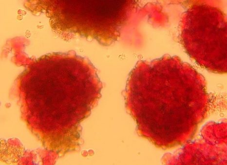
Pancreatic islets seen under the microscope ©François Pattou/Université de Lille

From the discovery of insulin in 1921 to the first ever pancreas transplants in the late 1960s, the recent history of type 1 diabetes research has brought scientific and medical advances which have transformed patient prognosis and quality of life.

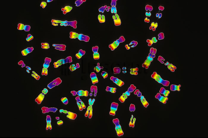
In France and worldwide, researchers continue to strive to improve the management of patients. Here at Inserm, eleven teams distributed among nine units contribute their own endeavors. Their work primarily involves the characterization of pancreatic cells and improving knowledge of the disease (risk factors, genetic susceptibility, pathophysiological mechanisms) and its complications.

One of these teams, based in Lille, is exploring islet transplantation, a promising technique discussed in a new publication in Diabetes Care[1] and in an update in The Lancet[2]. Other very interesting avenues currently being explored include immunotherapy and the development of artificial pancreases.


I. Type 1 diabetes research at Inserm

The teams at Inserm are involved in various collaborative projects to bring about advances in the treatment of type 1 diabetes. The findings of some already look promising.

  • EXALT (2014-2019)[3], with the participation of Christian Boitard’s Inserm team at Institut Cochin, was a project that aimed to evaluate the effects of an innovative immunotherapy on type 1 diabetic patients. Based on the administration of a peptide, the objective of this immunotherapy was to modify the autoimmune reaction directed specifically against the pancreatic beta cells. The first part of the project used experimental models to demonstrate that there is indeed an effect on the autoimmune reaction of diabetes. The results of the phase 1b clinical trial are currently being analyzed.


  • Hypo-RESOLVE (2018-2022)[4], a European project currently being conducted by Éric Renard in Montpellier (Inserm 1191/JRU 5203), aims to consolidate scientific knowledge on hypoglycemia. The idea is to create a permanent clinical database, conduct studies to further elucidate the underlying mechanisms of hypoglycemia and perform a series of statistical analyses to define its prediction factors and consequences. In addition, the researchers also wish to calculate the financial cost of hypoglycemia in the countries of Europe.


  • Artificial pancreas: Éric Renard and his colleagues have conducted research in collaboration with the University Of Virginia (Charlottesville, VA, USA) to create an artificial pancreas. The algorithmic system developed has been incorporated in the Tandem Control-IQ device, with a view to its commercialization (see photo below). Currently being trialed in 120 type 1 diabetic children in France as part of a national hospital-based clinical research program, interim-analysis data indicate that normal blood glucose levels are maintained for 71% of the 24-hour period, with a significant reduction of the time spent with abnormally high or low blood glucose levels. While its name might be confusing, the artificial pancreas is not actually a fake organ that will be transplanted into the patient but rather a technological device made up of three key elements: a sensor, a pump and an algorithm. The subcutaneous sensor continuously measures the patient’s blood glucose. The pump administers insulin through a fine cannula placed under the skin. The challenge of the artificial pancreas currently lies in the third part of the system: the algorithm capable of automatically making the link between the sensor and the pump.


  • Raphaël Scharfmann’s Inserm team at Institut Cochin has over the previous decade produced new cell models of type 1 diabetes, in the form of human beta-cell lines. His team is now seeking to develop innovative therapies based on the use of stem cells.

II. An alternative to insulin

a) Insulin-producing islets

In this context of ongoing innovation, the allotransplantation of islets of Langerhans, these specialized cells in the pancreas that produce insulin, has also emerged as a particularly interesting therapeutic avenue. Over the previous two decades, François Pattou, Marie-Christine Vantyghem and Julie Kerr-Conte in Inserm unit 1190 Translational Research in Diabetes, and their colleagues in the surgical and endocrinology/diabetes departments of Lille University Hospital have been developing this approach, in which over 50 people have received transplants.

Beyond the indisputable benefit for patients, their research is a perfect illustration of the contribution of translational research and of how laboratories and hospitals can join forces to improve knowledge and treatment of the disease.

In France, 3.9 million people have diabetes, around 5% of whom have type 1. This form of the disease is caused by a deficiency of the hormone insulin, which leads to prolonged high blood glucose levels (hyperglycemia).[5]  Type 1 diabetes is considered to be an autoimmune disease because it is caused by a dysfunction of the immune cells that identify the pancreatic islets of Langerhans as cells foreign to the body, and destroy them. These islets are therefore no longer able to fulfill their usual role of insulin production.

b) Principle of the transplant

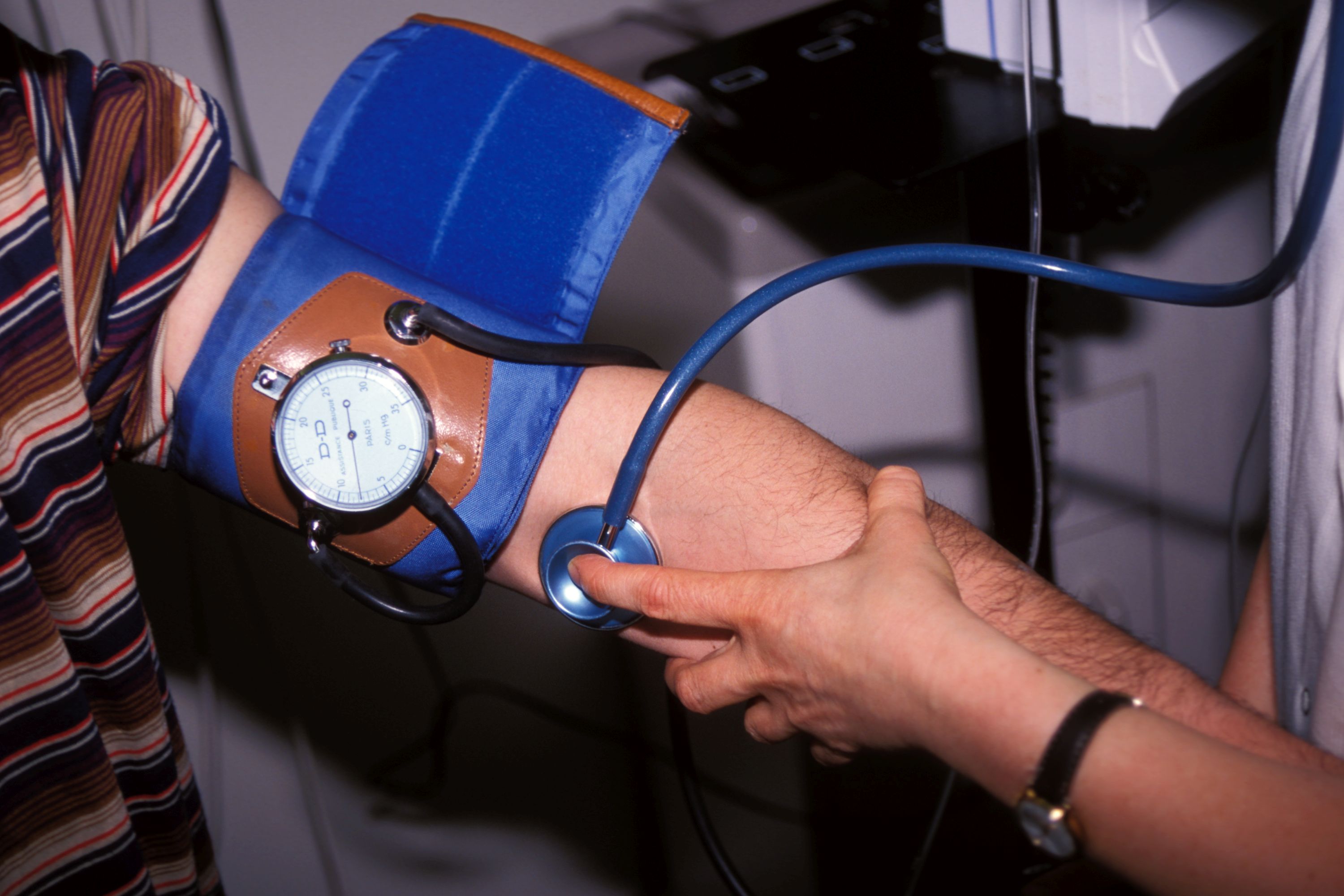
At present, the reference treatment for type 1 diabetes is based on the administration of insulin, either by multiple daily subcutaneous injections or by pump. Patients have the use of human insulin analogs  that make it possible to restore and maintain normal blood glucose levels.

Nevertheless, some patients find that their blood glucose levels are not properly controlled with this treatment, despite strict observance of dietary and therapeutic advice. Severe complications can then develop. Poorly-controlled blood glucose can be harmful to the organs, affecting the heart and vessels to begin with, but also the small arteries that supply the kidneys, the nerves of the lower limbs, and the retina.

Alongside the technological approaches (insulin pump, glucose sensors and – soon – the closed-loop pump), the biological approach involving the transplantation of islets represents an essential step for diabetes research. By making it possible to restore almost physiological insulin secretion, the transplantation of insulin-secreting cells transforms the lives of patients, who until now had run out of all therapeutic options.

The principle of islet transplantation or diabetes cell therapy is to replace the destroyed pancreatic cells in order to restore regulated insulin production. This makes it possible to normalize patient blood glucose control and even discontinue the use of insulin. “Islet transplantation is offered to two types of patient: those who have very unstable, often longstanding, type 1 diabetes, particularly with severe hypos and/or hypo unawareness, and kidney transplant patients who are already taking immunosuppressants which then just need to be adjusted”, points out Vantyghem.

II. Two decades of transplants

a) The early days of islet transplantation

In type 1 diabetic patients whose renal complications justify kidney transplantation, the simultaneous transplantation of a whole pancreas has for a long time represented the most effective therapeutic alternative to insulin. A trend which could nevertheless reverse in coming years, given the non-negligible risks associated with the procedure. The pancreas is a fragile organ, difficult to remove from donors.

The injection of islets – i.e. only the useful, insulin-secreting, cells – is a procedure that is less complicated but equally as effective.  “Pancreas transplantation is an effective procedure but carries a high risk of sometimes severe complications. In the beginning, pancreas transplantation did give better results in terms of blood glucose control. But great progress has been made in islet transplantation, which is also less risky, and can now be offered to patients who would not tolerate a pancreas transplant”, specifies Pattou.

The first tests and clinical trials of this technique date back to the late 1960s. However, the major turning point in the history of islet allotransplantation is considered to be the year 2000. This was the year in which Canadian researchers published the results of a clinical trial in the New England Journal of Medicine[6]. Thanks to pancreatic islet transplantation, seven type 1 diabetic patients became fully insulin-independent one year later, no longer requiring their insulin injections.

 “After the year 2000, research into this procedure took off, with other successes reported worldwide – particularly by our group. The problem is that very few studies went on to focus on patients over the long-term – and we are the first to present the results of a 10-year study”, highlight Pattou and Vantyghem.

b) 10 years of follow-up

The new study, published in Diabetes Care by Pattou, Vantyghem and their teams, retraces the paths of 28 patients who had received an islet transplant between 2003 and 2012, half of whom had already received a kidney transplant for renal failure.

Prior to this research, various groups had already published results on the outcomes of islet transplant patients, but none had gone beyond a follow-up period of five years. All highlighted the clinical benefits for the patients and their improved quality of life. However, in the absence of rigorous longer-term follow-up, questions remain unanswered. Are the benefits of islet transplantation maintained beyond five years? What about the complications associated with taking immunosuppressants?

The protocol for performing islet transplant surgery has been established over the previous two decades by the team in Lille, drawing attention not just to the quality but also to the quantity of the islets used. “We chose to start by transplanting a large number of islets. If they come from a particularly robust pancreas containing many islets, one transplant can be enough. However, this situation is exceptional and one or two additional transplants are generally needed. The particularity of our program is to rapidly schedule new transplants, independently of the results of the first, to give patients every chance of achieving normal insulin production”, explains Pattou.

Islet transplant: a few words about the procedure

The isolation of islets now takes place in a number of specialist laboratories, in Lille by Kerr-Conte’s team and also in Geneva, Paris, Montpellier and Strasbourg, from pancreases taken from donors with brain death.

The preparation is then injected into the liver through a small abdominal incision or percutaneously in order to infuse the islets in the portal vein of the liver. The main risk of this procedure is thrombosis, meaning that patients must be given anticoagulants.

Pancreatic islets seen under the microscope – François Pattou/Université de Lille

c) Promising results

As part of their study, the patients were seen at the hospital at least once a year for ten years to check the condition of the transplant. The majority of the patients were seen quarterly. At each visit, the patient’s clinical situation, blood glucose control, exogenous (originating from outside the body) insulin requirements and the diabetic and treatment-related complications were evaluated.

The research team had chosen to study, as primary endpoint, insulin independence with normal blood glucose levels. The doctors wanted to determine the proportion of their patients able to maintain good blood glucose control, without the exogenous provision of insulin. The study highlights that this is the case after five years for half of the 28 patients, and after ten years for almost one third (28%). These findings are similar to those obtained with whole-pancreas transplantation.

The researchers have also shown that 80% of the patients had a transplant that was still functional after five years, with no major hypoglycemic events. After ten years, this remained the case for two-thirds of them. Finally, there was no significant deterioration in kidney function, despite the possible renal toxicity of the immunosuppressants, which was probably compensated for by precision dose adjustment and the excellent blood glucose control obtained in the majority of the patients.

These results show that such a procedure can considerably improve the blood glucose levels of uncontrolled type 1 diabetic patients, protect them from the potentially fatal risk of severe hypos or hypo unawareness, avoid the serious complications associated with the disease and significantly improve their quality of life.

In order to understand why the procedure has greater long-term efficacy in some patients rather than others, several avenues are emerging. “It would appear that there is a signal in favor of women, with some experimental elements showing that the presence of estrogens is favorable to the islets’ survival. The patients with the best long-term results are particularly those who had regained good blood glucose control just after the transplant. To optimize this, attention needs to be paid to the quality and quantity of the islets initially transplanted”, state Pattou and Vantyghem.

An application to obtain reimbursement status for islet transplantation from France’s National Authority for Health is in progress. The response is expected for 2020.


III. What does the future hold?

Various difficulties remain, notably the potential immunological rejection of the transplanted cells, and the limited number of human insulin-secreting cells available for transplant given the current shortage of donors.

Indeed, to avoid rejection of their transplant, patients are required to take immunosuppressant treatment which has a certain number of side effects. Several teams are currently working on finding new solutions to avoid this phenomenon. In addition, in order to develop alternatives to human donors, a number of teams worldwide are currently conducting studies that aim to produce islets from pluripotent stem cells, and successfully transplant them[7].

These two issues could be resolved through a joint approach in order to disseminate diabetes cell therapy more widely: “The Holy Grail would be to produce islets from stem cells in the laboratory and then transplant them inside capsules of biomaterials that shield them from the immune system”, highlights Pattou.

In addition, various research is ongoing to develop fully autonomous artificial pancreases. At present, the blood glucose control obtained with these external devices remains inferior to that obtained with the transplant and the “biological” restoration of insulin secretion.  “These approaches nevertheless prove complementary and are offered in accordance with the profile of each patient”, specifies Vantyghem

The publication of the islet transplantation results after 10 years is a significant new step in the treatment of type 1 diabetes. These findings show that the cell therapy can work over the long term if sufficient islet mass is transplanted.   This research opens up very interesting avenues for the various teams working on new sources of insulin-secreting cells, especially those produced from stem cells and which will make it possible to compensate for the shortage of donors.

IV. What the patients say

Michèle, age 61, transplanted 14 years ago

©Alain Vanderhaegen – Communication Division of Lille University Hospital

“Your daughter won’t survive beyond the year 2000. She will never marry or have children”.  That was how an unsympathetic doctor informed Michèle’s mother that her child had type 1 diabetes. At the time, Michèle did not really understand what was happening. This was in the 1970s and for her the year 2000 was very far off. She did not feel ill. Yes, she had lost a lot of weight recently but she did not feel unwell.

Then, all of a sudden, everything changed. Her resulting stay in hospital, being the only child in a ward of elderly people suffering from major diabetes complications, including amputations, was genuinely traumatic. “Psychologically, it was very tough. I lived a life of untruths, to avoid other people’s questions”, she explains.

Administering the treatments was difficult. The needles had to be sharpened, the syringes boiled and the insulin injected with a constant feeling of shame, a desire to hide. Life went on for Michèle, she held on. She met her husband Jacques, had a son, both of whom support her. From a young age, her son knew what to do when his mom had a hypo, he knew to get sugar from the cupboard when it was needed.

But her severe hypos became increasingly frequent. Some days, up to five or six in succession, leaving Michèle and her loved ones exhausted. Her diabetes specialist, having met the Lille teams at a conference, decided to refer her for islet transplantation. It took her one year to decide to take the plunge, which she did in 2006. She then had three grafts in quick succession. Immediately after the third, Michèle was able to discontinue her insulin injections.  “The fact that the cells come from deceased donors bothered me. I was also afraid that the transplant would change me. In fact, it has changed my life but I’ve stayed the same, except that these three people are always with me. That shadow will never go away. Progress in transplants using stem cells would be a major step forward”, she emphasizes.

It has been 14 years now since Michèle stopped using insulin, since she has been taking immunosuppressants every day, with no major side effects. For both her and her loved ones, it is a liberation. “I’m a free woman once more – my husband and I can do things independently again. Sometimes I worry about the aging of the transplant, as going back would be unthinkable”, she says.


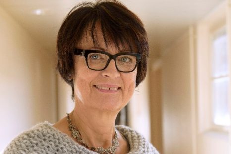
Carole, age 60, transplanted 4 years ago

©Alain Vanderhaegen

At 14 years of age, Carole was a freshman at boarding school. An experience that she did not enjoy, especially since she could not stop fainting. However, when she changed schools, things seemed to improve. She felt good, and no longer gave her fainting a second thought. Until a medical visit that revealed sugar in her urine, followed by a blood test confirming the diagnosis of type 1 diabetes.

Back at school, after a month in hospital, she was the subject of a lot of attention. “I could say anything I wanted in class, because people would say: it’s not her fault, poor thing, she’s diabetic.  So I made the most of it, I had fun”, she explains.

She tolerated her treatment well, ate more or less normally, got her high school diploma and became an elementary school teacher. At work, she always had bottles of orange juice with her in case of hypos.

But those hypos became increasingly severe, encroaching on her daily activities and deteriorating her quality of life. “When giving birth to my daughter, the doctors did not take my diabetes into account. I had a pulmonary edema and was hospitalized elsewhere. I didn’t see my baby for two weeks, so I went on hunger strike so that they would let me see her. When I left the hospital, I had a hypo one morning. All of a sudden, I couldn’t remember that I had a daughter – I’d forgotten her. Had I been alone, I would have been capable of going out and leaving her behind”, she says.

One day, a friend told her about the islet transplants in Lille which he had heard about on the radio. Carole discussed it with her diabetes specialist who promised to look into it. In the end, it was Carole who found the contact details of the team herself.

Transplanted in 2015 and 2016 on three occasions, Carole is keen to make the most of her life. She has been insulin-independent since the second transplant. Five years later, she continues to tolerate the immunosuppressants well, which she never forgets to take. Her hypos have stopped. She can now drive, do sport, go for walks by herself without having to keep her husband informed. “I can even eat biscuits when my family makes them. But I’m so used to not eating sugar that I actually don’t like it. The transplant changed my life at a time when I was no longer doing anything by myself. These islets are my friends, I’ve given them their own name and I celebrate their anniversary”, she smiles.


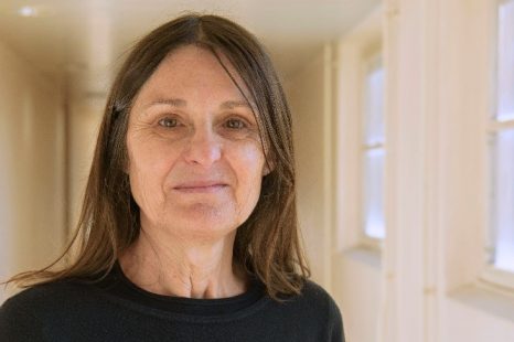
Béatrice, age 54, transplanted 1 year ago

©Alain Vanderhaegen

The adolescence of Béatrice was disrupted by the discovery of her diabetes. Originally from the countryside near Rennes, no-one she knew had heard of the disease until her diagnosis at the age of 11. It was difficult to begin with: at school, no-one made the effort to understand her situation. She was obliged to eat in the canteen kitchen and often found herself alone in the school yard. Her insulin injections improved things a little, although there were side effects, particularly weight gain, with each dose increase. Béatrice also suffered from discrimination, which continued into her professional life.

Over the years, new technologies emerged to help patients like Béatrice control their blood glucose better, such as insulin pumps with automatic shut-off and sensors that continuously monitor blood glucose. Nevertheless, there were also disadvantages: her hypos would trigger an alarm at any time of the day or night. “It was very difficult to live with. When I had the transplant my husband said that we could finally get some sleep“. Our quality of life improved markedly”, she emphasizes.

It was when reading an article in the journal of the French Association of Diabetics (AFD) that Béatrice discovered islet transplantation. She was tired, she had just lost her job. “I had the impression that I’d reached the end of the road, I needed a new solution in order to survive. Quite simply, to continue to live. As I was reading the article, I had the immediate feeling that the procedure was for me“, she explains.

It took two years before she was able to convince her diabetes specialist to refer her on, and that she was finally able to meet the Inserm-Lille University Hospital team. At the end of 2018, Béatrice received three transplants and became insulin-independent very soon afterwards. “For me the transplant was a huge gift. Out of respect for the donors, I need to keep fighting. I want to keep them alive, I call them my little angels”, she explains.

Although the immunosuppressants are not without their side effects, notably pins and needles in the legs, Béatrice acknowledges that they are in no way near as bad as the complications related to her type 1 diabetes. “My diabetes was always on my mind, I had the impression that my life revolved around it. We’ll see how I get on, and how the transplant holds up over the next few years, but right now I’m optimistic. For the first time in a long time, I have hope”, she says.

Glossary

Islets of Langerhans: the cells of the islets of Langerhans (or pancreatic islets) are endocrine cells of the pancreas whose primary function is to produce insulin. The islets of Langerhans are complex micro-organs disseminated in the exocrine pancreas.

Allotransplantation: the most common form of transplantation, in which the donor and recipient are the same biological species but strangers to each other.

Xenotransplantation: transplantation in which the graft comes from a biologically different species – for example, pigs.

Pluripotent stem cells: cells able to multiply infinitely and differentiate into the various types of cells that make up an adult organism.


[1] https://care.diabetesjournals.org/sites/default/files/care_upcoming/DC190401_ADVANCEDCOPY_STAMPED.pdf

[2]Advances in β-cell replacement therapy for the treatment of type 1 diabetes”.

Vantyghem MC, de Koning EJP, Pattou F, Rickels MR.

Lancet. 2019 Oct 5;394(10205):1274-1285.

[3] https://cordis.europa.eu/project/rcn/110445/reporting/en

[4] https://hypo-resolve.eu/

[5] https://www.inserm.fr/information-en-sante/dossiers-information/diabete-type-1

[6] Islet Transplantation in Seven Patients with Type 1 Diabetes Mellitus Using a Glucocorticoid-Free Immunosuppressive Regimen, New England Journal of Medicine, July 2000. https://www.nejm.org/doi/full/10.1056/NEJM200007273430401

[7] Https://www.ajd-diabete.fr/le-diabete/tout-savoir-sur-le-diabete/le-traitement/ (only available in French)

Atopic dermatitis: how allergens get on our nerves

Mast cells and sensory neurons cluster in “bunches” under the skin. ©Nicolas Gaudenzio

Atopic dermatitis, or eczema, primarily affects infants and children, and manifests itself in hypersensitivity to allergens in the environment. A skin disease characterized by flare-ups, it is often treated with topical anti-inflammatories. A new study led by Inserm researcher Nicolas Gaudenzio, from the Epithelial Differentiation and Rheumatoid Autoimmunity Unit (UDEAR – Inserm / UT3 Paul Sabatier), in collaboration with his colleagues at Stanford University (United States) shows that immune cells and sensory neurons interact in the skin to form units that can detect allergens and trigger inflammation. A discovery that provides an insight into how atopic dermatitis works, and points the way to new therapeutic possibilities. Their findings have now been published in the journal Nature Immunology.

Dry skin, pain, and itching… Atopic dermatitis affects the everyday lives of nearly 20% of children, and up to 5% of adults. The condition can have a significant impact on the quality of life of these patients.

Several studies have shown that genetic factors are involved in the development of this chronic inflammatory skin disease, and suggest that they result in impairment of the skin barrier. This enables the allergens present in the environment, from pollen to dust mites, to penetrate the dermis and stimulate the immune system, which reacts abnormally to this “threat” by triggering eczema.

However, the mechanisms of hypersensitivity to allergens and immune system hyperactivity in patients with atopic dermatitis are not yet fully understood. Led by Inserm researcher Nicolas Gaudenzio, the young “IMMCEPTION” group studies the way in which the immune system interacts with sensory neurons to regulate inflammatory processes in atopic dermatitis.

In particular, the researchers have taken a lead from existing clinical data which show that patients with this disease have numerous neuropeptides in their blood: chemical messengers that carry nerve messages, and whose level is correlated to disease severity. Identification of these neuropeptides in the blood indicates activation of the sensory neurons. These patients also have a number of enzymes in the blood indicating the presence of mast cells. Mast cells are immune cells present in the skin that play an essential role in modulating inflammatory and allergic processes.

Based on these observations, Gaudenzio and his team decided to focus on the interaction between sensory neurons and mast cells, and have now published their findings in the scientific journal Nature Immunology.

The researchers studied animal models of atopic dermatitis. Under the skin of mice showing signs of inflammatory reactions, they observed mast cells and sensory neurons clustering together in “sensory neuroimmune units” not dissimilar in form to a bunch of grapes. “The mast cells and neurons cling together in the dermis. We don’t yet understand the molecular interactions that bind them together, but we have quantified the distances between them, which are tiny,” highlights Gaudenzio.

The researchers then showed that when the mice were exposed to dust mites, these “sensory neuroimmune units” were able to detect the presence of these allergens, triggering allergic inflammation.

In the longer term, this discovery could have practical therapeutic implications.  Until now, patients could be treated with biological treatments (biological therapy), but these obviously treat the disease further down the line, after flare-ups have occurred. We believe we have put our finger on a trigger mechanism and now want to continue our research to identify new molecules that could block interactions between mast cells and sensory neurons, and thus have a beneficial therapeutic effect for patients,” explains the researcher.

To do so, the group will first need to characterize the molecular interactions within these units in more detail, and analyze the role they play in modulating the immune system.

“One of the questions we are now going to try and answer is what these mast cell-sensory neuron units are for. They must represent a defense mechanism for the body, since they are also found in healthy individuals. But it could be that they don’t work properly in people who have atopic dermatitis—that’s what we’re trying to understand,” concludes Gaudenzio.

This study was funded by the European Research Council (ERC).

Elaboration et validation de l’iBox, le premier outil universel de prédiction du risque de perte de rein greffé basé sur l’intelligence artificielle.


The natural lipids in buttermilk could contribute to reducing cardiovascular risk in vulnerable populations

A French consortium led by INRA, in collaboration with INSERM, the Universities of Lyon and Clermont Auvergne, Lyon Civil Hospitals and the Human Nutrition Research Centres (CRNH) of Rhône-Alpes and Auvergne, have now demonstrated that consuming certain lipids present naturally in dairy products (called “polar lipids”) could reduce the cardiovascular risk in overweight postmenopausal women by lowering their blood levels of LDL-cholesterol (“bad” cholesterol) and triglycerides. To reach this conclusion, the scientists included a cream cheese enriched in buttermilk polar lipids in the daily diet of volunteers for a period of one month. They were able to reveal certain mechanisms in the gut that might explain the effects observed. These natural dietary compounds could therefore form part of a new nutritional strategy designed to reduce the cardiovascular risk in vulnerable populations. This work was published on 12 June in Gut.

Dairy products contain a wide variety of lipids, both triglycerides and polar lipids, the latter being specific lipids that naturally stabilise fat droplets and are notably found in cream and buttermilk. Polar lipids play a crucial physiological role as they are essential components of cell membranes. Preliminary experiments in rodents had revealed the beneficial effects of milk polar lipids on liver metabolism and the regulation of blood cholesterol levels. However, until now, these effects had never been demonstrated in human volunteers with a high cardiovascular risk.

The research teams therefore tried to decipher the impacts of milk polar lipids on the cardiovascular risk profile of overweight postmenopausal women, a population that is particularly vulnerable to this risk. To achieve this, 58 such volunteers were asked to include cream cheese that was more or less enriched in milk polar lipids as part of their daily diet. After a month of consuming a specially-designed buttermilk concentrate enriched in milk polar lipids (a by-product of butter manufacture), the team observed a significant reduction in their blood levels of LDL cholesterol, triglycerides and other important markers of cardiometabolic risk. These milk polar lipids thus improved the cardiovascular health profile of these postmenopausal women.

The scientists then sought to unravel the mechanism underlying this effect. Their complementary studies suggested that certain milk polar lipids and cholesterol (of both dietary and endogenous origin) may form a complex in the small intestine that cannot be absorbed by the gut and is ultimately excreted in the stools.

These findings could ultimately provide foundations for new nutritional strategies to reduce cardiovascular risk factors in certain vulnerable populations.

Furthermore, they should contribute to a diversification of the ingredients used by the agrifood industry. Indeed, the functional properties of milk polar lipids relative to the texture of foods, alongside their potential protective effects on health, could offer a promising alternative to the soy lecithin that is currently used in a large number of foods, as well as providing an opportunity for the valorisation of buttermilk.

This work was carried out in the context of the ANR VALOBAB project (2012-2016) coordinated by Marie-Caroline Michalski, INRA Research Director in the Université Lyon 1-INRA-INSERM-INSA de Lyon Joint Research Unit for Cardiovascular, Metabolism, Diabetes and Nutrition (CarMeN). The project also involved the French Dairy Interbranch Organisation (CNIEL) and six other research laboratories and centres: CarMeN in Lyon, CRNH Rhône-Alpes, the Joint Research Unit for Human Nutrition and the INRA-UCA Joint Research Unit for Microbiology, the Digestive Environment and Health, both in Clermont-Ferrand, CRNH Auvergne, the INRA-AgroCampus Ouest Joint Research Unit for the Science and Technology of Milk and Eggs in Rennes, ENILIA (Surgères, with the participation of ACTALIA Produits Laitiers and Philolao) and ITERG (Bordeaux). The VALOBAB project received support from the ANR (ANR-11-ALID-007) and was accredited by the VALORIAL (Tomorrow’s Foods) Competiveness Cluster.

This clinical study, sponsored by Lyon Civil Hospitals, was also funded by PHRC-I VALOBAB.

A New Atherosclerosis Marker on the Way

Dépôts lipidiques dans une artère ©Inserm/Coutard, Michèle

A team of researchers from Inserm and the Université de la Réunion are proposing a new radioactive tracer to locate atheromatous plaques in arterial walls, which can cause cardiovascular events. The agent, called Darapladib, was tested in mice and ex vivo in humans. Darapladib binds specifically to an enzyme that is overexpressed in these plaques, allowing them to be located via medical imaging. The study, published in ACS Medicinal Chemistry Letters, opens up new perspectives to create tools for the clinical prevention of cardiovascular events.

Atheromatous plaques are a major risk factor for cardiovascular events. The plaques are clumps of lipids that bind to arterial walls, partially obstructing blood circulation. The condition is known as atherosclerosis. Plaques can also break apart and clog the artery remotely, which can cause a heart attack or cerebrovascular accident depending on which artery is affected.

At present, ultrasound technology is used to detect plaques in the carotid artery. Patients whose arteries are more than 70% obstructed generally undergo operations to remove the obstructed segment. But since this approach has its limits, clinicians are enthusiastic about developing complementary tools.

Researchers from Inserm and the Université de la Réunion are making progress on one of these potential tools: Darapladib. The agent was originally developed to reduce the risk of myocardial infarction and cardiovascular death. By exploiting its ability to specifically recognize the Lp-PLA2 enzyme − which is strongly overexpressed in unstable atheromatous plaques − the researchers hoped to be able to slow down or even reverse the plaque formation process. But the agent did not have the expected therapeutic effect. “This doesn’t call into question its very high affinity for Lp-PLA2,” explains Olivier Meilhac, the Inserm research director in charge of the study. “Using it as a tracer in the carotid artery could allow us to identify hazardous plaques and inform the decision of whether or not to operate.”

To convert the agent into tracer visible to nuclear imaging (positron emission tomography or PET), the research team had to make it radioactive.

The machine detects the radioactive signal, showing where Darapladib binds in the body.

 Inserm Transfert and the Université de la Réunion have filed for a patent for this new application of an obsolete drug.

The researchers then tested their Darapladib, radioactively marked with fluorine-18, by comparing it with radioactive glucose (18F-FDG), which is routinely used to track cancerous cells. They injected one or the other of these products into the bloodstream of mice with atherosclerosis, then observed the affected blood vessels with PET. Darapladib was highly concentrated in the plaques, but the signal from 18F-FDG was weak.

Figure 1. 18F-Darapladib and 18F-FDG in the aortas and hearts of ApoE KO mice. The hearts were removed to facilitate dissection of the aorta and are represented by a dotted orange circle.

The researchers then incubated these products with fragments of human carotid arteries presenting atheromatous plaques (recovered from surgical operations), and they observed the same results.

Figure 2. Ex vivo accumulation of 18F-Darapladib compared with 18F-FDG in human carotid arteries. 1a) macroscopic snapshots; 1b) PET images; a) uncomplicated plaque; b) complicated plaque.

The value of the Darapladib clinical diagnosis now needs to be confirmed in preclinical and clinical trials to be conducted in the future. It also remains to be proven whether its target, Lp-PLA2, is truly associated with a risk of rupture when concentrated in certain plaques.

Fertility and endometriosis: a research update from Inserm

©2019 Flore Avram/Inserm

Today, around 1 in 8 couples seek help because they are struggling to conceive. This is probably linked to the fact that couples are starting families later in life than before, or because they are setting aside the taboos linked to infertility and are more willing to seek help. Infertility has therefore become a public health problem, and the scientific community is rallying in response.

Where are we with research into this area, which lies at the heart of current societal problems? What are the prospects for the transfer of such research into clinical practice? Fertility research covers many different areas. The aim of this press kit is not to tackle them exhaustively, but to highlight the sectors in which research is making progress.

When research makes progress, everyone’s health benefits.

  1. Research into combating infertility

The term infertility is used when a couple are unable to conceive a child naturally after 12 months of trying. This term covers cases of total sterility, where there is no hope of natural conception, and subfertility, the majority of cases, in which couples have a reduced – but not zero – chance of achieving a pregnancy.

Cases of infertility are divided into 4 categories based on their cause:

– 30% are female-related;

– 30% are male-related. In men, azoospermia and oligospermia are the two leading causes of infertility identified to date;

– 30% are combined, meaning that they are caused by reduced fertility in both partners;

– 10% are unexplained.

In women, with the exception of mechanical causes affecting the fallopian tubes – when they are impaired or blocked (usually following an infection) – or uterus, endometriosis and abnormal ovulation are the most common causes of infertility.

Causes of abnormal ovulation include polycystic ovary syndrome (which affects around 10% of women around the world), hyperprolactinemia, and primary ovarian insufficiency (which may also be a side effect of chemotherapy).

Current research seeks to both improve understanding of the causes of infertility, and also to study new therapies or management methods that aim to increase the chances of conception.

  • Improving understanding of the causes
  • The genetic approach

Many researchers are studying the genetic causes of ovarian insufficiency. Several fertility problems are caused by certain genes not working, or not working properly. One rapidly growing area of research, due in particular to the improvement in high-throughput screening methods, is the study of genetic variants.

The Inserm laboratory led by Nadine Binart, for example, is working on primary ovarian insufficiency (POI), which is characterized by the inability of ovarian follicles to mature or by diminished ovarian reserve. Based on DNA analysis of women with POI, researchers are working to isolate the genes that are involved or altered in their genetic make-up. This approach is helping to improve understanding of the disease, but does not make it possible to provide specific treatment to these women, as sterility becomes definitive once there are no more eggs left in their ovaries. Preventive management can however be introduced if the genetic abnormality is found before the ovarian reserve is entirely depleted – for example, during family testing. This is the role of clinical research, which makes it possible to lessen the impact of these diseases when mutations are identified in affected families, to inform young patients about the risk of losing their eggs over time, and to introduce fertility preservation methods if appropriate.

  • The hormonal approach: the example of kisspeptin and prolactin

It is well-established that breastfeeding results in increased secretion of prolactin (PRL) by the pituitary gland, inhibiting a woman’s ability to ovulate. This prevents the onset of a new pregnancy. Some diseases also lead to an increase in PRL, including tumors of the pituitary gland from which this hormone is secreted. These cases of hyperprolactinemia, which result in period problems and infertility, are a leading cause of anovulation. In 2011, the Inserm team led by Jacques Young and Nadine Binart revealed the underlying mechanism that blocks ovarian function. Using a mouse model of the disease, the researchers showed that PRL inhibits secretion of a neurohormone called kisspeptin, which is the starting point for the entire hormone cascade responsible for ovarian cyclicity. In a mouse model, administration of kisspeptin made it possible to restore ovarian cyclicity despite the hyperprolactinemia.

This pathophysiological discovery explains the link between infertility and hyperprolactinemia for the first time, and points the way to developing innovative therapies. The basic concept has recently been validated in women,[1] which will make it possible to offer a therapeutic alternative for patients who do not respond to the drugs currently used.

1.2. Preserving fertility: areas of research and latest findings

Specialist “oncofertility” consultations have developed extensively in recent years and should now be an integral part of the care pathway for all young female patients with cancer. Several “fertility preservation” techniques designed to cryopreserve gametes, or preserve reproductive capacity, are now available, and others are currently in development. In France, since 1994, these methods have been included in various pieces of bioethics legislation. Article L.2141 11, modified by law 2011-814 of July 7, 2011, states that “All persons whose fertility is likely to be impaired by their medical care, or whose fertility risks being prematurely impaired, may have their gametes or reproductive tissue collected and preserved with a view to their later use of assisted reproductive technology, or with a view to preserving and restoring their fertility.” Fertility preservation methods are also included in the 2014-2019 Cancer Plan, which stipulates that “all patients must have access to cancer treatments, and innovative treatments in particular.”

  • Improving gamete preservation

Several techniques for cryopreserving female gametes are currently available. The standard method involves freezing mature eggs or embryos obtained from these eggs. It is not however suitable for prepubescent girls, who need to begin treatment urgently, and can also present problems in patients with hormone-sensitive cancers. Therefore, other techniques, although still considered experimental, may be offered in these situations.

Improving the available methods and developing new strategies is currently a major focus for oncofertility. This is one of the areas of research on which the Inserm team led by Nadine Binard and Charlotte Sonigo is working, in collaboration with Prof. Michael Grynberg.

  • Using anti-Müllerian hormone

Chemotherapy reduces fertility through a direct toxic effect on the ovaries. Cyclophosphamide, which is commonly used in cancer treatment, causes massive destruction of the germ cells contained in the ovarian follicles. In a mouse model, researchers have recently shown that treatment with anti-Müllerian hormone, which is normally secreted by the ovaries, can limit reduction of follicular reserve during chemotherapy. Use of anti-Müllerian hormone is therefore a promising approach to fertility preservation.

1.3. The role of new technologies: Using artificial intelligence in reproductive research

The store of germ cells contained in the follicles constitutes the ovarian reserve. Assessing the quantity of these germ cells is a common way of providing information on ovarian physiology and of measuring the impact of the environment on the ovaries. The standard method used in mice is time-consuming and tedious. In conjunction with a company specializing in artificial intelligence, Inserm researchers have recently developed an automated artificial intelligence method for follicle counting that uses a deep learning approach.[2] This new tool will be made available to the fertility research scientific community, saving a great deal of time and enabling better reproducibility of data.

  1. Research into combating endometriosis

Endometriosis is a complex disorder characterized by chronic inflammation due to the presence of tissue resembling the uterine lining outside the uterus. This “ectopic uterus” continues to respond to ovarian hormones, which in some women can cause severe pain and sometimes infertility. In response to increased visibility of the disease in the media, notably due to the work of patient organizations, the French health minister has announced an action plan to improve management of endometriosis. In terms of research, there has been a surge in studies of endometriosis over the last 5 years. Around 1,200 articles per year are being produced by researchers around the world, helping to advance understanding of this disorder.


©2019 Flore Avram/Inserm

  • An estimated 1 in 10 women have some form of endometriosis.
  • The locations of endometriosis lesions vary.
  • Endometrial cell reflux during periods occurs in 90% of women, but only 10% of them develop disease.
  • The disease is typically described as having 4 stages, based on the extent and depth of lesions; however, there is no correlation between disease symptoms and severity.
  • There are 3 forms of endometriosis: superficial peritoneal endometriosis, ovarian endometriosis (or endometrial cyst, or endometrioma), and deep endometriosis.

2.1. Improving understanding of the causes

 The epidemiological approach

At present, little is known about the causes of endometriosis, its natural history, and the factors affecting its progression. Epidemiological research plays a crucial role in advancing knowledge in this area. There are only a few large epidemiological cohorts around the world in which these aspects can be explored. The largest cohort for exploring endometriosis risk factors is currently a cohort of 116,430 American female nurses who were between 25 and 42 years old in 1989. The risk factors identified in the literature and confirmed in this cohort include: low birth weight, early menstruation, low body mass index, and short menstrual cycles (under 24 days).[3] However, beyond these factors, little information is available on the causes of the disease, and its natural history is largely unknown. The following table is based on a review of the literature published in August 2018:


*The positive association between smoking and reduced risk of endometriosis may be explained by the antiestrogenic effect of tobacco. This would confirm the therapeutic interest of estrogen blockers, which are available in drug form: far more suitable than cigarettes, whose harmful effects have been widely documented.

In a bid to improve understanding of this disease, several epidemiological studies are being launched in France by the team led by Marina Kvaskoff, Inserm epidemiologist and researcher. These include a recently formed patient cohort dedicated to the study of endometriosis: the ComPaRe-Endometriosis cohort. The study team’s objective is to have enough women in the cohort to obtain robust findings in relation to the many questions that are still unanswered about this disease. In less than 6 months, over 8,000 women have already taken part in the study. The team aims to recruit 15,000 to 20,000 participants, and a broad call for participation has gone out to women with endometriosis or adenomyosis (a form of endometriosis limited to the muscle wall of the uterus) to help speed up research into these disorders simply by completing online questionnaires about their experience of the disease (https://compare.aphp.fr/). The study initially looks to explore the natural history of the disease (change in the symptoms and characteristics of the disease over time), and to identify the factors that determine its progression and result in better response to treatment. This research will also make it possible to describe the circumstances of diagnosis and the patient care pathway, and to assess the impact of the disease on patients’ daily lives.

Endometriosis is also being studied within large French cohorts, such as the CONSTANCES cohort, a prospective study of 200,000 men and women (105,000 women) representative of the French population. Marina Kvaskoff’s team has developed an epidemiological research study to determine the prevalence and incidence of the disease in France, and to explore its risk factors within this cohort. Other studies are currently in development and will be conducted in other cohorts in due course.

  • The environmental approach

Several epidemiological studies have explored the associations between organochlorine chemicals (solvents, pesticides, insecticides, fungicides, etc.) and endometriosis, but their results have been inconsistent. A French meta-analysis of 17 studies[4] published in February 2019 attempted to draw more robust findings. The risk of developing endometriosis was 1.65 times higher in women exposed to dioxins, 1.70 times higher for those exposed to polychlorinated biphenyls (PCB), and 1.23 times higher for organochlorine pesticides. Although statistically significant, these estimates should be considered with caution due to the significant heterogeneity of the studies and the small estimated effect size. The level of evidence was judged to be “moderate” with a serious risk of bias, supporting the need to conduct further well-designed epidemiological research in order to fill the persistent data gaps.

  • Using the genetic and epigenetic approach for early detection

Detecting endometriosis in the early stages, before patients experience symptoms, would make it possible to improve patient care. Although the heritability of endometriosis has been estimated at 50%, it is highly complex and clearly highly polygenic. Numerous candidate genes have been studied from this perspective in analyses of disease predisposition. Initial results have shown that there is no gene for endometriosis, but that the existence of genetic variants characteristic of the disease could enable it to be diagnosed and to improve patient care. In 2017, efforts by the international community made it possible to identify a total of 14 variants (located on the genes WNT4, GREB1, ETAA1, IL1A, KDR, ID4, CDKN2B-AS1, VEZT, FN1, CCDC170, SYNE1, FSHB, and in the chromosomal regions 7p15.2 and 7p12.3). These 14 genes are involved in proliferation and the cell cycle, adhesion and the extracellular matrix, andinflammation, which makes sense in relation to endometriosis. However, each of the variants identified explain only a small part of the genetic variation in endometriosis. In future, the combination of high-risk alleles in a patient might provide a probability of being affected that could be used to diagnose patients and categorize them based on endometriosis type and severity.

The existence of specific epigenetic markers for endometriosis could also theoretically be used for early detection, with endometrial cells presenting specific epigenetic abnormalities that modify expression of the main transcription factors. However, it is not known how the interactions between the defective epigenomic cells and mutated epithelial cell genes contribute to the pathogenesis of endometriosis.

  • The microRNA approach

The full complexity of endometriosis cannot however be understood through genetics alone. Genes only influence phenotype through their expression. This expression is regulated by epigenetic molecular mechanisms. As such, most research focuses on studying the microRNA that could be “markers” for the disease. Several have been identified in patients’ plasma thus far, but with very poor reproducibility from one research team to the next. For example, a study published in 2013[5] identified just four miRNA (miR-199a, miR122, miR145*, and miR-542-3p) as enough to categorize patients, with very few errors. Confirmation of this article’s findings in independent cohorts has however been slow. One possible explanation for this is the fact that extraction of circulating RNA remains very heterogeneous from one study to the next, perhaps due to the technical tools used in extraction. In future, new, more comprehensive approaches could provide more consistent results.

  • The cellular approach: oxidative stress

Several studies have shown increased oxidative stress in the serum of women with endometriosis. Oxidative stress is a highly general mechanism that induces and is caused by inflammation. It would seem logical to find changes linked to oxidative stress in the context of a painful disease like endometriosis. In mouse models, treatment with antioxidants (N-acetylcysteine) has been seen to reduce endometrial lesions.

Research led by a team from the Institut Cochin has also identified several genes linked to glutathione metabolism within the gene cascades that are deregulated in endometriosis lesions. Glutathione is a peptide that plays a key role in detoxification of hydrogen peroxide, a central molecule in oxidative stress. Down-regulation, particularly of the GCLM and GCLC genes crucial to glutathione synthesis, could explain increased oxidative stress in endometriosis lesions.

  • The dysfunctional immune system: a possible approach?

The survival of endometrial cells outside the uterus could be linked to poor function of the immune system causing chronic local inflammation. Studies have shown an increase in some immune cells around endometriosis lesions.

2.2. Treatment: areas of research and latest findings

Changing diagnostic methods: phasing out surgery

Before considering treatment, the first stage is to reduce the diagnosis time for endometriosis, which is currently around 7 to 10 years after onset of the initial symptoms. With this in mind, doctors and researchers are working to develop a diagnostic score, based on a dozen questions, from which doctors will be able to provide a diagnosis with 85-90% reliability. This score may be accompanied if necessary by imaging, which can inform endometriosis diagnosis if carried out and interpreted by trained medical personnel.

Doctors and researchers agree that diagnostic surgery is contraindicated for endometriosis.

The 3 pillars of treatment

Drug therapy, surgery, and assisted reproductive technology (ART) are currently the only 3 methods for treating the symptoms of endometriosis and its potential impact on fertility. In the absence of new treatments, the key is to understand the role played by each component in this therapeutic arsenal, so that they can be used effectively.

Drug therapy is based on blocking ovarian function to bring about artificial menopause via continuous administration of contraceptives. These therapies (the combined pill, estrogen pill, or GnRH agonists) must be personalized and adapted to the needs of the patient. These therapies should be prescribed as the first-line treatment for women who are not seeking to become pregnant, in order to reduce the pain caused by the disease.

For patients who want to conceive, ART and surgery may be considered. ART should be used routinely before all surgical procedures in order to maximize the chance of conceiving a child for couples who want to do so. Surgery must not be used in women who do not want to conceive in whom drug therapy is effective. Endometriosis surgery can be highly invasive and debilitating (removing some parts of the colon, with a high risk of ovarian reserve depletion if ovarian cysts are removed, etc.), and does not prevent the disease from recurring, as it does not treat the cause. Doctors and researchers also agree that women who undergo surgery at a young age have a high risk of their endometriosis lesions recurring, and encounter further difficulties if they decide they want to become pregnant.

All efforts must therefore be made to move away from using surgery as the standard treatment for endometriosis, as has too often been the case in the past.

Some forms of endometriosis – particularly those affecting the ovaries – are now an indication for providing women with access to various fertility preservation techniques.

The role of new technologies: the example of high-intensity focused ultrasound

In Lyon, teams of research clinicians led by Prof. Gil Dubernard (Hospices Civils de Lyon and Inserm unit 1032 LabTAU) have developed an ultrasound-based treatment for bowel endometriosis. When endometriosis infiltrates the rectal wall, it causes debilitating rectal pain that may affect quality of life. After failure of medical treatment, a surgical procedure is often proposed that consists of removing all or part of the rectum and sometimes requires a temporary colostomy (artificial anus).

A phase I clinical trial carried out in 11 patients in 2017 demonstrated that high-intensity focused ultrasound may be a useful alternative to surgery. An ultrasound probe inserted into the rectal passage is able to “desensitize” the lesions within a few minutes. A follow-up trial of 12 patients seeking to confirm these initial results was completed on April 1, 2019. Data analysis is ongoing and will be available shortly.

Meanwhile, in collaboration with the company EDAP TMS (the clinical trial sponsor), the therapeutic ultrasound Inserm laboratory led by Cyril Lafon, LabTAU (Université Claude Bernard Lyon 1/Inserm), is working on optimizing the conditions of ultrasound delivery (insonification) and improving the ergonomics of the probe in order to increase the number of patients eligible for this new treatment.

It is highly likely that this innovative therapy will replace many of the rectal surgeries carried out in this functional disorder that resolves upon menopause.


[1]     Hypothalamic-Pituitary-Ovarian Axis Reactivation by Kisspeptin-10 in Hyperprolactinemic Women With Chronic Amenorrhea.

Millar RP, Sonigo C, Anderson RA, George J, Maione L, Brailly-Tabard S, Chanson P, Binart N, Young J.

[2] Sonigo C, Jankowski S, Yoo O, Trassard O, Bousquet N, Grynberg M, Beau I, Binart N. High-throughput ovarian follicle counting by an innovative deep learning approach. Sci Rep. 2018 Sep 10;8(1):13499. doi: 10.1038/s41598-018-31883-8.

[3] https://www.ncbi.nlm.nih.gov/pubmed/30017581

[4] https://www.ncbi.nlm.nih.gov/pubmed/30530163

[5] (Wang et al., JCEM, 2013)

Find out more:

Read more at Health and Research From A-Z on inserm.fr

Endometriosis

Fertility preservation techniques

Inserm magazine number 36: Fertility: Are our future generations in danger?

Artificial Intelligence Facilitates Chemical Toxicity Evaluation: the Case of Bisphenol S

©Photo by Philipp Katzenberger on Unsplash

A novel IT tool based on artificial intelligence methods has made it possible to identify the toxic effects of bisphenol S – a frequent substitute for bisphenol A in food containers – using existing published data. In more general terms, this tool developed by Inserm researchers led by Karine Audouze at Unit 1124 “Environmental Toxicity, Therapeutic Targets, Cellular Signaling and Biomarkers” (Inserm/Université de Paris), will make it possible to reveal the toxic effects of any chemical substance (or physical agent) provided that it is already present in databases or published studies. The stages of the development and use of this tool are described in Environmental Health Perspectives.

Artificial intelligence can now be used to simultaneously analyze databases and scientific literature in order to evaluate the toxicity of substances for humans. Such “in silico meta-analysis” has been made possible thanks to an IT program designed by Karine Audouze and her colleagues at Inserm JRU-S1124 (Environmental Toxicology, Therapeutic Targets, Cell Signaling and Biomarkers). It was validated by researching the toxicity of bisphenol S, a frequent substitute for bisphenol A – an endocrine disruptor that has already been banned from use in food containers.

In practice, the researchers incorporated various types of biological and chemical data in their IT program, including the 2,000 terms referenced in the AOP-wiki database (AOP: Adverse Outcome Pathways). “This database comprises precise descriptions of the various biological steps (molecules, signaling pathways) leading from molecular disruption to pathological effects, such as obesity, steatosis, cancer, etc. New toxicity processes are added regularly”, states Audouze. In parallel, with bisphenol S having been used to test the program, the authors incorporated the various designations and synonyms of this component that are found in the scientific literature. Thus equipped, the program scanned the abstracts of scientific articles submitted by the authors, searching for those pre-saved terms. “The aim was to establish links between those terms representing the chemical substance and those corresponding to the pathological processes”, clarifies Audouze. To achieve that, the researchers taught their system to read intelligently. This means that the program gives more weight to terms found side by side rather than far apart from each other, to those found in the results and conclusions sections at the end of the abstract rather than in the hypothesis section at beginning and, finally, by quantifying the words spotted. “The system goes beyond a rapid scan to offer genuine automated text analysis”.

In the end, the analysis revealed a correlation between bisphenol S and the risk of obesity, which was then manually verified by the authors. Then, to further increase the performance of their tool, the authors also incorporated the biological data from ToxCast, a US database that references the effects of many chemical and physical agents on various cell types thanks to robotic analysis. “As such, this strategy makes it possible to suggest mechanisms associated with the toxicity discovered by the program”, explains Audouze. This is how the researchers observed that bisphenol S promotes the formation of adipocytes.

“This IT tool can be used to establish a quick overview of the effects of a chemical agent, which is desirable when the latter is suggested as a substitute for an existing substance. It does not provide proof of toxicity as such but serves to rapidly incorporate a large quantity of data and rank the most likely harmful effects, thereby making it possible to design the most pertinent biological and epidemiological studies”, illustrates Audouze.

This tool can now be freely accessed on the GitHub platform. Any researcher wishing to test the toxicity of an agent can use it by developing a dictionary specific to that agent.

This project was funded by the European Human Biomonitoring Initiative, HBM4EU (https://www.hbm4eu.eu).

Hypertension: A New Drug Coming Soon?

Prise de la tension artérielle chez un patient ©Inserm/Depardieu, Michel

Firibastat is the first in a new class of antihypertensive drugs targeting the renin-angiotensin system in the brain. A phase IIa placebo-controlled clinical trial provided initial data on its efficacy in hypertensive patients and was coordinated by Michel Azizi from Inserm/Hôpital Européen Georges-Pompidou AP-HP Clinical Investigation Center 1418 and the Hypertension Department of the same hospital and by Catherine Llorens-Cortes, Inserm Research Director at Unit 1050 “Center for Interdisciplinary Research in Biology” at Collège de France. On the basis of these results, published in The Journal of Hypertension, the phase IIb trial was launched in the USA.

Hypertension is the most common chronic disease in France. Linked to an abnormally high pressure of the blood on the vessel walls, its general lack of symptoms can make it appear harmless. Yet if not brought under control, it constitutes one of the main causes of cardiovascular, cerebrovascular and neurodegenerative complications (myocardial infarction, stroke, Alzheimer’s disease…). Hypertension can be normalized by means of lifestyle measures, either alone or more often than not in combination with medication. However, up to 30% of patients either do not respond – or respond poorly – to the treatments currently available. To remedy this, interventional approaches and new therapeutic targets linked to the pathophysiology of the disease are being studied.

The development of the drug firibastat[1] in the treatment of hypertension continues. It is the first in a new therapeutic class targeting the brain renin-angiotensin system controlled by angiotensin III, a peptide that in various hypertension models exerts a stimulating effect on blood pressure via three mechanisms. It increases the activity of the neurons that favor vasoconstriction, it inhibits the reflex that adjusts the intensity of heart contractions to the blood pressure level and, finally, it contributes to the increased secretion of the anti-diuretic hormone vasopressin in the blood, reducing the volume of urine produced by the kidneys.

Firibastat counters these various mechanisms by specifically inhibiting aminopeptidase A, an enzyme in the brain that produces angiotensin III. This drug, taken orally, becomes active in the brain after crossing the blood-brain barrier. Its safety was already tested in healthy subjects in two phase I clinical trials. The recently-published results of the phase IIa trial confirm the safety data and provide the initial efficacy elements. Enrolled in this trial were 34 patients (73% of whom male) with a daytime ambulatory blood pressure of between 135/85 and 170/105 mmHg. They had an average age of 57 and were not obese (average BMI 26.8 kg/m2). One half of the patients received firibastat for four weeks followed by placebo for another four weeks and the other half received the same treatment but in the reverse order: placebo followed by firibastat.

The results demonstrate better control of systolic blood pressure (SBP) with firibastat after four weeks with an average -4.7 mmHg reduction in SBP versus +0.1 mmHg with placebo. A difference which, however, is not statistically significant. “This can be explained not just by the small number of subjects but also by the fact that they had moderate hypertension, explains Inserm Research Director Catherine Llorens-Cortes. Firibastat is an antihypertensive agent and not a hypotensive agent, meaning that it can act on hypertension but will have no effect on normal blood pressure. “Its efficacy should therefore increase with the severity of the hypertension”, clarifies the researcher. A phenomenon which appeared to be confirmed in this trial because the decrease in ambulatory SBP reached -9.4 mmHg in the event of strong baseline hypertension whereas the benefit was less marked for lower baseline SBPs. The authors also verified the safety of firibastat and observed that it did not interfere with the systemic renin-angiotensin system, controlled by angiotensin II.

“These encouraging findings gave the green light to the phase IIb study which has recently been completed in the USA. It has confirmed the efficacy of firibastat in 254 hypertensive overweight high-cardiovascular-risk patients after two months of treatment, including in African-American patients whose hypertension is most often resistant to the treatments currently available.” clarifies Llorens Cortes.

[1] In partnership with the company Quantum Genomics, and with the support of the French National Research Agency (ANR): ANR RPIB CLINAPAI and LabCom CARDIOBAPAI

Syndrome Temple and Silver Russell understanding of epigenetic mechanisms regulating fetal growth

© Inserm/Alpha Pict/Caro, Daniel

A team Sorbonne / AP-HP / Inserm, led by Professor Irene Netchine, physiology professor at Sorbonne University and pediatrician at the Hospital Armand Trousseau AP-HP, studied the molecular mechanisms of the clinical similarity between syndrome Temple and Russell Silver syndrome. Their study, published in Science Advances , highlights the importance of gene network concept “imprinted” in the diagnosis and treatment of patients with these rare syndromes.

In the human genome, genetic information is carried by two copies, each inherited from one parent. For most genes, both copies (maternal and paternal) are expressed equivalently. However, some genes are known as ”  subject to parental imprinting  ” when only one of the two copies is expressed; the other copy extinguishing. The expression of these genes corresponds to an epigenetic mechanism, that is to say biochemical modifications of the DNA molecule, which are different on each of the two copies. These imprinted genes are usually involved in growth, metabolism and development .

The genes expressed by the mother copy have a tendency to restrict fetal growth to preserve its own reserves, while the genes expressed by the paternal copy foster meanwhile fetal growth to ensure healthy offspring and pass on its genes to the next generation.

Syndrome Temple and the syndrome Silver Russell are associated with impaired fetal growth is reduced , and are caused by genetic or epigenetic abnormalities in two regions containing imprinted genes, specifically on chromosomes 11 and 14 , respectively.

These patients also have metabolism and food intake disorders, as well as a premature puberty or advanced.

In his study, the team of Prof. Netchine analyzes the molecular mechanisms of the clinical similarity between the two diseases. In Russell Silver syndrome, a decrease in the expression of a gene ( IGF2 ) usually expressed from the paternal chromosome 11, is the origin of stunting. As to Temple syndrome, changes in genes on chromosome 14 (particularly some RNAs normally expressed from the maternal copy of chromosome 14) also impact gene expression IGF2 , while epigenetic marks are unmodified on the chromosome 11.

These discoveries help advance understanding of epigenetic regulation of gene expression, and consider a new approach to the physiology of fetal growth.

MON 810 and NK603 GM Maize: No Effects Detected on Rat Health or Metabolism

Photo by Charles Deluvio ???? on Unsplash


A diet based on MON 810 or NK603 transgenic maize does not affect the health or metabolism of rats, under the conditions of the GMO 90+1 project1. This unprecedented study performed by a research consortium led by Inra brought together a number of partners2, including Inserm. The research was performed as part of the Risk’OGM program funded by the French Ministry of Ecological and Inclusive Transition. For six months, rats were fed a diet containing either GM maize (MON 810 or NK603) or non-GM maize, in varying concentrations. The researchers, using high-throughput biology techniques, did not identify any significant biological markers related to the transgenic maize diet. Neither did anatomic pathology examination reveal any alteration of the liver, kidneys or reproductive system of the rats fed diets containing these GMOs. This research, published on December 10, 2018 in Toxicological Sciences, does not reveal any harmful effects related to the consumption of these two types of GM maize in the rat even after lengthy periods of exposure.

The researchers used two well-known types of GM maize: MON 810, which produces the protein Bt rendering it resistant to certain insects, and NK603 in which the modification of a gene renders it resistant to glyphosate. For 6 months, the rats were fed a diet containing either transgenic maize or non-GM control maize.  This time period, which is double that of the test required by European regulations, is equivalent to one third of the average lifespan of rats.

The objective of the researchers was to test for early biomarkers of biological function alterations in rats fed GM maize over periods of 3 and 6 months. For that, they used two ultra-sensitive high-throughput techniques: transcriptomics (gene expression) and metabolomics (study of the compounds derived from the body’s functioning). These techniques were used to identify and measure metabolites (amino acids, sugars and other small molecules) and to characterize the expression of messenger RNA and cellular microRNA. These methods are capable of detecting a broad spectrum of metabolic variations. The researchers identified markers able to differentiate the MON810 and NK603 diets. However, following the six-month period of the experiment, no significant differences were identified between the GM and non-GM diets, from the biological point of view.

In addition, in the rats fed the GM diets, anatomic pathology techniques (macro- and microscopic study of the tissues to detect potential abnormalities) revealed no alteration of the organs, particularly the liver, kidneys and reproductive system.

As such, the researchers did not detect any harmful effects of the MON810 and NK603 maize diets on the health and metabolism of the rodents, even after a lengthy exposure period.


1 The GMO 90+ project In 2010, the French Ministry of Ecology launched the Risk’OGM program, in the context of the 2008 law on genetically modified organisms for the establishment of a new legal and regulatory framework based notably on the principle of a triple evaluation of the impact of GMOs – from the health, environmental and socioeconomic standpoints. To set this dynamic in motion and fulfil public authority requirements in terms of expertise, guidance and completed research on GMOs, two calls for research proposals were held, in 2010 and 2013, respectively. The GMO 90+ project was selected during the 2013 call for proposals, with the following scope: to test for biomarkers predictive of biological effects in the study of the subchronic toxicity (3 and 6 months) of GMOs in the rat. Driven by a consortium pooling the various scientific competences, the purpose of this research was to determine whether the feeding of rats with genetically-modified maize led to metabolic changes which could be linked to early effect biomarkers (measurable biological characteristic). The objective was to provide key data which could be used in risk evaluation processes. http://recherche-riskogm.fr/fr/page/gmo90plus  
2 List of project partners: 1-Toxalim (Research Centre in Food Toxicology), Université de Toulouse, INRA, ENVT, INP-Purpan, UPS, Toulouse, France. 2-INSERM UMR-S1124, Toxicologie Pharmacologie et Signalisation cellulaire, Université Paris Descartes, USPC, Paris, France 3- Centre de Recherche sur l’Inflammation (CRI), INSERM UMRS 1149, Paris, France. 4- Laberca, ONIRIS, UMR INRA 1329, Nantes, France 5- Université de Rennes, Inserm, EHESP, Irset (Institut de recherche en santé, environnement et travail) – UMR_S 1085, Rennes, France. 6- Methodomics, France. 7- Institut de Mathématiques de Toulouse, UMR5219 – Université de Toulouse, CNRS – UPS IMT, Toulouse, France. 8- Anses, Maisons-Alfort, France. 9- Profilomic, Saclay/Gif sur Yvette, France 10- UMR1332 Biologie du Fruit et Pathologie, INRA, Université de Bordeaux, Villenave d’Ornon, France. 11- UR 1264, MycSA, INRA, Villenave d’Ornon, France. 12- Laboratoire Reproduction et Développement des Plantes, University Lyon, ENS de Lyon, UCB Lyon 1, CNRS, INRA, Lyon, France 13- CRO CitoxLAB, Evreux.

fermer