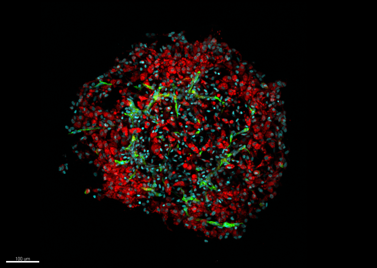
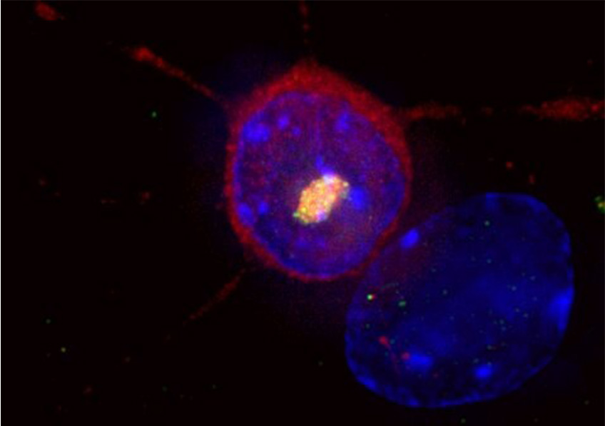
Menu

Restoring Sight: the Artificial Retina Shows Growing Promise

Adobe/Stock

Restoring sight to patients with age-related macular degeneration (AMD) or retinitis pigmentosa has become an increasingly likely prospect over recent years, with many researchers working to develop an artificial retina. In a new study, a team from Institut de la Vision (Inserm-CNRS- Sorbonne Université) led by Inserm researcher Serge Picaud has used animal models to show that a device made by the firm Pixium Vision could induce high-resolution visual perception. Their findings, published in Nature Biomedical Engineering, have paved the way for clinical trials in humans.

Age-related macular degeneration (AMD) is characterized by deterioration of the retina that can lead to central vision loss. This highly incapacitating condition is thought to affect up to 30% of people over the age of 75. For years, several groups of researchers have been working on the development of an artificial retina that could restore eyesight – not just to these patients but also to those with retinitis pigmentosa. 

The retina is made up of light-sensitive cells (photoreceptors) whose purpose is to transform the light signals received by the eye into electrical signals sent to the brain. It is these cells that are destroyed during the course of the aforementioned diseases, the outcome of which can be blindness. The principle of the artificial retina – which is implanted beneath the patient’s own retina – is simple: to act as a substitute for these photoreceptors. Its electrodes stimulate the retinal neurons to send messages to the brain.

Two devices of this type, Argus II (Second sight, USA) and Retina Implant (AG, Germany), are already in widespread use. “Nevertheless, these companies are gradually withdrawing from the market, particularly because the results seen in patients have been insufficient for targeting the device at those with AMD. The patients managed to see light signals but those able to distinguish letters were very much in the minority”, emphasizes Serge Picaud. 

Implantation in patients 

Reinventing the device to increase its performance: this is the aim of the Picaud and his colleagues. Supported by Pixium Vision, their artificial retina is wireless and less complex, unlike previous devices. In addition, this implant introduces a local return of the current, thereby enabling better resolution of the images perceived by the eye. Finally, the image is projected onto the implant by infrared stimulation that activates photodiodes connected to electrodes, enabling more direct stimulation of the retinal neurons.

In a study published in Nature Biomedical Engineering, Picaud and his colleagues tested this device on non-human primates, demonstrating that it can restore significant visual acuity. In vitro tests showed first of all that each pixel activates different cells in the retina. This selectivity is expressed in the form of very high resolution, such that implanted animals can perceive the activation of just one of the implant’s pixels in a behavior test.

The high resolution of these implants led to the device being fitted in five French patients with AMD, for whom initial results show a gradual restoration of central vision. They are able to perceive light signals and some can even identify sequences of letters, with growing rapidity over time.

“The objective now is to conduct a Phase 3 trial in a larger group of patients with AMD. If the artificial retina works for them, there is no reason why it should not work in patients with retinitis pigmentosa – a disease also related to photoreceptor degeneration”, concludes Picaud.

How People with Autism Might Avoid Socio-Emotional Situations

One hypothesis put forward to explain the repetitive behaviors of people with Autism Spectrum Disorder is a lack of cognitive flexibility. However, this may well not be the case. A recent study by a team of researchers from Inserm and Université de Tours used MRI to track the brain activity of autistic and non-autistic subjects faced with situations similar to those that cause problems in the daily lives of people with the disorder. Their findings, published in Brain and Cognition, suggest that the inflexibility of autistic individuals is actually the result of a strategy used to avoid socio-emotional situations. This research, which suggests now considering the cognitive and socio-emotional domains as closely linked rather than dissociated, opens up new avenues in the understanding and management of autism.

In their daily activities, people with Autism Spectrum Disorder (ASD) experience difficulty adapting their behavior to environmental changes. ASD is characterized by two main diagnostic criteria: the individual experiences persistent difficulties in social communication and is locked into repetitive behavioral patterns, restricted interests and/or activities. But while both criteria need to be present in order to diagnose ASD, little attention has been paid to how they interact.

In a study led by Marie Gomot, Inserm researcher at the Imaging and Brain laboratory (Inserm/Université de Tours), this question was explored by comparing the cognitive management of socio-emotional information and that of non-social information in people with ASD.

While the processes responsible for ASD symptoms have not yet been fully elucidated, one current hypothesis is a lack of cognitive flexibility – in other words, difficulty in alternating between multiple tasks and in analyzing one’s environment in order to adapt to these changes.

To evaluate this flexibility, the researchers used MRI to track the brain activity of ASD and non-ASD participants who underwent a test simulating situations similar to those that cause problems in the daily lives of people with the disorder.

The research team used a modified version of a test traditionally used in neuropsychology in order to test cognitive flexibility while processing non-social or socio-emotional information. Five cards were presented, each illustrated with a different face. The participants were asked to match the central card with one of four surrounding cards, according to one of the following three rules: frame color (non-social information), facial identity (social information) or facial expression (socio-emotional information). In order to evaluate their cognitive flexibility, the participants were asked throughout the test to make different matches (same color, same identity or same facial expression) by changing or maintaining one of the three rules.

The research team saw no significant difference between the ASD and non-ASD participants when it came to the behavioral parameters measuring cognitive flexibility alone – namely the capacity to adopt a new rule. However, the study did reveal the importance of information type for these cognitive flexibility processes in ASD. While the ASD participants needed more attempts than the non-ASD participants in order to adopt the rule linked to socio-emotional information, they had no particular difficulty in adopting those involving the processing of non-emotional information.

In parallel, the MRI revealed a significantly higher level of brain activity in the ASD participants when they were required to demonstrate cognitive flexibility. This brain activity only stabilized when the ASD participants received confirmation that they had found the correct rule to apply, thereby suggesting that people with ASD require a higher level of certainty in order to adapt to a new situation.

“These findings are important because they suggest that the implementation of routines and repetitive behaviors by people with ASD might not be due to a genuine lack of cognitive flexibility but rather to avoid being confronted with certain socio-emotional situations, specifies Gomot. The need for a high level of certainty combined with a poor understanding of the codes that govern socio-emotional interactions would thereby lead to the avoidance of tasks with a socio-emotional component.” And to conclude, “this research confirms the close link between cognitive and emotional dysfunction in ASD and the need for future studies to take them into joint consideration more often.

Human Adipose Tissue Reproduced in the Lab

©STROMALab

Can human adipose tissue be reproduced in a laboratory? It can now, thanks to a research team with members from Inserm, CNRS, Université Toulouse III-Paul-Sabatier, the French Blood Establishment (EFS) and the National Veterinary School of Toulouse (ENVT) working together at STROMALab. This team used 3D culture to develop adipose tissue organoids (or adipospheres) – small cellular units that mimic the characteristics and organization of adipose tissue as it presents in vivo. In their article, published in Scientific Reports, the researchers describe the various stages of the experimental conditions needed to obtain these adipospheres from human cells. An innovation that could make it possible not just to study diseases related to the impaired functioning of this tissue, such as obesity and type 2 diabetes, but also to develop new drugs to treat them.

Human adipose tissue, highly vascularized by a network of capillaries, is made up of fat cells known as adipocytes. Until now, laboratory researchers used 2D models that did not take into account the 3D architecture of this tissue as found in the human body.

Mini organs, which are known as organoids and capable of reproducing the cellular organization of a specific organ, have already been developed for some tissues, such as that of the intestine. However, none had been able to reproduce in 3D the cellular and vascular organization of the adipose tissue in a laboratory setting.

However, this is now possible thanks to researchers from Inserm, CNRS, Université Toulouse III-Paul-Sabatier, the French Blood Establishment (EFS) and the National Veterinary School of Toulouse (ENVT) working together at STROMALab. Thanks to the advent of the new 3D cell culture methods, the control of the selection and characterization of adipose tissue stromal cells (support cells), the team was able to develop organoids of this tissue, called adipospheres.

Generating organoids in 3D

From these stromal cells of the human adipose tissue, the researchers developed new 2D – followed by 3D – culture conditions, making it possible to obtain both adipocytes and endothelial cells from this tissue. The adipospheres obtained contained an intact vascular network organized around adipocytes in the same way as in actual human tissue. Better still, the adipocytes obtained were capable of differentiating into those of brown or white tissue (the two types of human adipose tissue) in the same way as those encountered in the human body.

Transplantation in mice

The research team then transplanted these adipospheres into mice in order to verify the functionality of their vascular network. They observed that not only was this network maintained in the body but also that it extended itself by establishing connections with the host’s circulatory system.

The researchers also observed so-called chimeric vessels, constituted of both mouse and human cells. “These are all signs that the host tolerates the transplanted organoids well, explain Isabelle Ader, Inserm researcher, and Frédéric Deschaseaux, from the French Blood Establishment (EFS), authors of the study. From this we can conclude that not only are these small structures faithful to the organization of human tissue, but also that they are capable of staying alive by establishing connections with the host circulatory system that provides them with the necessary oxygen and nutrients.

According to the researchers, this innovation will enable continued study of the functioning and properties of human adipose tissue. By working directly on this tissue, the use of animals will be reduced.

This innovation will also make it possible to test various drugs that could be used to treat certain diseases related to a pathology of adipose tissue, such as obesity or type 2 diabetes“, conclude Isabelle Ader and Frédéric Deschaseaux.

Read the article (in french) published in Magazine de l’Inserm, n°43, Juin 2019.

Wound Dressings to Regenerate Joints

Cartilage articulaire © Inserm/Chappard, Daniel

Researchers from Inserm and Université de Strasbourg at Unit 1260 “Regenerative Nanomedicine” have developed an implant which, when applied like a wound dressing, regenerates cartilage in the event of major joint lesions and incipient osteoarthritis. The details of this innovation, which has been validated in the preclinical setting, have been published today in Nature communication.

Increases in life expectancy and the number of accidental traumas call for the development of new types of surgery to replace defective joints. Among the chronic diseases, osteoarthritis – described as destruction of the cartilage affecting the various joint structures, including the bone and synovial tissue that lines the inside of the joints – represents a genuine public health issue. Depending on the clinical diagnosis, various therapeutic options are possible – ranging from microtransplant to joint replacement. Nevertheless, these procedures are all invasive, potentially painful, limited in efficacy and not without side effects. In reality, apart from joint replacement, current treatment strategies are limited to temporary cartilage repair and pain relief. Treatments mainly involve the injection of anti-inflammatories as well as hyaluronic acid to improve joint viscosity. Stem cells can also be used, particularly because they secrete molecules able to control the inflammation.

Within this area and with the aim of regenerating this supple and often elastic connective tissue that covers our joints and enables the bones to move and slide in relation to each other, a team of researchers from Inserm and Université de Strasbourg has recently developed a dressing for cartilage – inspired by the new-generation wound dressings that act as a second skin. With the dressings developed by Ms. Benkirane-Jessel and her team, the therapeutic response reaches a new milestone. We are no longer talking about repair but the actual regeneration of the joint tissue.

What Ms. Benkirane-Jessel’s team has developed is an innovative osteoarticular implant technique, able to reconstitute a damaged joint and whose application can be likened to that of wound dressings. “The implant we’ve developed is intended for two cases in particular: major cartilage lesions and incipient osteoarthritis.” she explains.

These dressings comprise two layers. The first – which acts as a support (reminiscent of everyday wound dressings) – is a membrane comprised of polymer nanofibers and supplied with small vesicles containing growth factors in quantities similar to those secreted by our own cells. The second is a layer of hydrogel loaded with hyaluronic acid and stem cells from the patient’s own bone marrow. It is these cells that – by differentiating into chondrocytes (cells that form the cartilage) – will regenerate the joint cartilage.

The scientists envision a promising future for their “cartilage dressing” which, in addition to the shoulder and knee joints, could also be used for the temporomandibular joint that connects the jawbone to the skull. Quite incapacitating, disorders in this area can cause pain, joint sounds and above all the inability to open and close the jaw completely. The research team has already conducted studies on cartilage lesions in small and large animals (mice, rats, sheep and goats), which are highly suitable models with cartilage comparable with that of humans. The objective is to launch a study in humans with a small cohort of 15 patients.

This project has received the support of Satt Conectus, ANR and the Grand Est region.

Miniaturized Chemical Sensors to Monitor Brain Function

©Stéphane Marinesco / Inserm, Photograph of an implantable chemical sensor (bottom right) made with platinized carbon fiber and coated with a recognition enzyme, placed next to a human hair (top).

A team of Inserm and CNRS researchers has succeeded in developing new-generation chemical sensors to monitor the brain’s metabolism, particularly during stroke, trauma or epileptic seizure. Measuring less than 15 µm in diameter, these minimally-invasive tools monitor what is happening in the brain in order to obtain data that are much more reliable and representative of the neurochemical exchanges. This research has been published in ACS Central Science.

Analyzing the interstitial fluid of the brain can reveal important chemical information about the state of the latter. In the clinic or in laboratory animals, the ability to detect, over time, the levels of metabolites characteristic of brain energy (such as glucose) can help detect the onset of brain lesions, enabling doctors to act before it is too late. In addition, the activation of neuronal networks leading to a release of neurotransmitters can be detected in interstitial fluid. However, up until now the size of the probes and the local injury caused by their implantation were parameters which disrupted the quality of the measurements obtained. In particular, the rupture of small cerebral blood vessels during implantation represents a major trigger for inflammation. Within the first hour after implantation, local chemical brain tissue composition can be affected.

The first innovation presented by the scientists in this research consisted of developing miniature sensors.

Invisible to the naked eye, they measure less than 15 microns in diameter (compared with 50 to 250 microns, currently), making them narrower than a strand of hair. The major advantage of being able to miniaturize the sensors to this extent is that implanting them no longer causes lesions in the nervous tissues. “Their size is smaller than the average distance between two brain capillaries, meaning that they are not damaged by the device” explains Stéphane Marinesco, Inserm researcher in charge of the study.

The second innovation was to coat the carbon fibers with platinum followed by a very thin layer of enzyme.

Up until then, electrochemical analysis using carbon fiber microelectrodes was limited to a highly-restrictive number of so-called “oxidable” molecules. Coating them with platinum makes it possible to attach enzymes and detect a potentially unlimited number of molecules. For Marinesco, “while platinum deposition is a commonly used technique in the field of microelectronics, it is usually performed with flat silicon substrates. Our results show that, despite their unusual cylindrical geometry, carbon fibers could be successfully covered with a platinum layer. The sensitivity achieved is similar or better than that of the thicker solid platinum wires which are commercially available.”

When these sensors were implanted in the brains of rats during laboratory testing, no injuries to the brain tissue or blood vessels were detected.

In addition, these microelectrodes supplied more precise and reliable evaluations of glucose, lactate and oxygen concentrations compared with conventional sensors (in which one sensor per parameter is necessary by implanting a multi-microelectrode “comb”). Numerous tests were performed on these new microelectrodes, in particular on their stability over time because they were also tested after 6 months of storage (room temperature in darkness).

Marinesco clarifies that: “This minimally invasive device represents a major advance in our ability to analyze the brain interstitial fluid, paving the way for the measurement of new physiological parameters and multiple applications. This novel tool could be used to test the effect of certain medicinal products on the brain. Finally, in the longer term, monitoring the human brain could provide invaluable information to doctors in order to better understand how a patient with brain lesions recovers after a head injury or stroke. This device could also help them to take the best therapeutic decisions depending on the patient’s condition”.

Cancer under pressure: visualizing the activity of the immune system on tumor development

Cancérogenèse : Surexpression de TRF2, marqué en vert, dans les vaisseaux tumoraux, marquage rouge, dans un cancer ovarien. ©Inserm/Wagner, Nicole, 2014

As tumors develop, they evolve genetically. How does the immune system act when faced with tumor cells? How does it exert pressure on the genetic diversity of cancer cells? Scientists from the Institut Pasteur and Inserm used in vivo video techniques and cell-specific staining to visualize the action of immune cells in response to the proliferation of cancer cells. The findings have been published in the journal Science Immunology on November 23, 2018.

Over time, the uncontrolled proliferation of tumor cells results in the accumulation of new mutations and changes to their genome. This gradual process creates significant genetic diversity among the cancer cells in any given patient. And although the cells in the immune system, especially T cells, are potentially able to eliminate these abnormal cells, tumor diversity can have a harmful effect, complicating the action of the immune system and rendering some therapies ineffective. Understanding this frantic race between tumor development and the immune response is key to the success of future immunotherapy techniques.

Scientists in the Dynamics of Immune Responses Unit (Institut Pasteur/Inserm), directed by Philippe Bousso, in collaboration with Ludovic Deriano, Head of the Genome Integrity, Immunity and Cancer Unit (Institut Pasteur), investigated how spontaneous immune responses to tumors influence this tumor heterogeneity. They demonstrated that the immune system can employ mechanisms to significantly reduce tumor diversity, favoring the emergence of more genetically homogeneous tumor cells.

In their study, the scientists marked each cancer cell subclone with a separate color in a mouse model. By monitoring these different colors they were therefore able to characterize the evolution of tumor heterogeneity in time and space. They were also able to observe the contacts between T cells and cancer cells and determine how some tumor cells are destroyed. Their research highlights the drastic impact the immune system can have on tumors by reducing their heterogeneity.


Visualizing the action of stained immune cells.
In this video, the tumor cells are shown in gray. The tumor-specific T-cells, in purple, come into contact with the cancer cells and destroy them. The killed cells are shown in blue. In green, the control cells circulate but do not kill the tumor cells. © Institut Pasteur / Philippe Bousso


Visualizing different clusters of cancer cell clones.
This video illustrates how tumor subclones, each marked by a different color (blue, orange and green), develop in the bone marrow. The vessels are shown in white. © Institut Pasteur / Philippe Bousso

The same impact on the heterogeneity of tumor cells has also been observed in response to immunotherapies that release the brakes on the immune system, an approach which was awarded the Nobel Prize in Physiology or Medicine this year.

This research shows that taking into account the interaction between immunotherapies and tumor heterogeneity could contribute to the development of optimum therapeutic combinations and sequences.

In addition to the organizations mentioned above, this research was funded by the Fondation de France, the French National Cancer Institute (INCa) and the European Research Council (ERC).

Tools: Sensory Organs in Their Own Right ?

©Photo by Adi Goldstein on Unsplash

What if by holding a tool we could perceive our environment through touch – using the whole tool, and not just the tip? A study by Inserm researchers at the Lyon Neuroscience Research Center (Inserm/Université Jean Monnet Saint-Etienne/Université Claude Bernard Lyon 1/CNRS) has shown just that – the capacity of the human brain to incorporate a tool as an actual sensory organ. This research, published in Nature, raises the question of a new paradigm concerning the sense of touch, its interpretation when developing our use of tools, and in its medical applications – particularly prosthetics.

The sense of touch is essential to the control we have over our hands and, by extension, over the tools we use to perceive our environment through touch.

Inserm researchers at the Lyon Neuroscience Research Center (Inserm/Université Jean Monnet Saint-Etienne/Université Claude Bernard Lyon 1/CNRS) examined the mechanisms which enable the brain to locate touch through tools. To do this, they used three complementary approaches which involved tapping a wooden rod held in the hand.

The first approach involved tapping different locations of a rod held by a volunteer whose vision was obstructed and then asking him or her to locate the impact. Irrespective of where the rod was tapped, the volunteer was able to sense the location of the impact with the same accuracy as when it was his or her own arm which was tapped.

These results demonstrate the human capacity to “incorporate” the entirety of a tool held in the hand as if it was part of the body, with the brain integrating it as a sensory organ in its own right.

The second approach involved recording the vibrations of the rod perceived at the base of the wrist and on the skin of the hand holding it. The researchers observed that the characteristics of the rod’s vibrations transmitted to the hand predictably depended on the location of the impact.

Finally, in the third approach, the characteristics of the vibrations recorded in the second approach were processed by a computerized simulator of skin responses, thereby modeling the responses, to the vibrations, of the mechanoreceptors (sensory neurons of the skin) in contact with the rod. The research team observed that the mechanoreceptors were able to very precisely decode the vibratory motifs of the rod. Since these motifs strictly depend on the location of the impact, the brain is able to interpret their “profile” sent by the mechanoreceptors and, as a result, locate the area of impact.

This study shows that the human brain treats the tools as sensory extensions to the body, a mechanism which the research team suggests calling “sensing with tools“. The phenomenon newly-described here represents a new paradigm which could improve knowledge of tool-incorporation phenomena in humans and the sensory perception of the visually-impaired, as well as the understanding of prosthesis use in amputees.

20% of reactions to radiologic contrast media are real allergies

©Adobestock

A team of Pole-Imaging Research Explorations-European Hospital Georges Pompidou AP-HP, Paris Descartes University and INSERM led by Professor Olivier Clément, and a team from Caen University Hospital and the University of Caen Normandy, led by Dr Dominique Laroche, conducted the first national prospective multicenter study on allergic reactions to contrast media in radiology. 31 centers in France bringing together radiologists investigators, allergists, anesthetists and biologists have investigated 245 cases of hypersensitivity to contrast media.

Promoted by the AP-HP, the study, funded by the Hospital Regional Program Clinical Research, 2003, shows that allergy is responsible for over 20% of hypersensitivity reactions to contrast media and recommends that patients diagnosed allergic, having a high risk of recurrence, are subject to monitoring based on skin tests performed in an allergist specializes in drug allergy.

This work was published in the journal EClinicalMedicine the Lancet in its issue of July 2018.

In radiology, patients may experience immediate hypersensitivity reactions to iodinated contrast media (for scanners) and gadolinés (for MRI) is injected them in the examination. The reactions such as hives, angioedema, bronchospasm, hypotension and anaphylactic shock. Severe reactions, rare, occur within minutes after injection and require from the imaging team a quick diagnosis and management.

For iodinated contrast agents, reactions have long been falsely labeled “iodine allergy” and mistaken reactions to seafood or skin disinfectants.

But the real allergy to contrast medium is diagnosed by elevated plasma markers tryptase and histamine in the first hour of reaction and intradermal skin tests to make between six weeks and six months after it. The few retrospective studies post on the performance of this type of skin test showed that between 13 and 65% of the responses were truly allergic in origin, according to the populations tested. However, these studies suffered from a lack of clinical data, in particular the name of the injected product, or incomplete or late tests performed, or they mixed the immediate reactions and delayed reactions.

A team of Pole-imaging research explorations-European Hospital Georges Pompidou AP-HP, Paris Descartes University and Inserm, led by Professor Olivier Clément, and a team from Caen University Hospital and the University of Caen Normandy, led by Dr Dominique Laroche studied prospectively immediate hypersensitivity reactions to iodinated products and gadolinés. This multicenter study was conducted in 31 French centers equipped to perform skin tests six weeks to six months after a reaction.

After receiving contrast media for radiology review, 245 patients with immediate reaction took a blood sample in the first hour after it to measure the levels of histamine and tryptase in their plasma. They are seen to offer six weeks after a visit to the allergist to test all existing contrast agents (10 gadolinés iodinated or 5).

Skin testing revealed three types of reactions: allergic (if positive test contrast diluted); potentially allergic (if positive test only to pure product) and nonallergic. They identified 41 patients allergic to iodine products and 10 patients allergic to gadolinés products.

The results showed that over the reaction was severe, the more allergic mechanism revealed by the skin test was frequent : 9.5% in the skin reactions; 22.9% in the moderate reactions; 52.9% in reactions involving life-threatening, and 100% when there was cardiac arrest. Similarly, the levels of histamine and tryptase plasma increased with the severity of the reaction. The presence of cardiovascular signs were also very strongly linked to allergic mechanism.

The group of potentially allergic patients showed clinical symptoms and histamine assays and tryptase intermediate between the group of patients allergic and non-allergic people. This suggests that some of them are truly allergic to the contrast material.

The teams also studied cross-reactions with other different contrast the one responsible for the reaction products: 62.7% of patients had allergic cross-reaction to one or more pure products tested.

This study shows that 21% of radiology hypersensitivity reactions are actually caused by an allergy to contrast media.

Allergic patients have a greater risk of recurrence if their is reinjected contrast agent giving a positive skin test.

Patients exhibited severe symptoms (anaphylactic or cardiovascular symptoms) should benefit from a dose of histamine and tryptase the waning of resuscitation and allergy testing in the six months to determine the allergic or not of their reaction, and especially to know which products will be shown against or authorized for future injections.

Myositis: A New Classification Representing a Decisive Step Towards Improved Diagnosis and Personalized Treatment

Coupe transversale de muscle humain, régénération de fibres musculaires après un traitement de la myopathie de Duchenne. Crédits: Inserm/Fardeau, Michel

Prof. Olivier Benveniste’s Inflammatory myopathies and innovative targeted therapies team at the Institute of Myology has produced a new classification of the different forms of myositis (rare inflammatory muscle diseases). Four new types of myositis taking into account the various clinical criteria of patients have now been defined. This research, involving teams from the Institute of Myology, Inserm, the Paris public hospitals system (AP-HP) and Sorbonne Université, was published in September in JAMA and paves the way for reliable diagnosis and personalized treatments.

Myositis (rare inflammatory muscle diseases) is a group of rare autoimmune muscle diseases in which the immune system, in charge of protecting the body from external aggressions (bacteria, viruses…), dysfunctions and attacks the body (in this case, the muscle). These diseases affect between 3,000 and 5,000 adults and children in France.

While the different forms of myositis all have an autoimmune component, each has its own specific triggering mechanisms. Until now, three types of myositis (polymyositis, dermatomyositis, inclusion body myositis) had been identified according to a classification system established in 1975 and updated in 2017 (ACR/EULAR rheumatologist criteria) based essentially on clinical and histological criteria. Prof. Olivier Benveniste, head of the Inflammatory myopathies and innovative targeted therapies team at the Institute of Myology who has been monitoring patients daily at Pitié-Salpêtrière Hospital AP-HP over the past 20 years, had identified major diagnostic errors related to this incomplete and, as a consequence, non-homogeneous, classification which would sometimes even lead to errors in patient treatment. Some patients mistakenly diagnosed with inclusion body myositis were given high-dose steroids which made their condition worse.

That is why, together with his team and the Center of Reference for Neuromuscular Diseases of the Institute of Myology, Prof. Benveniste launched a study on 260 patients in whom he collected and analyzed the various clinical characteristics –and particularly the presence of autoantibodies, which are sometimes causes or consequences of the disease. Using innovative statistical methods, without a priori, in which the mathematical algorithm works unsupervised to aggregate similar patients into subgroups (cluster analysis), the researchers revealed a new classification with four major types of myositis: inclusion body myositis, dermatomyositis, immune-mediated necrotizing myopathy, anti-synthetase syndrome (with polymyositis no longer forming a type of myositis as such).

Characteristics of the four forms of myositis:

Inclusion body myositis: This form of myositis more often affects men over 60 years of age. It progresses slowly but ultimately leads to a highly disabling motor deficit. It particularly affects the quadriceps (thigh muscles used to climb stairs, get up from a chair, maintain stability when walking…), the muscles used to close and shake hands and the muscles used for swallowing. This disease is resistant to standard immunosuppressive treatments such as steroids. It is due to the presence of an inflammatory reaction (myositis) in the muscle and a neurodegenerative process related to Alzheimer’s disease (presence of inclusions).

Dermatomyositis: This form more often affects women. Children can also be affected. There is an associated cancer risk in the most elderly subjects (usually after 60 years). In addition to myositis, which causes predominant muscle weakness in the shoulders, this disease is characterized by the presence of typical dermatological lesions. Dermatomyositis is due to an imbalance of the immune system involving type 1 interferon that helps protect against viruses. New therapies specifically targeting this interferon pathway are under development. Dermatomyositis-specific antibodies are anti-Mi2, anti-SAE, anti-NXP2, and anti-TIF1 gamma.

Immune-mediated necrotizing myopathy: This is characterized by muscular weakness affecting patients of all ages. In the absence of treatment, this type of myositis leads to the most severe and disabling muscle atrophy. This disease is related to the presence of two specific anti-SRP or anti-HMGCR antibodies which directly attack and destroy the muscles. Anti-HMGCR may appear after taking statins. Treatment aims to remove these antibodies.

Anti-synthetase syndrome: This disease affects muscles but also joints (leading to rheumatism), and the lungs (leading to shortness of breath that is sometimes severe). Here too, certain antibodies appear to be responsible: anti-Jo1, anti-PL7 and anti-PL12.

This new classification is decisive in establishing a diagnosis and offering personalized treatment to patients.

“We realized that the current myositis classification was unsuitable and could often lead to the failure of a potential treatment due to non-homogeneous patient groups within a given trial. So our aim was to define a classification based on phenotypic, biological and immunological criteria in order to better diagnose the different types of myositis and ultimately find suitable treatments for patients. This new classification is becoming a reference because even the FDA, which up until then was using the US classification, recommends using our research as a basis. ” explains Prof. Benveniste.

Predicting The Response To Immunotherapy Using Artificial Intelligence

Photo by Ken Treloar on Unsplash

A study published in The Lancet Oncology establishes for the first time that artificial intelligence can process medical images to extract biological and clinical information. By designing an algorithm and developing it to analyse CT scan images, medical researchers at Gustave Roussy, CentraleSupélec, Inserm, Paris-Sud University and TheraPanacea (spin-off from CentraleSupélec specialising in artificial intelligence in oncology-radiotherapy and precision medicine) have created a so-called radiomic signature. This signature defines the level of lymphocyte infiltration of a tumour and provides a predictive score for the efficacy of immunotherapy in the patient.     

In the future, physicians might thus be able to use imaging to identify biological phenomena in a tumour located in any part of the body without having to perform a biopsy.  

Up to now, no marker can accurately identify those patients who will respond to anti-PD-1/PD-L1 immunotherapy in a situation where only 15 to 30% of patients do respond to such treatment. It is known that the richer the tumour environment is immunologically (presence of lymphocytes) the greater the chance that immunotherapy will be effective, so the researchers have tried to characterise this environment using imaging and correlate this with the patients’ clinical response. Such is the objective of the radiomic signature designed and validated in the study published in The Lancet Oncology.

In this retrospective study, the radiomic signature was captured, developed and validated in 500 patients with solid tumours (all sites) from four independent cohorts. It was validated genomically, histologically and clinically, making it particularly robust. 

Using an approach based on machine learning, the team first taught the algorithm to use relevant information extracted from CT scans of patients participating in the MOSCATO study[1], which also held tumor genome data. Thus, based solely on images, the algorithm learned to predict what the genome might have revealed about the tumour immune infiltrate, in particular with respect to the presence of cytotoxic T-lymphocytes (CD8) in the tumour, and it established a radiomic signature.   

This signature was tested and validated in other cohorts including that of TCGA (The Cancer Genome Atlas) thus showing that imaging could predict a biological phenomenon, providing an estimation of the degree of immune infiltration of a tumour.  

Then, to test the applicability of this signature in a real situation and correlate it to the efficacy of immunotherapy, it was evaluated using CT scans performed before the start of treatment in patients participating in 5 phase I trials of anti-PD-1/PD-L1 immunotherapy. It was found that the patients in whom immunotherapy was effective at 3 and 6 months had higher radiomic scores as did those with better overall survival.     

The next clinical study will assess the signature both retrospectively and prospectively, will use larger numbers of patients and will stratify them according to cancer type in order to refine the signature.        

This will also employ more sophisticated automatic learning and artificial intelligence algorithms to predict patient response to immunotherapy. To that end, the researchers are intending to integrate data from imaging, molecular biology and tissue analysis. This is the objective of the collaboration between Gustave Roussy, Inserm, Université Paris-Sud, CentraleSupélec and TheraPanacea to identify those patients who are the most likely to respond to treatment, thus improving the efficacy/cost ratio of the treatment.

[1] Results of the MOSCATO study published in Cancer Discovery : http://cancerdiscovery.aacrjournals.org/content/early/2017/03/26/2159-8290.CD-16-1396

// About radiomics

In radiomics, it is considered that imaging (CT, MRI, ultrasound, etc.) not only reveals the organisation and architecture of tissues but also their molecular or cellular composition. The technique involves the use of algorithms to analyse a medical image objectively in order to extract from it information which is invisible to the naked eye, such as the texture of a tumour, its micro-environment, its heterogeneity, etc. For the patient this represents a non-invasive approach that can be repeated over the course of the disease to follow its progress. 

fermer