Menu

ERC: France, the leading country for life sciences in Europe

The European Research Council (ERC) has just awarded Consolidator Grants to 19 French projects in life sciences, thereby placing France at the head of European countries submitting proposals in this area. 

The specific “Consolidator Grant” call, which is part of the final ERC call in the EU’s Seventh Framework Programme for Research, rewards the best researchers with 7-12 years’ experience since completing their PhD. Recipients receive an average grant of €1.84 million, up to a maximum of €2.75 million, for a period of up to 5 years.

This new funding will enable promising researchers to build their own research teams and develop their most innovative ideas. “In light of these results, Aviesan members have proven their excellence in life sciences at international level. This announcement strengthens our hopes of success for our researchers in the next Framework Programme, ‘Horizon 2020,’ open since January 2014,” notes Prof. André Syrota, Chairman of Aviesan, with satisfaction.

These results confirm the excellent position of France in Europe with respect to life sciences, with France consistently placed in the top three: the number of recipients of ERC “Starting Grants” (young independent researchers), and “Advanced Grants” (established researchers) who have decided to conduct their project in France, for the entire FP7, is 108 and 72, respectively, for the life sciences.

See details of results on the European Research Council (ERC) website.

The 19 recipients of ERC Consolidator Grants in life sciences (LS) in France:

Eric Bapteste
Evolution Paris Seine, CNRS, Université Pierre et Marie Curie (UPMC), Paris
Sequence similarity networks: a promising complement to the phylogenetic framework to study evolutionary biology

Déborah Bourc’His
Developmental Biology and Genetics, Institut Curie, CNRS, Inserm, Paris
Epigenetic Control of Mammalian Reproduction

Pierre Bruhns
Antibodies in Therapy & Pathology, Institut Pasteur, Inserm, Paris
Role of myeloid cells, their mediators and their antibody receptors in allergic shock (anaphylaxis) using humanized mouse models and clinical samples

Olivier David
Grenoble Institut des Neurosciences (GIN), Inserm, Université Joseph Fourier, CHU Grenoble, Grenoble
Functional Brain Tractography

Sonia Garel
Biology Institute, Ecole Normale Supérieure (IBENS), Inserm, CNRS, Collège de France, Paris
Neural and Immune Orchestrators of Forebrain Wiring

Jean-Marc Goaillard
Ion Channel and Synaptic Neurobiology Laboratory (UNIS), Inserm, Aix-Marseille University, Marseille
Biophysical networks underlying the robustness of neuronal excitability

Mohamed-ali Hakimi
Adaptation and Pathogenesis of Microorganisms (LAPM), CNRS, Université Joseph Fourier (UJF), Grenoble
Toxoplasma gondii secretes an armada of effector proteins to co-opt its host cell transcriptome and microRNome to promote sustained parasitism

Olivier Hamant
Reproduction et développement des plantes (RDP), INRA, ENS Lyon, CNRS, Lyon
Mechanical signals in plants: from cellular mechanisms to growth coordination and patterning

Abderrahman Khila
Institut de Génomique Fonctionnelle de Lyon (IGFL), CNRS, ENS Lyon, Université Claude Bernard Lyon 1, INRA, Lyon
RNA-mediated Transcriptional Gene Silencing in Humans

Rosemary Kiernan
Institute of Human Genetics (IGH), CNRS, Montpellier
RNA-mediated Transcriptional Gene Silencing in Humans

Federico Mingozzi
Research Center for Myology, Université Pierre et Marie Curie (UPMC), Inserm, CNRS, Paris
Molecular Signatures and Modulation of immunity to Adeno-Associated Virus vectors

Antonin Morillon
Dynamics of Genetic Information: Fundamental Basis and Cancer, CNRS, Institut Curie, Université Pierre et Marie Curie (UPMC), Paris
Dark matter of the human transcriptome: Functional study of the antisense Long Noncoding RNAs and Molecular Mechanisms of Action

Hélène Morlon
Applied Mathematics Centre (CMAP), CNRS, Ecole Polytechnique, Palaiseau
From 01/01/2014: Biology Institute, Ecole Normale Supérieure (IBENS), CNRS, ENS Paris, Inserm, Paris
Phylogenetic ANalysis of Diversification Across the tree of life

Mario Pende
Research Centre for Growth and Signalling, Inserm, Paris
mTOR pathophysiology in rare human diseases

Benjamin Prud’Homme
Developmental Biology Institute of Marseille, CNRS, Aix-Marseille University
Evolution of a Drosophila wing pigmentation spot, a sexual communication system

Bénédicte Françoise Py
Centre International de Recherche en Infectiologie (CIRI), CNRS, Université Lyon 1 Claude Bernard, ENS Lyon, Inserm, Lyon
Regulation of inflammasome activity through NLRP3 ubiquitination level

David Robbe
Mediterranean Institute of Neurobiology (INMED), Inserm, Marseille
Neuronal Dynamics of the Basal Ganglia and the Kinematics of Motor Habits

Maria Carla Saleh
Virology, CNRS, Institut Pasteur, Paris
Dynamics of the RNAi-mediated antiviral immunity

Michael Weber
Biotechnology and Cell Signalling (BSC) CNRS, University of Strasbourg, Strasbourg
Identification of novel functions and regulators of DNA methylation in mammals

Efficacy of gene therapy demonstrated in canine and murine models of myotubular myopathy

A team of French researchers, led by Dr. Anna Buj-Bello (Genethon/Inserm) and teams at the University of Washington and Harvard Medical School in the United States, have demonstrated the efficacy of gene therapy in models of myotubular myopathy, an extremely severe neuromuscular disease in children. Transfer of the MTM1 gene, which is deficient in the disease, corrected the affected muscles in mice and dogs and prolonged the survival of treated animals. This work, published today in Science Translational Medicine, has been achieved thanks to donations from the French Telethon and the support of the Myotubular Trust.

Discover the images of treated dogs.

ADN

©fotolia

Myotubular myopathy is an X-linked genetic disease affecting 1 in every 50,000 newborn boys. It is caused by mutations in the gene MTM1 encoding myotubularin, a protein involved in the functioning of muscle cells. In its most serious form, it causes hypotonia, generalized muscle weakness and death in the first years of life. There is currently no effective treatment for this severe rare disease.

The study by the French team at Genethon, and the U.S. team at the University of Washington, aimed at evaluating the efficacy of a single intravenous injection of an adeno-associated virus (AAV) expressing myotubularin in the muscles of mice and dogs which carry an MTM1 mutation.

In 2009, the group directed by Dr. Anna Buj-Bello performed the first study of gene therapy on mice with this disease at Genethon. Their success led to the development of a study in dogs which naturally carry this genetic abnormality, in collaboration with U.S. teams from Boston and Seattle. The vectors used for gene therapy have been developed and manufactured at Genethon.

Exceptional results: normalization of muscle strength and respiratory function and prolonged survival

The results of the study indicate an increase in muscle strength and improved respiratory function as well as improved mobility, and prolonged survival.

This normalization is the first demonstration of persistent correction by a single injection of AAV intravenously in a large animal model of neuromuscular disease. A single dose of drug-vector permitted the long-term expression of myotubularin in muscles.

For Dr. Anna Buj Bello, principal investigator at Genethon: “These results are the culmination of four years of research and show how gene therapy is effective for this genetic muscle disease. We finally can envision a clinical trial in patients. These are very promising results. ”

For Dr. Martin Childers from University of Washington: « The implications of the pre-clinical findings are extraordinary for inherited muscular diseases. Two of our dogs treated with AAV-mediated gene therapy appear almost normal with little, if any, evidence, even microscopically, of disease caused by XLMTM. »

For Dr. Alan Beggs, director of the Manton Center for Orphan Disease Research at Boston Children’s Hospital: “Demonstrating that gene therapy is effective in prolonging the lives of these dogs is extremely exciting, providing us with the necessary information to start planning clinical trials in humans.

Fulvio Mavilio, Chief Scientific Officer Genethon and co-author of the study: “These results have a significant impact on the prospect of developing treatments neuromuscular diseases. They are indeed very promising.

Frédéric Revah, CEO Genethon: “For the first time, researchers have obtained a systemic therapeutic effect on neuromuscular disease in dogs with a single intravenous injection: the treatment does not act locally but throughout the body. Genethon is proud to have worked with the best teams in the world and our next goal is working on the implementation of a clinical trial in humans.

Laurence Tiennot-Herment, President of the AFM-Téléthon and Genethon : “This result achieved by our laboratory Genethon, in association with the best American teams, is a major step forward for families who constantly fight the disease. Our determination to defeat the disease is stronger than ever and, thanks to the support of donors from Téléthon, we move step by step toward new victories.

The AFM-Telethon in France, Muscular Dystrophy Association in the United States, Myotubular Trust in Britain, Anderson Family Foundation and Joshua Frase Foundation participated in the financing of this study.

Inserm is celebrating its 50th anniversary in 2014: first dates for your diary

The French national institute of health and medical research is celebrating its 50th anniversary in 2014. Below you will see a list of the first key events this year.

JANUARY

30: Publication of the book Au Cœur du Vivant (The Heart of the Living), published by Cherche-midi

FEBRUARY

14: Launch of 4-D film attraction Virus Attack at the Futuroscope, Poitiers.

MARCH

10: First stage of the Science Tour (Vannes). A fleet of educational entertainment trucks will tour France to meet the public.

14: Press conference to launch the 50th anniversary of Inserm at the Futuroscope, Poitiers.

21: The first of 4 “Researchers welcoming patients” gatherings, on the theme of kidney diseases. 22 research teams in Strasbourg, Nantes, Toulouse, Marseille, Nice and Paris will open their laboratory doors to patient associations.

8–17 March: Famelab, regional selection stages. 3 minutes to use your powers of persuasion and become the new face of science. This competition for young researchers will be held in France for the first time, in partnership with Inserm.

APRIL

3: Grand Colloquium to celebrate the 50th anniversary of Inserm at the Sorbonne, under the patronage of the President of France.

MAY

13: Clinical Research gathering in Lyon.

14: Inserm’s 50th anniversary is featured in the 2014 edition of Futurapolis, the forum for technological and scientific innovation in Toulouse.

16: Destination lab: 50 research laboratories will open their doors to the public all over France.

23: Second “Researchers welcoming patients” gathering, all over France, on the theme of rare diseases.

JULY

3: Launch of a stamp to commemorate the 50th anniversary of Inserm.

18: 50th anniversary of Inserm, created in 1964 by the amalgamation of the French Institute of Hygiene and the Claude Bernard Association.

illustration 50 ans

 Welcome of the President François MITTERRAND by Philippe LAZAR, CEO of Inserm and Professor Jean BERNARD during the creation of the National Ethics Committee. ©M Depardieu/Inserm

Heavy drinking in adulthood accelerates cognitive decline in men

According to the results of an epidemiological study conducted jointly by French and English researchers at Inserm and University College London, men who drink more than 36 grams of alcohol (3.5 drinks) per day may have a faster decline in memory, which is reflected by a reduction in their attention and reasoning skills.
This study is published in the January 15, 2014, online issue of Neurology.

This new study involved 5,054 men and 2,099 women whose regular drinking habits were assessed three times over 10 years. Alcoholic drinks consumed included wine, beer and spirits. Then, when the participants were a mean age of 56, they took their first cognitive tests. These tests were repeated twice, 5 and 10 years later.

The researchers studied their memory skills and executive function, i.e. their ability to use their attention and reasoning skills in order to achieve a goal. The memory test required the subjects to recall in one minute as many words as possible from a list of 20 words spoken to them just before. Executive function was assessed using 3 tests: a logical reasoning test made up of 65 questions, and 2 tests of verbal fluency during which the participants had to write as many words as possible beginning with “S,” and as many names of animals as possible, respectively, in one minute.

Most of the research on the relationship between drinking and memory and executive function has been carried out on older people. “Our study is based on individuals with a mean age of 56 when the first cognitive tests were conducted, which is relatively young compared with previous studies on this subject. It suggests that heavy drinking is associated with a faster decline in all cognitive areas studied,” reports study author Séverine Sabia, PhD, of University College London in the United Kingdom.

For men, whereas the study found no difference in the decline of memory or executive function between non-drinkers, ex-drinkers, and light to moderate drinkers[1], heavy drinkers showed a more rapid decline in memory and executive function than moderate drinkers.

According to cognitive tests, this difference represented 1.5 to 6 additional years of cognitive decline. For example, a 55-year-old heavy drinker might have a decline in memory comparable to that of a 61-year-old individual.

In the present study, it was not possible to study heavy drinking by women at levels comparable to men, because there were too few women who drank in such quantity. However, a more rapid decline in executive function is suggested for women consuming more than 2 drinks.

The mechanisms involved in the association between heavy drinking and accelerated cognitive decline are not straightforward. One of the main hypotheses relates to brain and cardiovascular mechanisms that might exert effects over long periods of time. Indeed, heavy drinking is a known risk factor for vascular disease, and there are many arguments that various vascular factors contribute to cognitive decline. Moreover, heavy drinking may have both short-term and long-term deleterious effects on the brain, via neurotoxic and proinflammatory effects, and indirect effects through cerebrovascular disease and vitamin deficiency.

For the authors of this publication, “It would be interesting if we could repeat this type of study in order to evaluate the impact of heavy drinking on cognitive decline in women in greater detail. Moreover, new measurements of cognitive function will soon be available for the same participants. These will enable us to study whether the effect of heavy drinking on cognitive decline in mid-life (40-60 years) is even more obvious when the people grow older.”

From a public health point of view, this study agrees with previous work, and suggests that it is unlikely that moderate drinking exacerbates cognitive ageing. However, the results also show that heavy drinking in mid-life might lead to faster cognitive decline in later years.


[1] i.e. those who drank less than 20 grams of alcohol, or less than two drinks per day.

Chlordecone exposure and risk of preterm birth: new finding

Scientists at the Research Institute for Environmental and Occupational Health (Inserm unit 1085) in Rennes and Pointe-à-Pitre – in collaboration with the gynaecology and obstetrics department at the university hospital of Pointe à Pitre/Abymes and the Center for Analytical Research and Technology at the University of Liège – are publishing an article this week on the impact of chlordecone exposure on pregnancy duration and the risk of preterm birth (birth occurring before the 37th week of amenorrhoea).

The data collected and analysed come from the TIMOUN mother-child cohort in Guadeloupe. They were published in the American Journal of Epidemiology on 8 January 2014.

bananeraie 

© fotolia

Chlordecone is a chlorinated pesticide that was used in the West Indies between 1973 and 1993 to prevent banana weevil. Its continuing presence in soil, river water and sediments has caused contamination of certain foodstuffs. The contamination of West Indian populations by this pesticide has been proven in previous studies. Chlordecone is now regarded as an endocrine disruptor.

To assess the impact of chlordecone exposure on pregnancy outcome, the team led by Sylvaine Cordier in Rennes and Luc Multigner in Pointe à Pitre set up a large mother-child cohort in Guadeloupe, called TIMOUN (which means ‘child’ in Creole ; see the insert on the next page).

From 2005 to 2007, over 1,000 women were enrolled in the study during the third trimester of their pregnancy, mainly at the university hospital of Pointe à Pitre/Abymes and the hospital in Basse Terre. Exposure to chlordecone was estimated from its concentration in the mother’s blood at the time of birth. The following factors were taken into account: age, parity, body mass index prior to pregnancy, the place of enrolment, the mother’s place of birth, marital status, level of education, gestational hypertension, gestational diabetes and other pollutants such as PCBs.

Maternal exposure to chlordecone was found to be associated strongly with shorter pregnancy duration and an increased risk of preterm birth, regardless of the method of onset of labour (spontaneous or induced). This could be explained by the hormonal, oestrogenic and progestogenic properties of chlordecone.


The consumption of contaminated foodstuffs is now the main source of exposure to chlordecone in the West Indies. While we have a relatively accurate understanding of the types of foodstuffs involved in chlordecone exposure, researchers believe that food sourced, produced, distributed and sold through unregulated channels, as well as food grown in contaminated soil in family gardens, is now a decisive factor in exposure levels.

Therefore, researchers argue that “all appropriate measures should be taken to reduce maternal exposure during pregnancy” and are encouraging the introduction of mechanisms to inform pregnant women about the types of food they should be avoiding during pregnancy (due to risky supply circuits).

Preterm birth can affect postnatal development. The children in the TIMOUN cohort are now being monitored, thus enabling us to better understand these effects, as well as those resulting from prenatal exposure to chlordecone.

The TIMOUN study is a cohort study (see definition in Repères en épidémiologie) run jointly by Inserm Unit 1085 (formerly U 625) and the gynaecology/obstetrics and paediatric departments at the university hospital in Pointe à Pitre, with the participation of the research centre at the university hospital of Quebec (CHUQ, Canada), the School of Psychology at the University of Laval (Quebec) and the Centre for Analytical Research and Technology (CART) at the University of Liege (Belgium). The general purpose of the study is to assess the impact of chlordecone exposure during pregnancy, and on pre- and postnatal development. The cohort is made up of around one thousand women, monitored with their children from pregnancy (which occurred between 2005 and 2007).

FameLab, competition of scientific communication

Do you want to share your passion for science? FameLab France needs you!

FameLab, the international science communication competition, is launching in France for the first time in 2014 in partnership with AMSCTI, Inserm, CNES, CERN and Paris Diderot – Sorbonne Paris Cité University. Previous candidates have answered questions such as « Will we ever live on Mars? », « Can maths explain the universe? » and «Is 3D printing the second industrial revolution ? ». Candidates, whether researchers or doctoral students, must present their chosen subject in 3 minutes. A panel comprising of science, media, research and public relations professionals will judge each presentation according to the golden 3Cs rule : Content, Clarity and Charisma.

The closing date for all applications is 28 February 2014 (information on http://www.britishcouncil.fr/en/famelab). Regional selections will take place in Lille in association with the Forum Départemental des Sciences, Annecy with the Turbine and the LAPP/LAPTh, Toulouse with Science Animation, and Paris with Paris Diderot – Sorbonne Paris Cité University. The national final will take place in Paris at the end of April at Paris Diderot – Sorbonne Paris Cité University.

FameLab gives candidates the opportunity to:
– meet and connect with other science enthusiasts and become part of the FameLab France and FameLab International network
– share their passion for science with the public

The finalists will win a 2-day masterclass in science communication at CERN in Geneva, working with UK and French science communicators and media trainers to develop their media and presentation skills.

The national finalist chosen in Paris will represent France in the FameLab International final at the Cheltenham Science Festival in the UK in June, competing with candidates from 25 countries.

What the participants say…..

FameLab introduced me to the world of science communication. Inspired by the competition, I started my own video blog, in which I discuss computer science topics, and have received thousands of views so far. I wouldn’t have done this without FameLab

Ohad Barzilay, FameLab Israel 2009 Runner-up

“I had a chance to practice and improve my communication skills, meet unbelievable people, learn, laugh, be inspired and most of all have an amazing time.”

Myrtani Pieri, International Winner 2011

FameLab
Since its birth at the Times Cheltenham Science Festival in 2005, FameLab® has grown into arguably the world’s leading science communication competition. A partnership with the British Council since 2007 has seen the competition go global with more than 5000 young scientists and engineers participating in over 21 different countries. Cheltenham Festivals and the British Council co-produce together the FameLab International Final held at the Cheltenham Science Festival each June.

Leukemia: mode of action of a targeted treatment clarified

The mechanism of senescence – or premature cell ageing – can have an anticancer effect. This new work, conducted by Hugues de Thé and his team (Paris Diderot University/ Inserm/ CNRS/ AP-HP), was published in Nature Medicine on 12 January 2014. It reveals that targeted treatments for acute promyelocytic leukaemia, a rare form of blood cancer, cause a cascade of molecular events leading to cellular senescence and recovery. This action model could be activated in other types of cancers. 

The PML/RARA* protein causes the proliferation of cancer cells in patients affected by acute promyelocytic leukaemia. Existing targeted treatments combining a hormone – retinoic acid – and a poison – arsenic – result in permanent recovery for the majority of patients, without us having a precise understanding of their action on cancer cells. Previous work by Prof Hugues de Thé’s team has shown that the combination of arsenic and retinoic acid causes destruction of the PML/RARA protein and the elimination of leukaemic stem cells. It remained to understand the link between these two events.

This new research contributes the factors needed to understand the recovery. It demonstrates the unexpected involvement of a cascade of events leading to senescence. The aim of the treatment is to reach this final ageing stage of the cells in order to render them incapable of multiplying.

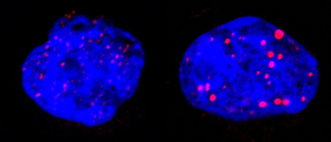
During this targeted treatment researchers showed that the p53** protein, arbiter between cell death and survival, triggers senescence through the involvement of PML nuclear bodies. These spherical structures are present in normal cells but are disorganised by PML/RARA in leukaemia. The treatment reorganises them (see figure below), activating p53 and triggering senescence. In this cascade of events (treatment, PML/RARA degradation, reformation of nuclear bodies, p53 activation), only one link has to be missing to block all the therapeutic effects.

2_cellules

Leukaemic cells before (left) and after treatment (right). The blue represents DNA in the nucleus; the red is nuclear corpus PML. These one are reorganised by the treatment targeting PML/RARA. © Photos provided by Prof Hugues de Thé

It is this phenomenon that enables the elimination of diseased cells and leads to total recovery of the patient, using only combined retinoic acid/arsenic treatment. The absence of chemotherapy avoids many severe side effects.

This understanding of the cellular and molecular mechanism of recovery from acute promyelocytic leukaemia opens prospects for activating this same PML/p53 pathway in other types of cancers.

This work was financed by the French Ligue contre le cancer [cancer research charity], the French Fondation ARC pour la recherche sur le cancer [ARC Foundation for Cancer Research] and the European Research Council (ERC).

* Acute promyelocytic leukaemia is caused by the modification of two genes, RAR and PML, leading to the development of cancer cells;
** the gene coding for p53 protein plays an essential role in cell proliferation under normal conditions and in maintaining the integrity of the cell genome.


Parkinson’s disease: an immense step forward thanks to gene therapy

A French and English team (AP-HP, Inserm, UPEC, CEA/Mircen, Oxford Biomedica, Cambridge University) has conducted a clinical phase 1/2 gene therapy study among patients suffering from an evolved form of Parkinson’s disease. Fifteen patients were able to benefit from this new treatment, which involves injecting a vector expressing the genes of three enzymes that are essential for the biosynthesis of dopamine, which is lacking in Parkinson’s disease. Thanks to this therapy certain cells in the brain begin to produce and secrete dopamine again. In all the patients, the motor symptoms of the disease were improved for up to 12 months after administration of the treatment. After a period of four years, this study is at this stage demonstrating innocuousness and tolerance of the lentiviral vector used for the first time in human beings. This study was coordinated by Prof. Stéphane Palfi, head of neurosurgery at Henri-Mondor Hospital (AP-HP) within the framework of the neurolocomotor research cluster directed by Prof. Césaro.
It is the subject of a publication in The Lancet

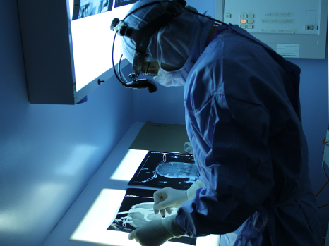
Palfi opération © APHP

Parkinson’s: a common neurodegenerative disease

With about 120,000 patients in France, Parkinson’s disease is the most common neurodegenerative disorder after Alzheimer’s disease. It essentially manifests itself through motor symptoms that steadily grow and become more severe such as trembling, rigidity of the limbs and diminished movement of the body. This pathology is due to the degeneration of neurons that produce dopamine, a neurotransmitter that participates in motor control .

Currently, the treatment of people affected by this disease consists of taking medication that mimics the action of the dopamine missing in the brains of these patients. While this treatment makes it possible to improve motor activity considerably during the first stages of the disease, severe undesirable effects appear at the end of this time such as fluctuations in the effect of the treatment and abnormal involuntary movements, called dyskinaesia.

Developing a new treatment that permits the physiological restoral of missing dopamine

For several years, experts on Parkinson’s disease, researchers and doctors, have held the hypothesis that the intermittent intake of medication during the day alters the functioning of the brain by stimulating neurons in an excessively irregular manner. This phenomenon would constitute the origin of the complications connected with dopaminergic treatment.

The currently most pressing issues in the treatment of Parkinson’s disease thus concern the development of a technology that would make it possible to induce:

  • sustained dopaminergic stimulation;
  • local dopaminergic stimulation in order to induce beneficial motor effects while avoiding the complications that follow stimulation in other regions of the brain not affected by Parkinson’s disease .

This is why researchers today are turning to gene therapy, which consists of causing a therapeutic gene to be expressed directly by brain cells.

Gene therapy consists of introducing therapeutic genes in vivo so that they express directly in the targeted cells.

It rests on the use of viral vectors such as lentiviruses, adenoviruses and AAVs (adeno-associated viruses), which have the ability to introduce their genetic material into the nucleus of host cells.

Some requirements must be absolutely satisfied for a wild virus to be able to be transformed into a vector with the ability to ensure the transfer of genes of therapeutic interest in complete security. These viral envelopes are stripped of their properties for multiplication and rendered non-pathogenic.

Increasing the synthesis of dopamine through gene therapy

In the majority of cases, Parkinson’s disease does not have a genetic origin. However, the biochemical modifications responsible for the symptoms can be corrected by using a gene therapy strategy of the ‘replacement or restoral of function’ type in order to increase the synthesis of dopamine (by expressing genes involved in the biosynthesis of dopamine) and restore the function of dopaminergic cells partially.

It is this approach that was adopted in the phase I/II biomedical study coordinated by Prof. Stéphane Palfi (Henri-Mondor Hospital, AP-HP), the results of which have just been published.

Fifteen patients were operated on by Prof. Palfi, coordinating investigator, in two centres of excellence in neurosurgery – Henri Mondor Hospital (AP-HP) in France and Addenbrookes Hospital in Cambridge, UK.

For the first time in human beings, the team used a lentiviral vector which expresses the genes of three enzymes – AADC (decarboxylase of aromatic amino acids), TH (tyrosine hydroxylase) and CH1 (GTP-cyclohydrolase 1) – essential in the biosynthesis of dopamine. The product was administered in the area of the brain called the striatum during a heavy surgical operation.

Once in the right place, the genes contained in the lentivirus can express themselves and reprogramme cells, which begin to produce and secrete dopamine in the extracellular environment.

Three increasing dosage levels (1×, 2× and 5×) were tested.

‘This biomedical gene therapy study shows innocuousness over the long-term transfer of genes by the lentiviral vector when it is injected directly into the brain of patients suffering from Parkinson’s disease’, explains Prof. Stéphane Palfi. ‘The clinical analysis suggests that the vector used enables a reduction in motor symptoms depending on the vector dose administered, with the strongest dose being the most effective .

The objective of future clinical developments of the vector will be to confirm an improved viral construction that would make it possible to induce an increased release of dopamine (phase 2a). This phase will be followed by a study of the therapeutic effect of ProSavin® by comparing a group of patients receiving the treatment and another group not receiving the treatment (phase 2b). This study, which is pioneering the use in gene therapy of a lentivirus injected in situ, will definitely open up new therapeutic perspectives for diseases of the nervous system.’

Architecture of phase I/II clinical trial

The local and sustained production of dopamine in vivo was restored in 15 patients suffering from an evolved form of this disease. The long-term monitoring of these patients (4 years) evidenced undeniable innocuousness, tolerance and signs of the therapeutic effectiveness of the viral vector depending on the administered dose, with the strongest dose of the vector inducing the most substantial therapeutic effects.

Key figures

15 patients treated

1 lentiviral vector used for the first time in humans

3 dosage levels tested

Research initiated in 2009

This clinical trial follows on from a preclinical study published in 2009, which showed for the first time the effectiveness and innocuousness of the medication in an animal model. Carried out within the framework of the MIRCen translational platform of the CEA, it has opened the door to the clinical study of ProSavin®.

How fiber prevents diabetes and obesity

Scientists have known for the past twenty years that a fiber-rich diet protects the organism against obesity and diabetes but the mechanisms involved have so far eluded them. A French-Swedish team including researchers from CNRS, Inserm and the Université Claude Bernard Lyon 1 (Unité Inserm 855 “Nutrition et Cerveau”) has succeeded in elucidating this mechanism, which involves the intestinal flora and the ability of the intestine to produce glucose between meals. These results, published in the journal Cell on 9 January 2014, also clarify the role of the intestine and its associated microorganisms in maintaining glycaemia. They will give rise to new dietary recommendations to prevent diabetes and obesity.   

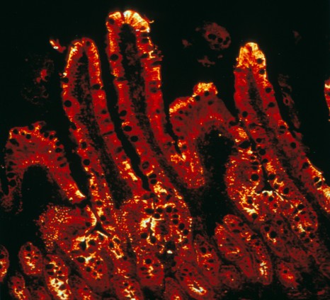
inserm_17416

© Inserm / MITHIEUX G. & BESNARD P
The enzyme responsible for the final reaction in intestinal glucose production is highlighted by immunofluorescence (red) using confocal microscopy. 

Most sweet fruit and many vegetables such as salsify, cabbage or beans are rich in so-called fermentable fibers. Such fibers cannot be digested directly by the intestine but are instead fermented by intestinal bacteria into short-chain fatty acids such as propionate and butyrate, which can in fact be assimilated by our bodies. The protective effect of these fibers is well known to researchers: animals fed a fiber-rich diet become less fat and are less likely to develop diabetes than animals fed a fiber-free diet. Nevertheless, the mechanism behind this effect has until now remained a mystery.

The team headed by Gilles Mithieux, CNRS researcher in the “Nutrition et Cerveau” unit (Inserm / Université Claude Bernard Lyon 1), wondered whether this mechanism could be linked to the capacity of the intestine to produce glucose. The intestine is in fact capable of synthesizing this sugar and releasing it into the blood stream between meals and at night. However, glucose has particular properties: it is detected by the nerves in the walls of the portal vein (which collects the blood coming from the intestine), which in turn sends a nerve signal to the brain. In response, the brain triggers a range of protective effects against diabetes and obesity: the sensation of hunger fades, energy expenditure at rest is enhanced and, last but not least, the liver produces less glucose.

In order to make the connection between fermentable fibers and the production of glucose by the intestine, the researchers subjected rats and mice to diets enriched with fermentable fibers, or with propionate or butyrate. They then observed a strong induction of the expression of genes and enzymes responsible for the synthesis of glucose in the intestine. They showed that the intestine of these animals used propionate as precursor to increase the production of glucose. Mice fed a fat- and sugar-rich diet, but supplemented with fibers, became less fat than control mice and were also protected against the development of diabetes thanks to significantly increased sensitivity to insulin.

The researchers repeated the experiment with mice whose intestine’s ability to produce glucose had been suppressed by genetic engineering. No protective effect was then observed: these mice became fat and developed diabetes like those fed a fiber-free diet. It is therefore the production of glucose by the intestine from propionate and butyrate that is behind the positive effects of fermentable fibers on the organism.

Apart from this previously unknown mechanism, this work sheds light on the role of the intestinal flora which, by fermenting dietary fiber, provides the intestine with precursors to produce glucose. It also demonstrates the importance of the intestine in the regulation of glucose in the body. Finally, these findings should make it possible to propose nutritional guidelines and to highlight new therapeutic targets for preventing or treating diabetes and obesity.

A good outcome for the CHILD-INNOVAC project: successful test in humans of a nasal vaccine against pertussis

The CHILD-INNOVAC European research programme, coordinated by Inserm, has enabled the development of an innovative vaccine that can be administered intranasally, to combat pertussis, which has shown a resurgence in developed countries in recent years. The research consortium, headed by Camille Locht, Director of the Centre for Infection and Immunity of Lille (a joint Unit involving Inserm, CNRS, Institut Pasteur de Lille and University of Lille Nord de France), today published promising results from Phase I clinical trials of the vaccine in human subjects in the online journal PLOS ONE

Researchers from the CHILD-INNOVAC European project, which brought together 10 European partners*, evaluated the efficacy and safety of a new concept in intranasal vaccination against pertussis. They also carried out clinical trials in humans, which provided conclusive results. 

Pertussis is a wrongly “forgotten” disease, according to Camille Locht, a Research Director at Inserm and Director of Scientific Affairs of the Institut Pasteur de Lille. The disease currently affects several tens of millions of individuals, and kills approximately 300,000 children annually worldwide. The associated morbidity and mortality are increasing throughout the world. Its resurgence has become a matter for major concern since 2010 in some developed countries, such as the USA, Australia, the UK, the Netherlands and France. 

The CHILD-INNOVAC project is more specifically focused on combating two major respiratory pathogens: Bordetella pertussis (the bacterium that causes whooping cough) and respiratory syncytial virus (which causes bronchiolitis in infants). These pathogens mainly affect infants aged 0 to 6 months, who are poorly protected by the vaccines presently available. The project also provides proof of concept that this vaccine may be applied to other respiratory infections.

The researchers from the CHILD-INNOVAC project have succeeded in testing in humans, for the first time, a live bacterial vaccine, genetically attenuated and specially designed for intranasal administration to combat major respiratory pathogens. “This original method of administration will make the vaccine accessible to greater numbers of people at a smaller cost,” explains the project coordinator, Camille Locht.

CHILD-INNOVAC: a European success
The main success of this European project is the achievement of a vaccine for which the immunogenicity and safety could be tested in humans in only two and a half years (compared with 5-7 years for most projects of this type). This is a very short time, which Camille Locht explains is due to “the skills and motivation of the consortium, which brought together experts in their respective areas of specialisation from seven European countries. It was possible to relay the data in a flexible and efficient manner at the different stages of the project.” The project received a budget of €5 million, awarded by the European Commission under FP7. 

The Phase I trials in humans allowed the immunogenicity and safety of the vaccine to be measured in comparison with a placebo, under double-blind conditions. They took place in Sweden, which has the most “naive” population with respect to vaccination against pertussis, given that vaccination was abandoned in that country for several years, for reasons of inefficacy. 
The main objective of these trials was to record all possible adverse events, namely cough, sneezing, nasal discharge, effects on general health, etc. These measurements were examined by an independent data monitoring committee.
The second objective was to assess colonisation of the nasal mucosa by the vaccine, and the triggering of an immune response. 
It was possible to test three different doses of the vaccines: a low, intermediate and high dose. 
Results obtained after monitoring the vaccinated subjects for 6 months, and analysing 60,000 data points, showed that the vaccine induced no adverse events compared with the placebo, even at the high dose. The vaccine colonised the nasal mucosa best at the high dose. Moreover, immune responses were triggered in all subjects whom the vaccine had colonised. “It is of special interest that a single nasal administration was able to induce an immune response that was maintained for at least 6 months, i.e. for the duration of the study,” comments Camille Locht. 

The next step will involve administering higher volumes in an effort to increase the level of colonisation of the nasal mucosa by the vaccine. Camille Locht and his collaborators also hope to improve the stability of vaccine over time, with a view to industrial development in the near future. 

Inserm Transfert, in charge of the valorization of the IP portfolio related to the BPZE-based technology, recently entered into an agreement with a biotech partner to further develop the technology.

Further information

CHILD-INNOVAC

The CHILD-INNOVAC project was aimed at developing innovative intranasal vaccines against the two main respiratory pathogens, pertussis and respiratory syncytial virus (RSV). The project has provided prototypes for polyvalent vaccines that can be administered intranasally, based on attenuated B. pertussis. The immunity induced by the BPZE1 vaccine was studied in detail, together with its genetic and biological stability and safety.
CHILD-INNOVAC began in 2008, and was supported by the EU (FP7) for 4 years. It was coordinated by Inserm, along with 27 other European projects. The project involved 10 partners, including 2 private companies and 8 laboratories, based in 7 European countries:

Inserm (coordinator), France: http://www.inserm.fr/
Inserm Transfert, France: http://www.inserm-transfert.fr/
Université Libre de Bruxelles, Belgium: http://www.ulb.be/
Innogenetics, Belgium: http://www.innogenetics.com
National University of Ireland, Maynooth: www.immunology.nuim.ie
Istituto Superiore Di Sanità, Italy: http://www.iss.it/
Swedish Institute for Communicable Disease Control: http://www.smittskyddsinstitutet.se/in-english/
Netherlands Vaccine Institute
National Institute for Public Health and the Environment, the Netherlands: http://www.rivm.nl/
Imperial College of Science, Technology and Medicine, England: http:/www3.imperial.ac.uk/

fermer