Menu

Early morning waking, a winning trend among the French

Setting one’s alarm an hour or two earlier to run, meditate, read or even cook is a trend that is attracting many followers in France, also known as “morningophiles.” Recommended by Hal Elrod in his book The Miracle Morning, this lifestyle, which started in the United States, extols the virtues of waking up very early in the morning, as a source of well-being and productivity.

Are there health benefits involved? Are we all equal when it comes to sleep? What are the risks associated with lack of sleep? Can we do anything about our internal clock?

Such are the questions that are often asked of Joëlle Adrien, neurobiologist and Inserm Research Director, in the Sleep and Alertness Management Workshops that she runs at Hôtel-Dieu Hospital in Paris. In her book Mieux Dormir et Vaincre l’Insomnie (Sleep Better and Overcome Insomnia), she explains the mechanisms of sleep (the ideal duration, the biological clock), explores the various sleep disorders and their remedies, and offers advice to improve the quality of sleep.

couple doing yoga

(c) Fotolia

Unemployment and health

In 2015, a study conducted by Pierre Meneton, entitled “Unemployment is associated with high cardiovascular event rate and increased all-cause mortality in middle-aged socially privileged individuals,” was published in the International Archives of Occupational and Environmental Health.

The results suggest that there may be an association between unemployment and poorer cardiovascular health, unlinked to other conventional risk factors, such as age.

Consult the scientific article to find out more

L'embauche

(c) Fotolia

Genetics and the gut microbiota together contribute to IBD

Modifications of the gut microbiota in chronic inflammatory bowel disease (Crohn’s disease and ulcerative colitis) are both the cause and consequence of these internal disorders. This has been shown a team of French researchers from Inserm, INRA[1], UPMC and AP-HP, who describe these mechanisms and propose new therapeutic approaches. Their work is published in Nature Medicine on 9 May 2016.

Harry Sokol

(c) Harry Sokol – Inserm

Transfer of the microbiota from Card9−/− mice is sufficient to induce the increased susceptibility to colitis observed in Card9−/− mice.

A histology section of axenic, genetically normal mice given the microbiota of genetically normal mice (left) or of Card9−/− mice (right), 12 days after induction of colitis. The colitis is much more severe in mice with the microbiota of Card9−/− mice.


Inflammatory bowel disease (IBD) is characterised by inappropriate inflammation of the digestive tract. It is characterised by inflammatory flare-ups of variable duration and frequency, depending on the patient. This group of diseases usually affects young adults, and its incidence is highest in industrialised countries. Researchers have already discovered susceptibility genes like NOD2, ATG16L1 or CARD9, but also suspect environmental factors and modifications of the gut flora, although when and how these factors are involved is not known.

The CARD9 gene encodes a protein involved in the immune system, and especially in the recognition of microorganisms. “The association between this predisposition gene, the immune system and bacteria deserved to be explored, given that all these factors are involved in IBD,” explains Harry Sokol, leader of this work.


To do this, his team used mice lacking this gene. The researchers found increased sensitivity in the gut of these mice when it was inflamed, with defective healing of the mucosa associated with an interleukin (IL) 22 deficiency and disruptions in the bacterial flora. Observations that did not really surprise them, given that “CARD9 protein expressed by cells of the immune system contributes to the production of IL22, involved in healing and protection of the gut mucosa, and in the recognition of microorganisms,” recalls Harry Sokol.

Except that on transplanting the gut flora from these genetically modified animals to other genetically intact mice but lacking a gut flora, the latter in turn became hypersensitive to gut inflammation. In addition, they also showed a defect in IL22 production. “In other words, the genetic defect in itself is not sufficient to induce the observed malfunctions. Alterations in the composition of the gut flora arising from the absence of CARD9 play a major role in gut hypersensitivity, and the functional defect in the IL22 pathway,” explains Harry Sokol.


The researchers therefore wanted to understand how this altered gut flora could confer these abnormalities on the recipient animal. They then observed that the bacteria present could not, or could only poorly convert tryptophan, an amino acid obtained from food, into an indole derivative that binds to lymphocytes and stimulates IL22 production. An observation that led the researchers to conclude that “Mutation of the CARD9 gene causes a modification of the gut flora mediated by a malfunction of the immune system. The flora loses its ability to produce indole derivatives, helping to exacerbate immunological abnormalities, especially in the IL22 pathway, conducive to inflammation.

These results show how all these mechanisms are interlinked—genetic, immune system and microbiota,” summarises Harry Sokol. “Thus, the abnormalities of the microbiota in IBD are both the cause and consequence of inflammation.”

But most importantly, the researchers showed that these mechanisms were reversible. By giving drugs that can mimic indole derivatives to mice lacking the CARD9 gene, they observed a remission in symptoms, and a return of the IL22 pathway to normal. Exciting results, but it remains to be shown that the same is true of humans. The researchers have already analysed the stools of about a hundred patients with IBD, and have observed a general decrease in the production of indole derivatives by the gut bacteria compared with healthy subjects. By combining this work with a genetic analysis to search for variants of susceptibility genes, they observed that this defect was particularly severe in patients showing a mutation in the CARD9 gene. The idea is now to compensate for this defect in patients. “We can already very easily find patients with a defect in the production of indole derivatives using a simple stool analysis. It may therefore be sufficient to supplement these patients with bacteria producing these derivatives, or directly administer the derivative in question to them.” Work already underway in the laboratory.


[1] “Interactions of commensals and probiotics with the host,” team, MICALIS Institute (Inra-AgroParisTech), Jouy en Josas

Confirmation that the Ebola virus persists in the semen of survivors of the epidemic

An international study, conducted by researchers from the Institute for Development Research (IRD), Inserm and Institut Pasteur and their Guinean partners (Donka University Hospital, Macenta Hospital, National Institute of Public Health, and University of Conakry, confirms that Ebola virus persists in the semen of survivors of the epidemic in Guinea, for up to 9 months after their recovery. These results, which recall the importance of monitoring survivors in order to prevent the risks of new epidemic outbreaks, are published in the Journal of Infectious Diseases on 3 May 2016.

Epidémie d'Ebola en Guinée

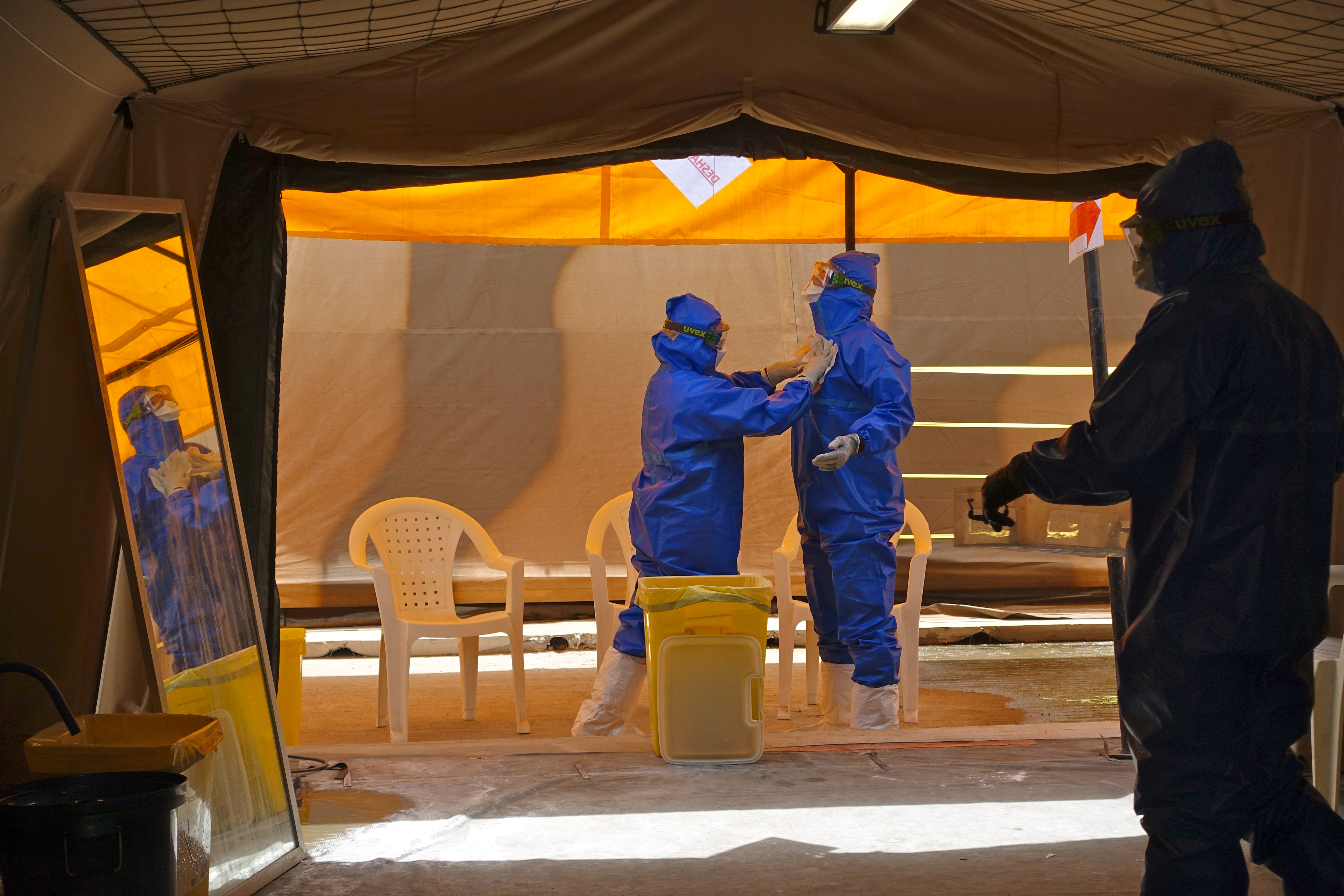
(c)  IRD/ Eric Delaporte

PostEboGui: multidisciplinary monitoring of a cohort of Ebola survivors

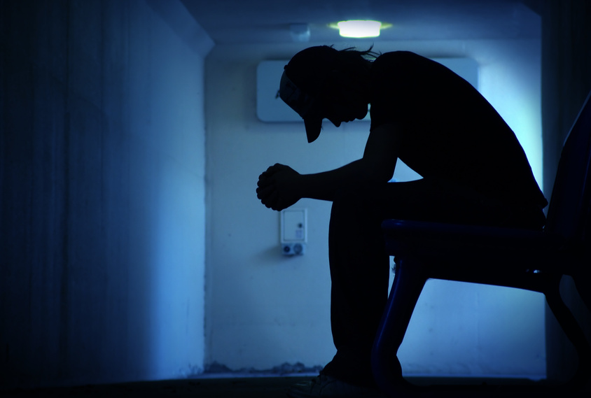
The objective of the PostEboGui1 programme, which has been conducted in Guinea since November 2014, is to monitor, for 2 years, a cohort of over 700 adults and children who survived2 the most serious Ebola epidemic in West Africa, in 2014. The researchers are developing a multidisciplinary approach (clinical, virological, immunological, social, and public health) in order to identify the clinical and social sequelae of the epidemic, as well as the potential risks of reactivating the virus, or transmitting it sexually

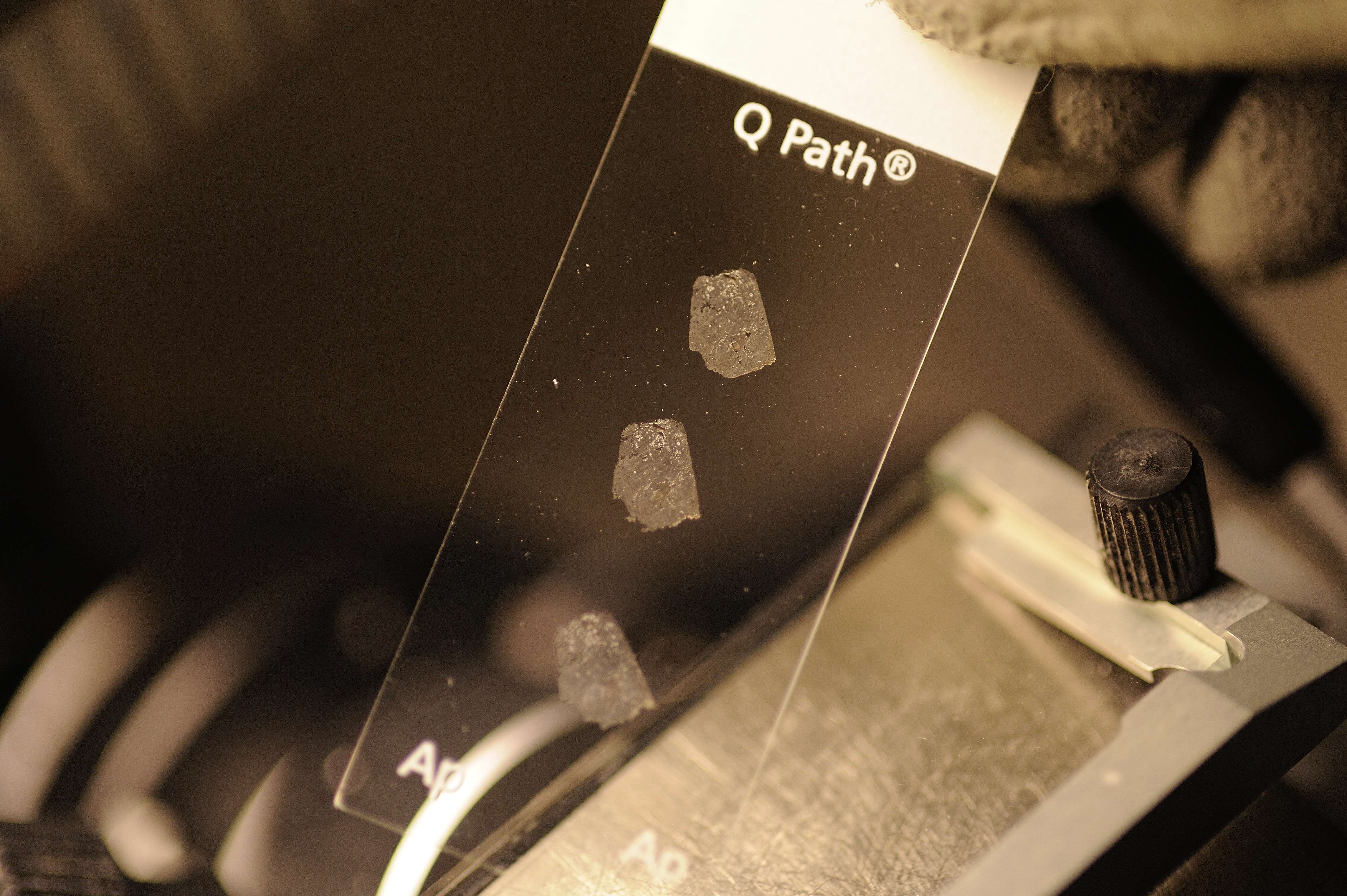
In this study, the researchers monitored the first 450 patients from the PostEboGui programme, both men and women, for 1 year. They took specimens of body fluids (tears, saliva, faeces, vaginal fluids and semen), on the first day of the study, and every 3 months thereafter. In order to detect the presence of the Ebola virus in these fluids, the researchers used molecular biology techniques employing the polymerase chain reaction (PCR) and detection of ribonucleic acid (RNA), in hospitals in Guinea.


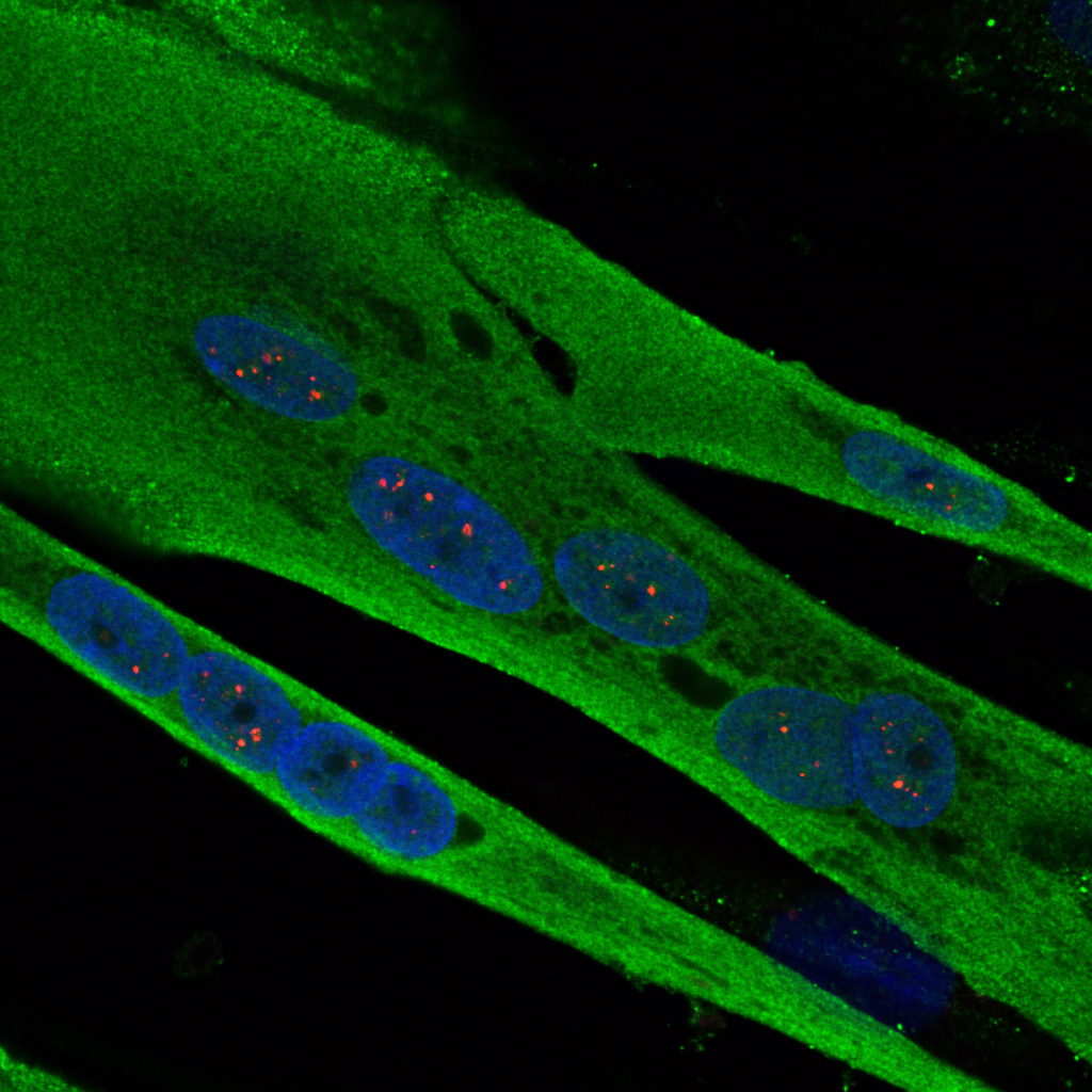
Presence of the virus in the semen for up to 9 months after recovery

The results relate to 98 specimens taken from 68 different people. Ebola virus was detected in 10 specimens taken from 8 men, for up to 9 months after recovery. In addition, the researchers showed that the persistence of the virus in semen decreases with time: the virus, present in 28.5% of samples taken between the 1st and 3rd months, was subsequently detected in only 16% between the 4th and 6th months, in 6.5% between the 7th and 9th months, 3.5% between the 10th and 12th months, and finally 0% after 12 months.


Improve survivor monitoring to limit resurgence of the epidemic

These results confirm those published in October 2015 in the New England Journal of Medicine on a cohort of survivors in Sierra Leone. They emphasise the need to recommend, at international level, the use of condoms by survivors in the months following their recovery.

Furthermore, the researchers insist on the importance of developing survivor monitoring, or even making it systematic, in order to limit the risks of a recrudescence of the epidemic.


Under the Ebola Task-Force, researchers are involved in monitoring survivors, especially in Guinea, from different aspects: surveillance of clinical and psychological sequelae, and risks of virus reactivation in patients who have recovered. They also focus on viral reservoirs in humans (the sites of “immune privilege” constituted by the eyes, brain and gonads).


In 2016, other research programmes will complete the scheme:

  • FORCE: This is a therapeutic trial conducted by Inserm in men showing traces of virus in the semen (treatment based on the antiviral agent favipiravir).
  • ContactEboGui: The objective of this project is to monitor people who have had contact with people who have been infected and declared cured (monitored under the PostEboGui programme), and who could have developed largely asymptomatic undiagnosed infections, in order to improve knowledge on the dynamic of the epidemic, and to identify the risks of secondary transmission and understand the routes of transmission.
  • Réservoir: This project is focused on the source of the epidemic, particularly the animal reservoir for the virus, in Guinea, Democratic Republic of the Congo, Cameroon, Congo and Gabon, in order to prevent future epidemics.

Research programmes initiated at the start of the epidemic in 2014 under the aegis of the French National Alliance for Life Sciences and Health (Aviesan) are also being pursued, in the areas of disease diagnosis, clinical trials, and human and social sciences.


1 Funded by the Interministerial Ebola Task-Force, IRD and INSERM, PostEboGui is conducted by the TransVIHMI joint international unit for translational research on HIV and infectious diseases, in partnership with the University of Conakry, the infectious disease department at Donka University Hospital, Macenta Hospital, INSP, and the Socio-Anthropological Analysis Laboratory of Guinea (LASAG) at Sonfonia University.

2   I.e. over half of the patients declared cured in the country.

Control of fertility: a new player identified

Individual small RNAs are responsible for controlling the expression of gonadoliberin or GnRH (Gonadotropin-Releasing Hormone), a neurohormone that controls sexual maturation, the appearance of puberty, and fertility in adults. This has just been demonstrated by the “Development and Plasticity of the Neuroendocrine Brain” team led by Vincent Prévot, Inserm Research Director (Jean-Pierre Aubert Research Centre, Lille). The involvement of microRNAs, transcribed from DNA, occurs around birth, and marks a key step in postnatal development. Failure of these microRNAs to act leads to the disruption or even total cessation of GnRH production by the hypothalamic neurons that synthesise it, and hence to infertility. In the most serious cases, sterility may result. Details of this work in mice are published in the 2 May 2016 issue of the journal Nature Neuroscience.

Figure images Anglais

(c) Inserm/Prévot Vincent

Images showing the expression of the neurohormone GnRH (green) by the hypothalamic neurons that synthetize it in mice in which GnRH neurons have been genetically tagged by the Tomato gene (gene coding for a red fluorescent protein).


Reproductive function is determined by events that take place in the brain. Gametogenesis (the production of spermatozoa and oocytes) and the secretion of hormones by the ovaries and testes are heavily dependent on the hypophysis, a small gland located below the brain, to which it is connected by a capillary network. The latter is in turn controlled by a glandular “orchestra conductor” located at the base of the brain, the hypothalamus. During postnatal development, activation of a small number of highly specialised neurons (the GnRH neurons) in the hypothalamus leads to the synthesis of a hormone, gonadoliberin or GnRH (Gonadotropin Releasing Hormone), and this process leads to the appearance of puberty.

This step, known as “mini-puberty” constitutes the first activation of the reproductive axis by the brain. It occurs between the first and third months of life of the infant, and is important to the correct course of sexual maturation*. At puberty, GnRH stimulates the synthesis by the hypophysis of other hormones, which in turn enter the bloodstream to promote the growth of the gonads (ovaries and testes), and to subsequently ensure reproductive function.

The appearance of puberty remains one of the greatest scientific enigmas of the 21st century. In the last 30 years, the discovery of mutations in various parts of the genome in patients with disorders of puberty has made it possible to identify some genes involved in this process.

However, physicians and scientists believe that these genes are responsible for only a third of the disorders of puberty encountered in patients. The discovery of the involvement of microRNAs opens up considerable prospects for the medical management of these patients, from both a diagnostic and therapeutic point of view.

MicroRNAs are small non-coding RNAs transcribed from our DNA. In contrast to messenger RNAs (mRNA), they are not translated into proteins. Because of this, microRNAs are not part of the “coding genome,” but constitute what some people call the epigenome. Regulation of gene expression, e.g. expression of the GnRH gene, by microRNAs is therefore considered “epigenetic” regulation.

Research conducted in mice by Vincent Prévot’s team shows that birth induces a radical change in the expression of microRNAs in the hypothalamic GnRH neurons. This modification of the microRNA expression profile is essential to the inhibition of the expression of transcription factors (proteins that activate or inhibit gene expression) that have a repressive effect on GnRH expression. This inhibition of inhibitory factors allows increased production of GnRH, which is indispensable to infantile and juvenile sexual maturation, and the occurrence of puberty. Indeed, in the absence of microRNAs, the expression of transcription factors that inhibit GnRH expression increases, and leads to the extinction of GnRH synthesis in the brain, leading to the arrest of sexual maturation, absence of puberty, and complete sterility in adult individuals. Analysis of the GnRH gene in humans shows that analogous phenomena might occur in our own species. The mechanism elucidated by this team might therefore explain the absence of puberty and the occurrence of infertility in some patients for whom no mutation or polymorphism (variation in DNA sequence) has been identified in the coding genome.

Figure Anglais_fusion (c) Vincent Prévot Inserm

(c) Andrea Messina/Inserm

In terms of diagnosis, the study carried out by Vincent Prévot’s team in Lille shows the interest of analysing DNA segments from which microRNAs are transcribed, as well as the genome segments that encode their binding sites on the target genes.

“The work published today shows the importance of studying the genome sequences that will be transcribed into mRNA molecules, to which microRNAs bind in order to regulate their translation into protein,” add the researchers.

From a therapeutic standpoint, the interaction of microRNAs with the genes they regulate may be prevented or mimicked by the administration of small analogous molecules, for which the study done by Vincent Prévot’s team provides proof of concept.

This research received financial support from the French Medical Research Foundation (FRM).   

* This mini-puberty is seriously compromised in premature infants, who are more likely to develop disorders of puberty and adult infertility than infants born at full term.

AIRE, a key factor in the unequal impact of autoimmune diseases on men and women

Nadine Dragin, a researcher from an Inserm/UPMC/CNRS/AIM team codirected by Sonia Berrih-Aknin and Rozen le Panse at the Institute of Myology, based at Pitié-Salpêtrière Hospital, AP-HP, has demonstrated the central role of AIRE, a key factor in immune tolerance, in the unequal impact of autoimmune diseases on men and women. This work, published in The Journal of Clinical Investigation on 1 April 2016, was funded by AFM-Téléthon.

Projet FIGHT-MG

(c) Inserm / Delapierre Patrick

Autoimmune diseases result from a malfunction of the immune system that attacks normal constituents of the body known as “autoantigens.” They affect 5–8% of the population, and affect women more than men. They thus represent the fifth leading cause of death in women of childbearing age.

To explain this inequality, the researchers from the Institute of Myology focused on thymic tolerance mechanisms, i.e. this state of immune non-response to an antigen. The research teams observed that the Auto-Immune REgulator (AIRE), a key factor in immune tolerance, is less strongly expressed in women than in men.

AIRE controls the expression of tissue-specific antigens on the epithelial cells of the thymus (a lymphoid organ that produces components of the immune system in humans). On contact with epithelial cells expressing these specific antigens, potentially pathogenic cells receive signals that lead to their destruction. A decrease in AIRE expression leads to reduced expression of these specific antigens, and hence to less efficient elimination of the cells. This phenomenon is observed after puberty, when the thymus in both women and female mice expresses less AIRE than that in males, leading to poorer immune tolerance, and hence to greater susceptibility to autoimmune disease. The researchers also showed that oestrogen was the hormone responsible for this effect, since oestrogen treatment of thymic epithelial cells from humans and mice resulted in a decrease in AIRE expression in these cells.

Taken together, these results therefore indicate that, in females, oestrogen induces changes in the expression of the AIRE gene, thereby increasing the sensitivity of women to autoimmune diseases. AIRE expression levels may therefore indicate a predisposition to an autoimmune disease, and make the oestrogen level a potential therapeutic target.

How can the onset of psychosis be explained?

A research team from Paris Descartes University, Inserm and Sainte-Anne Hospital, led by Professor Marie-Odile Krebs, has demonstrated that epigenetic modifications accompany the onset of a psychotic episode in a cohort of young at-risk people aged 15–25 years. These modifications compromise systems for responding to oxidative stress and inflammation. Through this new work, the researchers have shed new light on this disease, for which the main biological explanation, before now, was based on disruption of dopamine secretion in the brain.

The study was published in Molecular Psychiatry on 26 April 2016

desperate

© fotolia

Psychotic disorders preferentially affect a young population, and have a major social impact. Several years before the onset of a true psychotic episode, certain changes in behaviour (isolation, aggressiveness), or certain non-specific (anxiety, problems with concentration or sleep) or more specific symptoms (perceptual abnormalities, fixed ideas, etc.) are generally present. Certain assessment tools have make it possible to define “at-risk mental state” criteria. Approximately one third of people with an “at-risk mental state” will develop a psychotic disorder within three years. There is therefore strong clinical relevance in understanding the physiopathological mechanisms that accompany this change, in order to better define monitoring strategies and, especially, therapeutic interventions.

To study the onset of psychosis, the team of researchers adopted an original approach: they studied modifications in the methylation profile[1] (measured using a blood sample) of young at-risk subjects (ICAAR cohort), monitored for a period of one year. They compared the profiles of individuals who had experienced a psychotic episode with profiles of those who had not become ill. Their conclusions indicate that epigenetic modifications are involved in the onset of a psychotic episode. The modifications preferentially occurred in promoters of genes involved in protection against oxidative stress, in axonal guidance and in the inflammatory response.


Dynamic epigenetic changes accompany the onset of psychosis

The study was carried out on 39 young subjects, 14 of whom developed a psychotic transition in the year following their entry into the cohort. Analyses were concerned with over 400,000 methylation sites, distributed throughout the entire genome (also known as the “methylome”). They included the temporal dimension (comparison before and after the onset of psychosis), but also required the constitution of an appropriate control group (made up of young people who sought care and/or psychological support, but who did not meet the criteria of at-risk subjects). From the start of monitoring, people who went on to develop psychosis showed hypermethylation of the GSTM5 gene promoter[2]. During monitoring, hypomethylation of the GSTT1 gene promoter was observed, and hypermethylation of the GSTP1 gene. These three genes protect against oxidative stress. Other significant modifications were found in genes associated with inflammation and axonal guidance of neurons.


Approaches to developing molecular tools for early detection and targeted therapeutic agents

These results will lead to improved understanding of the biological upheavals that accompany the onset of psychosis. Until now, disruptions in dopamine secretion at brain level were the main physiopathological explanation for psychosis. With the help of these new data, its onset may be linked to an inflammatory or oxidative stress that disrupts the balance (homeostasis) that has already been weakened by a genetic, environmental or neurodevelopmental vulnerability. These results pave the way to the development of tests for the early detection of the illness and monitoring of its progress in these at-risk populations, since this disruption of homeostasis can be easily detected via blood samples, on a repeat basis if necessary. They also point to new therapeutic strategies aimed at preventing psychotic conversion.

[1] Epigenetic modifications are represented by biochemical tags present on the DNA. They do not modify the DNA sequence, but do, however, induce changes in gene activity. The best characterised are methyl groups (CH3: one carbon atom and three hydrogen atoms) attached to the DNA.

[2] A member of the glutathione transferase family, this gene encodes key enzymes that protect against oxidative stress.

Researchers provide guidance on criteria to identify Endocrine Disruptors in the context of European legislation

The European Commission is legally required to provide criteria identifying Endocrine Disrupting Chemicals (EDCs), a process that has been blocked for almost three years, allegedly because of a lack of scientific consensus and because an impact assessment study was deemed necessary. Now, a group of 7 independent researchers from universities and research institutions from Europe and the United States* show how little controversy there is around the definition of EDCs, and that the simple logic used for the identification and regulation of carcinogens can be used for EDCs. The study is published this Monday as a commentary in the scientific journal Environmental Health Perspectives.


First, the authors demonstrate that there is wide acceptance of the World Health Organization (WHO) definition of an EDC as an exogenous substance or mixture that alters function(s) of the endocrine system [i.e. the hormonal system] and consequently causes adverse health effects in an intact organism or its progeny, populations or subgroups of the population.

Second, the authors describe the approach used for the identification of other health hazards of equivalent concern, such as carcinogens or reproductive toxicants. This identification relies on a simple categorization with 3 levels not referring to the toxicological concept of potency[1]. A similar approach not relying on potency should be used for endocrine disruptors; the 3 categories proposed by the European Commission as one of the options considered correspond to “endocrine disruptors”, “suspected endocrine disruptors” and “endocrine active substances” (substances altering the endocrine system with no evidence of the induction of an adverse health effect); these categories are judged sufficient by the researchers. Inclusion of potency or dose-response considerations would modify the spirit of the pesticides and biocides laws, which call for a hazard-based (and not a risk-based) management of pesticides if exposure is not negligible.

Finally, they state that it is not defensible to conduct an impact assessment study to determine scientific criteria. This would be a dangerous precedent, since impact assessment studies are not meant to define hazards, but to quantify the health, social and economic impacts of regulation. This is in line with the decision of the European Court of Justice (2015), which stated that “the definition of scientific criteria to identify properties disrupting the endocrine system can only be done in an objective manner based on scientific data relative to the endocrine system, independently from any other consideration, and in particular from any economic consideration”.

Authors recognize that scientific uncertainty remains with regard to the finer detail of mechanisms, the exact extent of health and environmental effects of EDCs and their impact at the population level. There are also questions around the number of substances likely to be identified as EDCs. However, filling these knowledge gaps is not required to provide scientific criteria defining EDCs.


Several years have been spent trying to issue scientific criteria defining a hazard that was actually defined some time ago (in 2002) by a state-of-the-science report from WHO. For this reason, the scientists consider that the claim of a lack of consensus among scientists was forged to justify delays in the publication of the scientific criteria. Deferring the publication of the scientific criteria can be seen as a way to postpone full application of the 2009 pesticide and 2012 biocide laws. Authors insist that impact assessment studies should not be used as an argument for postponing the publication of a scientific definition. They express concern that scientific definitions might be distorted in order to modify the spirit of a law, thereby muddling science and policy, and postpone the application of existing laws. This postponement is all the more worrying since these scientific criteria are but one of the first steps towards identifying EDCs and providing more efficient protection of public health in the European Union.


Background information

Endocrine Disrupting Chemicals (EDCs) can be very diverse in terms of chemical nature, origin or occurrence. Suspected EDCs include metals (e.g., mercury), organochlorine pesticides such as DDT or triclosan (used e.g. in tooth paste or soaps), other pesticides, dietary contaminants such as bisphenol A, phenols such as parabens (used as preservatives in cosmetics) or phthalates, which can be found in perfumes, cosmetics, medical devices, polyvinyl chloride (PVC) plastics, rainwear… Effects on health outcomes such as congenital malformations, neurodevelopment, behaviour, breast cancer, have been reported for some of these substances in animal models or human studies. The resulting health-related costs in the European Union is estimated to be in the €100-200 billion range (Trasande L et al., JCEM, 2015).

Europe is the only large economy in the world with an ambitious legislation on EDCs. In addition to other health hazards such as carcinogens, mutagens or reproductive toxicants, the European Parliament has identified EDCs as a new type of hazard for health and the environment.

A strategy on endocrine disruptors exists in the European Union since 1999, and in 2009 and 2012, the European Parliament passed two laws on pesticides and biocides (the Plant Protection Products [pesticides] Regulation in 2009 and the Biocide Products Regulation in 2012). These laws stipulated that, for compounds for which population exposure is not negligible, pesticides and biocides containing endocrine disruptors should be regulated on a hazard-based logic (as opposed to a risk-based logic). This implies that there is no need to characterize in detail the dose-response function, identify all possible health effects induced, or possible thresholds in the effects of the compound on health. This approach allows faster management of health risks and more limited resort to test animals, and avoids the debate on the plausibility of the existence of thresholds in the action of these compounds. It is also the logic planned for the management of pesticides and biocides containing carcinogens, mutagens and reproductive toxicants.

The application of the part of these laws regarding endocrine disruptors required the European Commission to publish criteria for identifying endocrine disruptors not later than 2013. This has not been done. Instead, a Roadmap listing 4 options to identify criteria was published by the European Commission in 2014 and the start of an impact assessment study aiming at supporting the choice between the 4 options announced. In December 2015, the European Court of Justice ruled that an impact assessment study was irrelevant, and urged the Commission to publish criteria without delay. The assessment done by the researchers point to the “Option 3” of the European Commission roadmap as being scientifically relevant and operational.

Since 2013, the pesticide and biocide laws (for their part relative to endocrine disruptors) could not be applied because of the lack of publication of criteria for identifying endocrine disruptors. The European Commission indicated in February 2016 that two documents, including one providing criteria for the identification of EDCs, would be published by summer 2016.

Note that, after submission of this publication, a distinct consensus workshop was held by scientists on April 11-12, 2016 in Berlin on a similar topic. The workshop was focused on more fundamental scientific notions, and concluded on the relevance of the WHO definition of EDCs, and the lack of relevance of the concept of potency to identify EDCs. More information can be found on the BfR website.

APPENDIX

Distinction between hazard-based and risk-based regulation of chemicals (copyright RemySlama-Inserm)

distinction between hazard-based and risk-based regulation of chemicals (c) Remy Slama-Inserm

* Authors are affiliated with Inserm- Université Grenoble Alpes and CNRS-Museum national d’histoire naturelle (France); CHU Liège (Belgium) ;University of Nottingham and Brunel University London (UK), University of Torino (Italy); University of Massachusetts (USA)

[1] According to the International Union of 
Pharmacology, potency is « an expression of the activity of a drug [or toxic substance], in terms of the concentration or amount needed to produce a defined effect; an imprecise term that should always be further defined » (Neubig, Pharmacological Reviews, 2003).
Researchers point to the vagueness of this definition and recommend to use the more informative and related concept of dose-response function.

Involvement of Inserm and its Aviesan partners in research on the ZIKA virus

In the last two years, nearly 2 million people have been infected with the Zika virus in Latin America and the Caribbean. At the end of 2015, REACTing and members of the Aviesan alliance immediately became involved, particularly Inserm, Institut Pasteur, the Institute for Development Research (IRD), the French Blood Transfusion Service and the associated university hospitals. Initial contact was made with Brazilian researchers from Fiocruz in November 2015. The international Zika Summit conference, taking place at Institute Pasteur on the 25 and 26 April 2016, will review ongoing research projects:


Several observational and clinical research projects on the monitoring of symptomatic pregnant women, monitoring of children with malformation of the nervous systems or born of infected mothers, construction of biobanks of biological specimens and samples, assessment of the penetration of the virus in populations, and mathematical modelling of its dynamics, have already begun. Teams working in the neuroscience area have also been assembled to begin projects on this theme in a coordinated manner, given the neurotropic nature of the Zika virus.
More specifically, projects strongly involving the French Departments in the Americas are concerned with the following:

  • analysis of the consequences of infection during pregnancy for about 5,000 pregnant women in Guadeloupe, French Guiana and Martinique
  • expansion of the CARBO cohort, a cohort of patients with acute arbovirus infection
  • epidemiological analysis of cases of microcephaly and Guillain-Barré syndrome in French Polynesia (with strong involvement from Institut Pasteur)
  • research on sexually transmitted infection, and the persistence of the virus in semen.


In terms of diagnostics, the aim is to rapidly develop a reliable method at individual and population scale, particularly aimed at pregnant women and newborns, taking into account the variety of pathogens, particularly arboviruses, that co-circulate in countries where the Zika virus epidemic is rife.

Based on these initial elements, Inserm and its partners in the Aviesan alliance have responded to a European call for proposals. For the Aviesan partners, this means:

  • mobilising all French research forces working in the area, as well as involving all European and Latin American partners in a common approach
  • rapidly deploying innovative research, particularly on
    • Zika and the nervous system,
    • modelling the virus and its spatial configuration,
    • methods for controlling vectors,
    • the socio-economic dimension of the epidemic’s spread;
  • including partners that have already worked with European funding in similar situations, associated with dengue fever or emerging infections.

The origin of heart dysfunctions in myotonic dystrophy identified

An international team, including researchers in France at Inserm, CNRS and the University of Strasbourg, brought together at IGBMC[1] is lifting the veil on the molecular mechanisms causing heart dysfunctions in myotonic dystrophy, a genetic disease affecting one person in 8,000. This new study, published this week in Nature Communications, could contribute to discovering a treatment.

DM muscle cells

(c) Inserm/IGBMC

Myotonic dystrophy, also known as Steinert disease, is the commonest adult form of muscular dystrophy. Patients affected by this genetic condition suffer from wasting of skeletal muscles as well as arrhythmia and other cardiac dysfunctions. This is a particularly debilitating disease, for which there is currently no treatment.

Myotonic dystrophy is due to a mutation leading to the expression of RNA containing long repetitive sequences of the CUG trinucleotide. These mutated RNAs accumulate and alter regulation of alternative splicing[2] of numerous genes. Despite the significance of work already done on this disease, many points remain to be elucidated. This is true for the origin of arrhythmia and other cardiac dysfunctions, which represent the second most common cause of death in this disease.

In this new study, researchers have identified new splicing alterations in messenger RNA from heart samples of affected patients. Among these many alterations, biologists have established that those relating to the cardiac sodium channel (SCN5A) were fundamental to understanding the cardiac dysfunctions of these patients.

Scientists there clarified the molecular mechanisms leading to the alteration of SCN5A in these patients. Collaboration with Denis Furling’s team at the Institut de Myologie in Paris has enabled these cardiac alterations to be reproduced in a mouse model.

“The next step would be to see if, by restoring correct splicing of SCN5A, we can also successfully restore normal heart function”, explains Nicolas Charlet-Berguerand, Inserm Research Director, who coordinated this work. The researchers are hoping that this breakthrough will give a fresh boost to research into this rare disease.

Modèle d'épissage alternatif du canal sodique cardiaque

Alternative splicing model of the cardiac sodium channel (SCN5A) in myotonic dystrophy. (c) Inserm/IGBMC

This work was financed by the French Myopathy Association (AFM), the European research council (ERC), the European E-rare programme (ANR), Inserm and Labex-INRT (ANR).

[1] Institute of Genetics and Molecular and Cellular Biology (Inserm/CNRS/University of Strasbourg)

[2] In eukaryotes, this is a process by which RNA transcribed from a gene can undergo different cutting and splicing steps leading to the loss of various regions. This process enables proteins having distinct properties to be produced from the same gene.

Recycling an anti-hypertensive agent to fight brain tumors

Treatments available for glioblastoma—malignant brain tumors—have little effect. An international collaboration[1] led by the Laboratoire Neurosciences Paris-Seine (CNRS/ INSERM/UPMC)[2] tested active ingredients from existing medications and eventually identified one compound of interest, prazosin, on these tumors. Not only did it seem to be effective in this type of cancer, but it also acted on a signaling pathway that is common with other cancers. These promising findings are available online (advance publication) in EMBO Molecular Medicine.

arzneimittel

(c) Fotolia

Turning old into new is what recycling is all about—and what is being attempted by an international collaboration of research scientists coordinated by Marie-Pierre Junier and Hervé Chneiweiss at the Laboratoire Neurosciences Paris-Seine (Paris). The researchers chose to study the most common malignant tumors that develop from brain cells, glioblastomas, which represent the fourth most frequent cause of cancer deaths among adults and the second in children. This is due to the inefficacy of current treatments. Indeed, a glioblastoma can resist treatment and reawaken from a very small number of tumor cells called glioblastoma-initiating cells (GIC). It is these cells—whose characteristics and properties resemble those of stem cells—that were targeted in the study.

Rather than trying to discover new compounds, the team opted for repositioning existing drugs. In other words, they tested a collection of substances used for so long to treat other conditions that their patents have now fallen into the public domain[3]. This method makes it possible to develop new active ingredients cheaply and very rapidly. Twelve hundred compounds were thus tested on normal human neural stem cells and on glioblastoma-initiating cells from different aggressive tumors.

Twelve of these compounds showed a toxic effect on GIC—and none on the normal neural stem cells. The most effective was prazosin. Tested in mice carrying glioblastoma-initiating cells, prazosin significantly reduced the size of tumors and prolonged survival of the mice by more than 50%.

This compound, which has been used for many years to treat hypertension, is an alpha-adrenergic receptor antagonist. The researchers nonetheless made a surprising finding: glioblastoma-initiating cells are devoid of these receptors. The compound therefore acts via an “off-target” mechanism, or in other words through another pathway than standard interaction. The scientists thus identified an intracellular signaling molecule, PKCδ, which is over-expressed in GIC when compared with normal neural stem cells. In the presence of prazosin, it is only cleaved in GIC, which leads to their death.


Clinical trials will be initiated this year to confirm these findings. If they are conclusive, the compound could rapidly be introduced to complement existing therapies and improve the management of patients with brain cancer. Already, the scientists have discovered that other cancer cells display altered PKCδ signaling, including those in colorectal, pancreatic and liver cancer. Understanding the mechanism of action of prazosin may therefore pave the way for the development of new potential treatments for other cancers.

[1] Including scientists from the Laboratoire d’Innovation Thérapeutique (CNRS/Université de Strasbourg), the Stanford University Institute for Stem Cell Biology and Regenerative Medicine (USA) and the Instituto Estadual do Cérebro Paulo Niemeyer in Rio de Janeiro (Brazil).

[2] This laboratory forms part of the Institut de Biologie Paris-Seine.

[3] Pharmaceutical compounds are protected by a patent for 20 years after their discovery. Because of the length of the clinical trials that are necessary before a drug can be put on the market, the duration of their patent protection does not normally exceed 10-15 years after a Marketing Authorization (MA) is granted.

fermer