Menu

29 october 2017: world stroke day

A Cerebrovascular Accident results from the interruption of blood flow to the brain. The deprivation of oxygen and essential nutrients causes brain cells to die, leading to permanent damage (speech or writing difficulty, memory problems, bodily paralysis to a greater or lesser extent), or even sudden death.

World Cerebrovascular Accident Day is organised on 29 October each year and helps raise public awareness regarding the importance of immediate care for victims from onset of initial symptoms (confusion, speech problems, trouble understanding, dizziness, etc.).

Consult our last news about this topic :

Interaction between brain and heart may be new indicator of state of consciousness

©Fotolia

How do we know whether a patient is conscious when he or she is unable to communicate? According to an Inserm study conducted in 127 patients aged 17 to 80, changes in heartbeat in response to sound stimulation is a good indicator of state of consciousness. This is what Inserm researcher Jacobo Sitt and his team, based at the Brain & Spine Institute (ICM) at Pitié-Salpêtrière Hospital, AP-HP, demonstrate in an article published in Annals of neurology. This easy-to-perform examination complements the existing tests and enables finer predictive diagnosis, useful for both doctors and the patient’s loved ones.

The study of consciousness disorders distinguishes the vegetative state, in which the patient is awake but not conscious, from the minimal state of consciousness, which involves a certain degree of consciousness. Distinguishing between these states is very important in order to establish a prognosis on the patient’s neurological outcome, inform the patient’s loved ones, and implement suitable treatment. The tools developed up until now to determine state of consciousness, such as the electroencephalogram (EEG), functional MRI or PET scan, all concentrate on the brain. These methods require either heavy equipment or complex analysis.

Researchers from Inserm have used a novel approach, which consists of exploring the interaction between the heart and the brain.

Previous studies revealed that the “unconscious” processes of the autonomic nervous system, such as breathing or heartbeat, could be modulated by conscious cognitive processes. The perception of external stimulation, auditory stimuli, for example, could therefore be expressed by an effect on cardiac activity, and all the more easily if the subject is conscious.

By studying the data of 127 patients in vegetative or minimally conscious states, researchers observed that cardiac cycles were indeed modulated by auditory stimulation only in conscious or minimally conscious patients. They also showed that these results complemented those obtained with an EEG. Combining these tests (cardiac and EEG) markedly improves performance when predicting a patient’s state of consciousness.

These findings open up new perspectives for an overall approach in evaluating patient state of consciousness. The researchers now wish to extend the scope to include other physiological signals modulated by conscious processes, such as breathing or pupil dilation, in order to develop a comprehensive tool for better bedside evaluation of patient state of consciousness.

What does the auditory test involve?

The test involves playing sound sequences to the patient, which are repetitive to begin with and then, at random and infrequent intervals, present variations. During these variations, the researchers are able to determine whether there is a change in heartbeat, which would mean that the patient is aware of ambient sounds.

Beware of Clichés Related to Gender and Health!

©Picta productions

In terms of health, men and women are not painted with the same brush. This is due not just to their biology but also to social, cultural and economic reasons, which are not always taken into account. Gender stereotypes influence medical practices, research, teaching and patient behaviors, leading to situations of gender inequality and discrimination in the management of and access to healthcare.

The Inserm Ethics Committee and its “Gender and Health Research” Working Group have decided to try to raise the awareness of a wide audience on health inequalities through a series of short films, called Gender and Health: beware of clichés!, co-produced by Inserm, CNRS, and Université Paris-Diderot. CNRS and Université Paris-Diderot are the only higher education establishments in France to have set up structures dedicated to gender equality. The project is also supported by the French Ministry of Higher Education, Research and Innovation.

The 6 one-minute films[1] warn of the stereotypes in six domains of medicine and research: depression, life expectancy, cardiovascular diseases, osteoporosis, pain, and brain imaging. These short films have been designed to raise the awareness of healthcare professionals and patients with a view to promoting greater equality in medicine for the benefit of both sexes.


These videos have been released in the run-up to the “JRS Inserm” international conference, to be held in Paris on November 23, 2017, on the theme “Sex and Gender in Health Care and Research : An Innovative Articulation[2]


[1] The films, produced by Véronique Kleiner and available in French, English, and with subtitles, can be found on Inserm’s YouTube channel, at: https://lc.cx/pqHW.

[2] https://jrsgenre-recherche-sante.dakini.fr/

Is There a Long-term Link Between Childhood Environmental Harshness and Health?

©Fotolia

 A study, conducted by Coralie Chevallier, Inserm researcher at Unit 960 “Cognitive Neuroscience Laboratory” (Inserm/ENS), suggests that an unfavorable childhood environment leads to earlier reproduction and a decreased effort in looking after health during the course of life. These findings have been published in Evolution and Human Behavior.

Current models derived from evolutionary biology show that it can be advantageous for an organism living in a harsh environment (with low levels of resources or high levels of stress or violence) to adopt behaviors favoring benefits in the short term. While these benefits are more modest than those obtained over the long term, they are at least more certain. As such, the receipt of signals by a juvenile organism indicating that the environment is dangerous leads it to adapt its strategy in favor of reproducing sooner – to the detriment of longer-term investments in body maintenance and repair.

On the basis of this observation in animals, Coralie Chevallier, Inserm researcher at the “Cognitive Neuroscience Laboratory” of the École Normale Supérieure (ENS) in Paris, conducted a study to determine whether this model could be applied to humans. To do this, she and her team selected a panel of 1,000 men and women, between the ages of 19 and 87, and representative of the French population.

The panel members answered a series of questions concerning, for example, their age, gender, household composition, socioeconomic status and level of education. They then received a second questionnaire addressing three main themes:

  • childhood environment: parental involvement, upbringing, personal experiences, family difficulties, etc.;
  • reproductive strategy: number of children, age at first pregnancy, age of first sexual intercourse, and number of short-term partners;
  • state of health: body mass index (BMI), how they feel about their health, their desire to remain in good health, and tobacco consumption.

Analysis of these data shows a link between childhood environmental harshness and health and reproductive strategy, with sexual activity starting earlier, the first child being born at a younger maternal age, and poorer health in adulthood (overweight, smoking, etc.). In other words, reduced investment in health is indeed associated with a reproductive strategy that is advantageous in the short term, and these behaviors are associated with an unfavorable childhood environment.

These results therefore suggest that behaviors we could have a priori considered unrelated are actually part of more general strategies that adapt according to the environment. According to Coralie Chevallier, this research is important for the public authorities: “the exposure to harsh environments during childhood has major consequences throughout life and should be the subject of targeted public policies.”

A Novel Animal Model to Speed Up the Fight Against Alzheimer’s Disease

For the first time, an animal model can express the two biological characteristics of Alzheimer’s disease. Researchers from CEA, Inserm, Paris-Sud and Paris-Descartes Universities, and CNRS have developed an animal model that reproduces the progression of the human disease. These results offer new possibilities for testing drugs and developing a diagnostic method involving a simple blood test. Their findings were published in Cerebral Cortex on October 18, 2017.

Alzheimer’s disease remains incurable. The medicines currently available alleviate certain symptoms only partially and the scientific community is experiencing major difficulties in developing effective therapies.

One of the obstacles encountered is the impossibility of diagnosing the disease before it reaches an advanced stage. Yet the biological characteristics of Alzheimer’s appear at least 20 years before the onset of the symptoms (memory loss, etc.). That is why it is necessary to understand this silent phase if we are to hope to treat patients while the brain effects can still be reversed. However, until now, in vitro or animal models making it possible to study this long period preceding the apparition of cognitive symptoms did not exist.

What is more, Alzheimer’s disease, at an advanced stage, is characterized by two types of brain degeneration: the aggregation of Tau proteins in the neurons and the apparition of Aβ42 peptide plaques on the outside of the neurons. Current animal models only express one or the other of these two forms of degeneration.


The challenges facing Alzheimer’s research

► Worldwide, around 45 million people suffer from Alzheimer’s disease. A figure that could reach 52 million in 2020, 81 million in 2040 and 115.4 million in 2050.

► The therapeutic strategies currently available are considered insufficiently effective.

► Clinical trials in Alzheimer’s fail in 99.6% of cases (according to Jeffrey L Cummings, Travis Morstorf and Kate Zhong (2014) Alzheimer’s disease drug-development pipeline: few candidates, frequent failures; Alzheimer’s Research & Therapy20146:37).

► Patients are treated too late to be cured and the current animal models are not representative of the human disease.


A collaboration (1) which began in 2013, involving teams from CEA, Inserm, Paris-Sud and Paris-Descartes Universities and CNRS, led to the development of rodent models (2) which present very early stages of the disease as well as the two types of degeneration. Their findings have been published in the journal Cerebral Cortex (REF). For the first time, an animal model presents disease characteristics comparable to those of human patients.

The researchers now intend to use this animal model, called AgenT, in order to:

► Test potential drugs to determine their efficacy and effects on the two types of degeneration;

► Study the early phase of the disease, during which its development could be reversed;

► Search for blood markers of the earliest stage of the disease, making its diagnosis in living patients (ante-mortem diagnosis) possible from 45 or 50 years of age.


A patent was filed for this model by Inserm Transfert in Europe (WO/2015/067668) in November 2013 which was then extended internationally (WO/2015/067668). The granting of this patent is currently being examined in Europe, the United States and Japan.

Jérôme Braudeau (Doctor of Neuroscience, inventor of the model and last author of this paper) and Baptiste Billoir (a graduate of HEC Paris) are currently creating a start-up that will implement this model.

It will conduct preclinical testing for third parties, research laboratories and pharmaceutical industries, as well as its own research to develop the first early diagnostic blood test in humans.

The objective is to be able to screen for Alzheimer’s at least 10 years earlier than we can right now, thereby having a window for the early treatment of the disease before it progresses to an incurable stage.


InfracliniqueSubclinical
CliniqueClinical


Comparison between the progression of Alzheimer’s disease: 1/ in patients, 2/ in AgenT models (the progression of cognitive disorders and formation of senile plaques is very similar), and 3/ with the transgenic animal models available up to present. © AgenT


BioPrint: Inserm Launches its Second Accelerator of Technological Research

Machine à imprimer 3D laser NOVALASE, technologie LIFT (Light Induce Fast Transfer). ©Inserm/François Guénet. Prise de vue : septembre 2017

The creation of accelerators of technological research (ART) is a priority of Inserm’s strategic plan. On October 12, 2017, Inserm launches its second ART, this time devoted to 3D Bioprinting, in Bordeaux. It is the only tech unit to use the three principal 3D biological printing technologies: laser, ink jet and micro-extrusion. Within the ART, researchers and engineers work together to as quickly as possible transform the fruits of research into technological innovation.

A true pioneer, Inserm is one of the first research institutes to venture into 3D bioprinting. As of 2005, its researchers were printing cells and extracellular matrices using laser printer prototypes. Some years later, this technology sparked worldwide enthusiasm, with Inserm continuing on its trajectory. The BioPrint ART aims to apply its knowledge to various domains, such as tissue engineering, oncology or pharmacology, in order to succeed in creating tissues and complex organoids in the near future.

The BioPrint ART is located in the BioTis Joint Research Unit (Inserm/Université de Bordeaux). Within the next 2 years it will comprise a team of 10 people, assigned to units for cell production, printing, and bioprinted products evaluation.

The ultimate ambition of the BioPrint ART is to develop a structure for training and 3D printing biomanufacture, for use by Inserm’s various laboratories. This new organizational model represents technological progress at the service of fundamental research and the pharmaceutical industry.

For this launch day, a visit of the ART’s premises is proposed. Inserm Chairman and Chief Executive Officer, Yves Levy, will be accompanied by Manuel Tunon de Lara, President of the Université de Bordeaux and Jean-Christophe Fricain, Director of the ART.

For more information on 3D bioprinting:

http://presse.inserm.fr/bio-impression-laser-du-vivant-une-approche-innovante-a-bordeaux/13009/ [Laser Bioprinting in Bordeaux: An Innovative Approach (in French)]


You may remember that on October 16, 2016, Inserm launched its first ART, dedicated to Biomedical Ultrasound

“Canal Détox”

As part of this inauguration, Inserm is launching its new series “Canal Détox”, whose first episode is dedicated to 3D bioprinting. The aim of this series of short films is to decipher current affairs and verify the information that is circulating in the domain of life and health sciences. This new series, in the spirit of evidence-based medicine, will attempt, thanks to its researchers, to discuss a certain number of issues that regularly feature in the media/social media. It will use the most recent data, validated by scientific research that is convergent, robust and exists in sufficient quantities.

Episode 1: Des organes imprimés en 3D…vraiment ? [Organs printed in 3D… really? (in French)] Is waiting to be discovered on Inserm’s YouTube channel.

First clinical proof of concept for functional ultrasound imaging of brain activity in newborns

Coronal image of the brain vasculature of a premature neonate, obtained non-invasively using ultrafast Doppler ultrasound imaging.  Photo credit: Inserm U979 “Wave Physics for Medicine”, Langevin Institute – Waves and Imaging.

Physicians from Inserm Unit 979 “Wave Physics for Medicine” at the ESPCI Paris together with clinician researchers from the neonatal intensive care unit of Robert-Debré AP-HP pediatric hospital and Inserm Unit 1141 have just made a scientific and medical breakthrough: the non-invasive imaging of brain activity in newborns using ultrasound. This will open up new avenues for bedside neurological diagnosis in full-term and premature babies. The details of their research have been published in the October 11, 2017 issue of Science Translational Medicine.

 The technique used, called functional ultrasound imaging of brain activity, was invented in 2009 at the ESPCI Paris in Inserm Unit 979 “Wave Physics for Medicine”, led by Mickael Tanter, Inserm Research Director. Its originality lies in the use of ultrasound technology which, unlike other methods of brain imaging, is simple and portable. Physicians generally use magnetic resonance imaging (MRI) or positron-emission tomography (PET) to image brain activity. Although major technical progress has been made with these methods, they remain restrictive and costly, with long-waiting times for patients.


Similar in appearance to the ultrasound scanners used in obstetrics or echocardiography, the research prototype used has the particularity of being able to acquire images at very high speed. When combined with cutting-edge data processing algorithms, it is possible to map, with very high sensitivity, the subtle variations in blood flow in the small vessels of the brain, variations that are linked to neuronal activity. This new method combines ultrafast image acquisition with very high spatial resolution and a great depth of image. Until now, this had been applied only in pre-clinical studies, using animal models.


Therefore, the research published today establishes the first proof of concept of non-invasive functional ultrasound brain imaging in humans, performed in the neonatology and neonatal intensive care unit of Prof. Olivier Baud at Robert Debré Hospital, AP-HP, currently directed by Prof. Valérie Biran. The brain activity of premature neonates has been recorded in large regions of the brain, at rest and during seizures, at 1,000 images/sec and with a spatial resolution of 150 µm. These hitherto unheard-of data show propagation of cerebral blood flow between and during seizures, and make it possible to locate where they are coming from. Thanks to an ultrafast ultrasound prototype used at the patient’s bedside, images are acquired non-invasively by placing an ultrasound scanner on the baby’s head, above the fontanelle.


For Mickael Tanter and his colleague Charlie Demené, “this first proof of concept of a non-invasive form of neuroimaging that makes it possible to record neuronal activity across an extensive area of the brain, marks the entry of ultrasound into the world of clinical neurosciences with a method that is highly sensitive, portable and can be used directly at the patient’s bedside”.

This study demonstrates the potential of functional ultrasound imaging for the monitoring of premature neonates, who are tricky to examine and in whom it is difficult to diagnose neurological disorders. This technology is not heavy to handle and no patient transportation, contrast agents or ionizing emissions are needed. For Olivier Baud, “functional ultrasound brain imaging could represent a genuine revolution in the field of medicine by bringing new knowledge of neurovascular dynamics, brain development and neuroprotection mechanisms, as well as more early diagnosis of brain functional connectivity alterations”.


This study is part of the FUSIMAGINE project funded by the European Research Council (ERC) for the development of functional ultrasound brain imaging (http://fultrasound.eu)

Voies EEGEEG channels
DroiteRight
GaucheLeft


Identification of a molecular mechanism associated with schizophrenia and bipolar disorder

©Fotolia

A research team in psychiatry at CEA-Neurospin, together with the Mondor Institute of Biomedical Research (INSERM) and the Henri-Mondor (AP-HP) university hospitals, has shown that a genetic variant associated with several psychiatric conditions alters a prefrontal-limbic network, which may increase the risk of developing schizophrenia or bipolar disorder. The results of this study were published online on October 2, 2017 in Journal of Neuroscience.

The authors studied an allelic variation of the SNAP25 gene, which is involved in neurotransmission and associated with schizophrenia, bipolar disorder and attention-deficit/hyperactivity disorder.

The researchers combined a genetic association study in 461 patients with schizophrenia, an in vitro genetic construction, and an approach known as “genetics imaging[1]” involving two cohorts: one with 71 subjects (including 25 bipolar patients) and the other with 121 healthy subjects. They also interpreted the post mortem genetic expression of SNAP25 using the brain tissue of schizophrenic patients.

The results reveal that the variation of the SNAP25 gene changes the expression of the associated protein in the brain, which may impact the processing of information between the brain regions involved in regulating emotions. In line with this mechanism, the genetics imaging study, combining structural and resting state functional magnetic resonance imaging, shows that in both cohorts the at-risk variant is associated with a larger amygdala (a region of the brain) and altered prefrontal-limbic connectivity.

This study confirms the existence of a risk factor shared by schizophrenia and bipolar disorder: variation of the SNAP25 gene. These very common and debilitating diseases each affect 1% of the adult population. In addition to elucidating their mechanism, the results of this study point to the existence of symptoms potentially present in patients with various diseases in which the gene is implicated.


Frontal-limbic connections associated with the SNAP25 variation visualized by diffusion MRI tractography

(Credit: Stéphane Jamain (data from diffusion-imaging.com)


Amygdala-prefrontal network associated with the SNAP25 variation visualized by diffusion MRI tractography

(Credit: Josselin Houenou /BrainVisa/Connectomist 2.0)

[1]Genetics imaging uses imaging (here, magnetic resonance imaging (MRI)) to compare two populations of subjects which differ only by their genetic predisposition (here, SNAP25 variation).

Maternal and neonatal health : preliminary results of the French National Perinatal Survey 2016

The French National Institute for Health and Medical Research (Inserm) and the Directorate of Research, Studies, Evaluation and Statistics (DREES) have published the preliminary results of the French National Perinatal Survey 2016. This report, based on a sample of births representing all births in France for the year 2016, presents the changes in the main perinatal indicators of health, medical practices and risk factors. It also describes the organization of healthcare in the country’s 517 maternity units.

The National Perinatal Surveys are conducted at regular intervals (1995, 1998, 2003 and 2010). The latest, performed in March 2016, was jointly led by the Ministry of Health and Solidarity (DREES, Directorate-General of Health (DGS) and the Directorate-General of Health Care Provision (DGOS)) together with the National Public Health Agency (Santé publique France) and the Obstetrical, Perinatal and Pediatric Epidemiology research team (EPOPé) at Inserm (see boxed text).

In metropolitan France, certain risk factors increased in 2016

  • Advanced maternal age (35 years or older), overweight and obesity were more common in 2016 than in 2010 (respectively 21%, 20% and 12% in 2016 vs. 19%, 17% and 10% in 2010).
  • Between 2010 and 2016, there was no reduction in the rate of tobacco consumption during pregnancy (17%) and exclusive breastfeeding prior to maternity unit discharge fell (from 60% to 52%): two indicators for which France was already faring less favorably than its European neighbors in 2010.
  • The rate of anti-influenza vaccination of pregnant women was very low (7%), despite the high risk of complications represented by the disease.

Certain perinatal health indicators worsened

  • The preterm delivery rate has been on the increase since 1995 (from 4.5% in 1995 to 6.0% in 2016 for live-born singletons).
  • The proportion of infants with a low birth weight (for their gestational age) increased between 2010 and 2016 (from 10.1% to 10.8% for live-born singletons).

In the overseas departments and regions (DROM), the risk factors differ from those of metropolitan France and the perinatal health indicators are less positive overall

  • The socioeconomic characteristics of women in the overseas departments and regions (DROM) are less favorable than in metropolitan France.
  • 25% of pregnant women in the DROM declare that they do not live with a partner (vs. 5% of women in metropolitan France).
  • Pregnant women are younger (6% between 18 and 19 years of age vs. 2% in metropolitan France).
  • Pregnant women in the DROM have a higher rate of obesity (21% vs. 12% in metropolitan France).
  • Tobacco consumption is lower (5% vs. 17% in metropolitan France).


  • The frequency of hospitalizations during pregnancy is higher (27% vs. 18% in metropolitan France).
  • The preterm delivery rate for singleton live births is 10.1% (vs. 6.0% in metropolitan France).

Deliveries took place in larger maternity units, offering increased safety and a better response to women’s needs

  • The number of maternity units continues to decrease: with 517 in March 2016 (including 20 in the DROM).
  • In 2016, births most often took place in large, specialist, public maternity units, although there was no decrease in the number of small units (fewer than 500 births per year).
  • The percentage of units permanently staffed with an obstetrician (from 54% in 2010 to 61% in 2016), an anesthetist (from 75% to 81%) and a pediatrician (from 34% to 40%) increased.
  • Refusal of admission for lack of space was less common in 2016.
  • Pain management improved through more frequent use of patient controlled epidural analgesia (PCEA) and 88% of women were satisfied with what they were offered to manage pain and contractions.


Increased compliance during childbirth with professional recommendations on good practice

  • The rate of cesarean sections was stable (20.4% in 2016 vs. 21.1% in 2010).
  • The rate of episiotomy fell, from 27% in 2010 to 20% in 2016.
  • The care women received during childbirth was less medicalized, with less frequent use of oxytocin (drug used to speed up contractions and which presents a risk to maternal health) during labor.
  • Prevention of post-partum hemorrhage became widespread.


About French National Perinatal Surveys

The French National Perinatal Surveys cover all births (live and stillborn infants) during one week in all French maternity units. The data is collected from medical records and interviews with postpartum women. Data are also collected on the characteristics of maternity units and their organization of healthcare. The latest survey conducted in March 2016 covered 14,142 births and 13,894 women, including 13,384 births and 13,148 women in metropolitan France and 758 births and 746 women in the DROM. The data obtained are used to build reliable indicators and monitor their changes in relation to previous surveys. The survey was coordinated at departmental level by the Mother and Child Protection Services (PMI), the perinatal health networks and Inserm.

The 2016 survey was funded by DREES, DGS, DGOS and Santé publique France.

Biomedical Research Mobilized Around Thomas Pesquet

©Inserm/ Patrick Delapierre


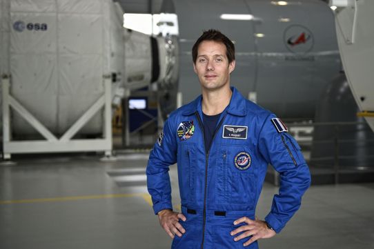
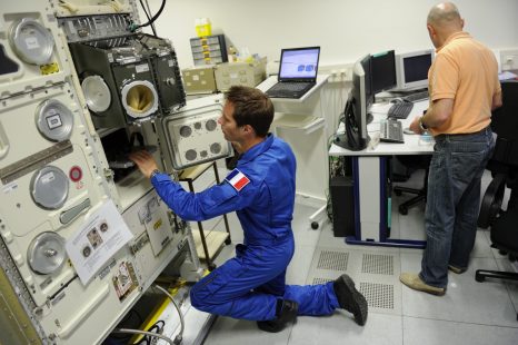
Space is an extraordinary laboratory for medicine and medical research on Earth. That is why Inserm and the CNES have been working together for more than a year[1] to accelerate research progress to improve the health of all of us. The presence of Thomas Pesquet, French astronaut from the European Space Agency (ESA), on board the International Space Station (ISS) for 6 months gave rise to many experiments, the preliminary results of which support the researchers’ hypotheses and sometimes even surprise them.

Thomas Pesquet will present these discoveries at an exceptional session organized by the French Academy of Sciences, to be held under the dome of the Institut de France on October 10, 2017.

In space, the muscles, bones, arteries, sensory organs, nervous system and immune system suffer under conditions of weightlessness and acceleration to which they are not adapted. Why? Evolution has optimized the human body to respond to Earth’s gravity and to live beneath the protective layer of the atmosphere. When the restriction of gravity is suddenly removed and when the body is exposed to cosmic radiation and heavy elements, it can experience specific and hitherto unknown problems. Furthermore, the body must deal with stressful conditions, restricted movement, little dietary variation and disruption of the internal body clock.

In the face of these observations, researchers from Inserm and CNES have rallied to:


  • Protect astronauts’ health, diagnose their conditions in real time, and send and process this information on Earth.


  • Use space as a field of investigation to improve our understanding of physiology and common diseases. In fact, many common conditions benefit from intensified medical monitoring of the astronauts, such as osteoporosis, circadian rhythm and sleep disorders, inner ear and balance disorders, attention and concentration difficulties, as well as arterial aging.


 The first post-flight results:

 On medical and physiological data

On board the station, monitoring the astronauts is a daily challenge. The EveryWear system, developed by CNES health subsidiary MEDES, offers a new approach with a personal assistant for astronauts to use in the form of a simple touchscreen tablet application. It comprises a set of wearable biomedical sensors with Bluetooth connection to a mobile terminal (in this case a conventional tablet).

The use of a tablet to collect a series of different types of information is a major advance in terms of the medical, physiological and personal data of an astronaut. The main advantages of EveryWear are its capacity to aggregate data from various tools and its simplification of the astronauts’ procedures. It is an adaptable system that can cover a broad spectrum of needs: nutrition, sleep, study of the cardiovascular system, with data collection adapted for harmonized processing. Here also, the time the personal assistant saves for the team is considerable. For example, in the past, the astronaut’s nutritional monitoring meant he had to answer questionnaires each time he consumed food. Now, the data are transmitted directly.


On sleep evaluation

Inserm Unit 1075 in Caen is working on miniature actimetric and temperature sensors with the company BodyCap. These sensors and the corresponding analysis software have been incorporated in the on-board assistant of Thomas Pesquet’s flight, particularly for the evaluation of sleep and circadian rhythms. The initial findings, which must be taken with caution as they are derived from the analysis of just one person’s data, are quite unexpected. They show that, despite the strong gravitational influence on body movements – and therefore on the actimetry – the relationships between the actimetric data and the state of vigilance (waking/sleeping) are the same on board the ISS as they are on Earth. Confirmation of this result would facilitate the medical and physiological monitoring of astronauts.

Since the monitoring of physiological and environmental data is a key determinant in the new domain of personalized medicine, improving these devices could have many impacts. For example, these results could be used to help improve connected watches whose sensors (sleep trackers) evaluate sleep in accordance with movement and the reorientation of the wrist during the night, themselves dependent on gravity.


On bone fragility

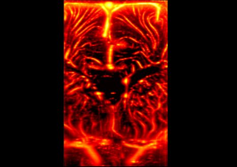
The team of Laurence Vico, Research Director at Inserm Unit 1059, is studying the changes that space flights induce in bone structure. To analyze the structure of the radius and tibia, the experiments use a high-resolution scanner (developed with the help of the European Space Agency). In addition to measuring bone mineral density, this equipment is also used to perform non-invasive “virtual bone biopsies”, enabling a very precise 3D view of the bone architecture.

While it is forbidden to disclose the individual results of the measurements taken from Thomas Pesquet, those from a larger cohort of 13 astronauts not only show that the weight-bearing bones (in the legs) fail to regain the same quality as prior to the flight into space but -and this is a new finding- it would seem that the non-weight-bearing bones (in the arms), which are preserved at the time of return to Earth, progressively deteriorate.

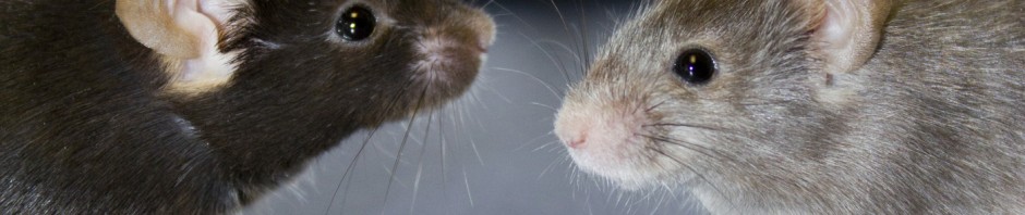
Comparison of these results with those obtained in mice shows that the cells responsible for orchestrating bone formation and resorption activities (osteocytes), and which normally live for several years, die prematurely when subjected to conditions of weightlessness. A countermeasure able to act on this premature aging, and which could be used in osteoporosis patients, is being tested.


On the heart

Pierre Boutouyrie conducts research at Inserm Unit 970. It is one of the first laboratories to have worked on the consequences of the conditions of microgravity or hypergravity, especially on the accelerated aging of our arteries. Space flights are good models in which to study this. With aging, we know for example that arteries lose flexibility, they lose their ability to act as shock absorbers of blood flow and that they are exposed to atherosclerosis which can lead to harmful effects on the heart, brain and kidneys.

For Thomas Pesquet’s flight, the changes related to microgravity were studied by fitting flexible piezoelectric sensors[2] that were linked to the astronaut’s onboard assistant. Again, the individual data cannot be disclosed here but the initial results of a bed-rest study conducted for 2 months in 10 patients, in addition to astronaut data published by an international team show that very rapid and very substantial arterial system remodeling occurs, reflecting its accelerated aging. These consequences are not just suffered by astronauts but also long-term bedridden patients.


Right now, researchers are testing countermeasures related to diet and physical activity in order to attenuate these effects in bedridden patients.


Watch the presentation of these discoveries by Thomas Pesquet, broadcast live from under the dome on the site of the Academy of Sciences!

On December 6, 2016, the Academy of Sciences opened its doors to over 300 high school students and their teachers. A live link-up from the ISS was organized in their honor, with Thomas Pesquet.

On October 10, 2017, Thomas Pesquet will return, in person, beneath the Dome of the Institut de France to tell us about his life in a research laboratory in space, and present the results of the scientific experiments developed by CNES and Inserm under the auspices of NASA and ESA.

This session is also the opportunity to award the prizes of the Academy of Sciences to the valedictorians of the Grandes Écoles, the winners of the national and international Olympiads, the winners of the Rogissart bursaries, and the winners of the Grandes avancées françaises en biologie présentées par leurs auteurs” (for major French advances in biology).

To view the live broadcast online on October 10 from 3 p.m. (French time): http://www.academie-sciences.fr/fr/Ceremonies/seance-thomas-pesquet-2017.html 

[1] http://presse.inserm.fr/espace-et-sante-le-cnes-et-linserm-signent-un-accord-cadre-a-lelysee/25039/ [Space and Health: In the Presence of the President of France, CNES and Inserm Sign a Framework Agreement at the Elysée Palace (in French)]


[2] Developed in collaboration with ESIEE Paris, ESYCOM – EA2552 Laboratory, Université Paris Est (ITMO Technologies de la santé) and the company BodyCap.

fermer