Menu

20% of reactions to radiologic contrast media are real allergies

©Adobestock

A team of Pole-Imaging Research Explorations-European Hospital Georges Pompidou AP-HP, Paris Descartes University and INSERM led by Professor Olivier Clément, and a team from Caen University Hospital and the University of Caen Normandy, led by Dr Dominique Laroche, conducted the first national prospective multicenter study on allergic reactions to contrast media in radiology. 31 centers in France bringing together radiologists investigators, allergists, anesthetists and biologists have investigated 245 cases of hypersensitivity to contrast media.

Promoted by the AP-HP, the study, funded by the Hospital Regional Program Clinical Research, 2003, shows that allergy is responsible for over 20% of hypersensitivity reactions to contrast media and recommends that patients diagnosed allergic, having a high risk of recurrence, are subject to monitoring based on skin tests performed in an allergist specializes in drug allergy.

This work was published in the journal EClinicalMedicine the Lancet in its issue of July 2018.

In radiology, patients may experience immediate hypersensitivity reactions to iodinated contrast media (for scanners) and gadolinés (for MRI) is injected them in the examination. The reactions such as hives, angioedema, bronchospasm, hypotension and anaphylactic shock. Severe reactions, rare, occur within minutes after injection and require from the imaging team a quick diagnosis and management.

For iodinated contrast agents, reactions have long been falsely labeled “iodine allergy” and mistaken reactions to seafood or skin disinfectants.

But the real allergy to contrast medium is diagnosed by elevated plasma markers tryptase and histamine in the first hour of reaction and intradermal skin tests to make between six weeks and six months after it. The few retrospective studies post on the performance of this type of skin test showed that between 13 and 65% of the responses were truly allergic in origin, according to the populations tested. However, these studies suffered from a lack of clinical data, in particular the name of the injected product, or incomplete or late tests performed, or they mixed the immediate reactions and delayed reactions.

A team of Pole-imaging research explorations-European Hospital Georges Pompidou AP-HP, Paris Descartes University and Inserm, led by Professor Olivier Clément, and a team from Caen University Hospital and the University of Caen Normandy, led by Dr Dominique Laroche studied prospectively immediate hypersensitivity reactions to iodinated products and gadolinés. This multicenter study was conducted in 31 French centers equipped to perform skin tests six weeks to six months after a reaction.

After receiving contrast media for radiology review, 245 patients with immediate reaction took a blood sample in the first hour after it to measure the levels of histamine and tryptase in their plasma. They are seen to offer six weeks after a visit to the allergist to test all existing contrast agents (10 gadolinés iodinated or 5).

Skin testing revealed three types of reactions: allergic (if positive test contrast diluted); potentially allergic (if positive test only to pure product) and nonallergic. They identified 41 patients allergic to iodine products and 10 patients allergic to gadolinés products.

The results showed that over the reaction was severe, the more allergic mechanism revealed by the skin test was frequent : 9.5% in the skin reactions; 22.9% in the moderate reactions; 52.9% in reactions involving life-threatening, and 100% when there was cardiac arrest. Similarly, the levels of histamine and tryptase plasma increased with the severity of the reaction. The presence of cardiovascular signs were also very strongly linked to allergic mechanism.

The group of potentially allergic patients showed clinical symptoms and histamine assays and tryptase intermediate between the group of patients allergic and non-allergic people. This suggests that some of them are truly allergic to the contrast material.

The teams also studied cross-reactions with other different contrast the one responsible for the reaction products: 62.7% of patients had allergic cross-reaction to one or more pure products tested.

This study shows that 21% of radiology hypersensitivity reactions are actually caused by an allergy to contrast media.

Allergic patients have a greater risk of recurrence if their is reinjected contrast agent giving a positive skin test.

Patients exhibited severe symptoms (anaphylactic or cardiovascular symptoms) should benefit from a dose of histamine and tryptase the waning of resuscitation and allergy testing in the six months to determine the allergic or not of their reaction, and especially to know which products will be shown against or authorized for future injections.

A molecule could relieve patients suffering from chronic diarrhea with familial amyloid neuropathy

The teams of hepato-gastroenterology department of adult Bicetre Hospital, AP-HP, in collaboration with the department of neurology adult, Inserm and the Université Paris-Sud, show that a derivative of somatostatin may be effective to treat chronic diarrhea suffered by patients with family amyloid neuropathy (NAF). This side effect concerns a quarter of patients after 5 years of follow-impairing their quality of life.

The results of this study, which highlight once again the expertise of the AP-HP in the treatment of rare diseases, are published in the journal Plos One .

Professor Franck Carbonnel and Dr. Michael Collins, Inserm researcher of Hepato-Gastroenterology department of adult Bicetre Hospital, AP-HP conducted a retrospective study of four patients with familial amyloid neuropathy (NAF). This is a rare progressive genetic disorder that causes multiple organ dysfunction and loss of autonomy.

The teams analyzed particularly gastrointestinal side effects such as diarrhea, nausea, vomiting, abdominal pain and incontinence. A quarter of patients after 5 years of follow suffer from chronic diarrhea. Diarrhea is a symptom very debilitating for patients.

The researchers show that similar to somatostatin, octreotide and lanreotide, appear effective for treating chronic diarrhea. Nine of the fourteen patients who received this treatment had a remission of chronic diarrhea after 6 months follow-up. These molecules are already available for other medical indications, including the treatment of neuroendocrine tumors, hemorrhagic complications of portal hypertension and some endocrine disorders. They are also used off-label in the treatment of severe dumping syndrome.

Their use in the refractory diarrhea had already been studied in the context of HIV, chemotherapy-induced diarrhea or diarrhea secondary to digestive suffering from scleroderma.

Induced diarrhea amylose is a multifactorial diarrhea, but involves disorders of the secretion of digestive hormones (including somatostatin) and of intestinal motility disorders.

Often humps transit aggravate constipation in these patients, associated with dysmotility. Sandostatin acts on intestinal secretion and intestinal motility in complex ways. Treatment with octreotide should be monitored and patients with amyloidosis warned of the risk of hypoglycemia.

This work was conducted in collaboration with the teams of Adult Neurology Service – reference center for family amyloid neuropathy and other neuropathies few devices – Professor Adams that showed the effectiveness of the drug candidate Patisiran, Interferent RNA, promising to treat and improve familial amyloid neuropathy transthyretin in July 2018.

To date, in France there are between 40,000 and 50,000 people with neuromuscular disease (NM).

The conditions under national chain of neuro muscular diseases FILNEMUS include muscle diseases (myopathy), diseases of the neuromuscular junction, rare diseases of the peripheral nerve and spinal muscular atrophy infantile.

Deciphering the link between skin allergies and the gut microbiota

Crédits: AdobeStock

Over the last few years, scientists have discovered connections between gut microbiota imbalances and various diseases. Now, in a study using mice, biologists from the CNRS, INSERM, and Claude Bernard Lyon 1 University—together with colleagues from the Institut Pasteur de Lille and the NIH (USA)—have revealed a surprising relationship between a viral detection system, the composition of the gut microbiota, and the development of skin allergies.[1] Their findings, published in PNAS (September 24, 2018) suggest potential new therapies.

The number of microorganisms hosted in our digestive tracts is 10 to 100 times greater than that of all the cells that make up our bodies, and the delicately balanced ecosystem they constitute may be modified by our diet and medication. Epidemiological data of various kinds suggest a link between changes in gut microbiota composition and the development of allergic diseases, like eczema, at body sites far removed from the intestine. But an explanation for this association had been lacking until now.

At the International Center for Infectiology Research (CNRS / INSERM / Claude Bernard Lyon 1 University / ENS de Lyon)—or CIRI—a team led by two researchers from the CNRS focused their attention on mice deprived of the MAVS gene, which plays a key role in the detection of viruses by the immune system. They noted an altered gut microbiota and severe allergic skin reactions in these mice. To demonstrate a relationship between the two phenomena, the researchers transferred the altered microbiota to normal mice. The latter in turn developed severe allergic reactions, showing that the transplanted gut bacteria were responsible.

Furthermore, the biologists revealed that such modification of the gut microbiota led to greater intestinal permeability, which allowed certain intestinal bacteria to migrate to the spleen and lymph nodes and increased the severity of allergic skin reactions.

These findings shed light on the unexpected role played by an antiviral protein (MAVS) in the maintenance of gut microbiota equilibrium. By showing that changes in the gut microbiota exacerbate the allergic response in the skin, this research sets the stage for the development of new therapies. In the not so distant future, might we treat eczema, or enhance already existing treatments, by acting on the microbiota? This approach is already being investigated for other diseases, like cancer.

[1] NIH: National Institutes of Health. This research was conducted at the International Center for Infectiology Research (CNRS / INSERM / Claude Bernard Lyon 1 University / ENS de Lyon), in collaboration with the Center for Infection & Immunity of Lille (CNRS / INSERM / Lille University / Institut Pasteur de Lille) and with the participation of the Cancer Research Center of Lyon (CNRS / INSERM / Claude Bernard Lyon 1 University / Centre Léon Bérard / Hospices Civils de Lyon).

Liver Cancer: When the Cell Environment Plays a Role in Tumor Development

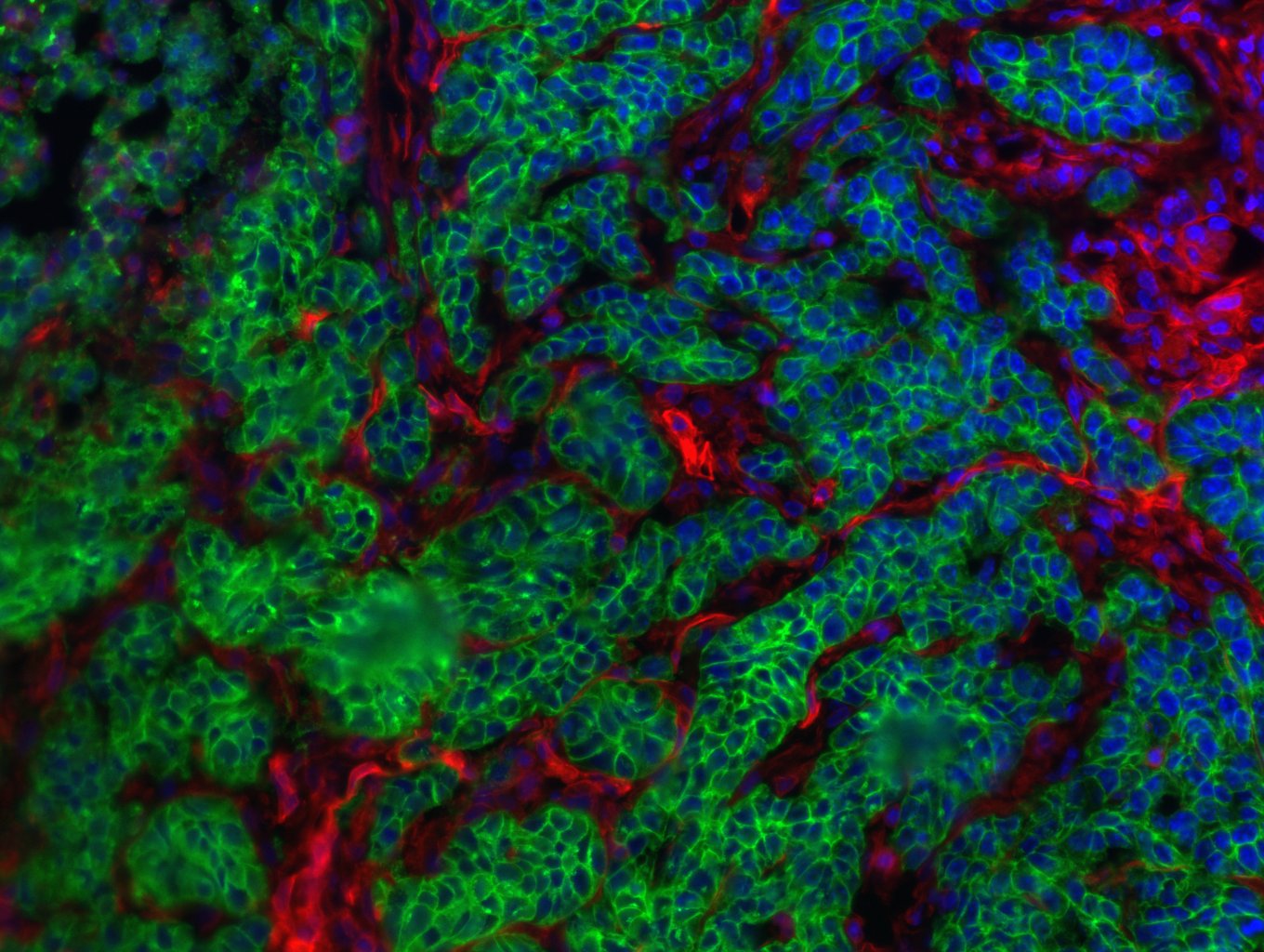
Liver cancer is the second most deadly form of cancer worldwide. While the majority of patients develop hepatocellular carcinoma, 10 to 20 % develop the second type of primary liver cancer: intrahepatic cholangiocellular carcinoma – a highly-invasive cancer of the liver bile ducts. And while both these tumor types have some risk factors in common, the number of patients presenting with intrahepatic cholangiocellular carcinoma has seen a marked increase in recent years. A team of researchers led by Prof. Lars Zender from University Hospital Tübingen (Germany), in conjunction with researchers from the National Cancer Institute (Bethesda, United States), Institut Pasteur, Inserm and CNRS, has recently demonstrated that the cell environment, with its dying liver cells, determines the path taken by the tumor cells. These findings were published in Nature on September 12, 2018.

Despite the progress made in the early detection and treatment of many types of cancer, and the resulting fall in death rates, a marked increase in mortality has been observed in patients with liver cancer. Hepatic steatosis (fatty liver), which is on the rise in western countries, often involves chronic liver damage, and represents a risk factor for liver cancer. Surprisingly, patients with the same predisposition, or risk factors for liver damage, develop hepatocellular carcinoma (HCC) or intrahepatic cholangiocellular carcinoma (ICC), indiscriminately. Yet these two cancers differ in their behavior and how they are treated.

The environment of the cancer cells, and particularly the type of cell death occurring in this environment, proves decisive to the development of the cancer (HCC or ICC).  Indeed, researchers from University Hospital Tübingen, NIH, Institut Pasteur, Inserm and CNRS have recently demonstrated that cell death type is a key factor in the development of tumor cells into a specific hepatic tumor.

In a same cell environment, when the cells die by apoptosis (classical cell death process), the precursors of the cancer cells develop into HCC. However, when the cells die by necroptosis (a form of necrosis), the precancerous cells transform into ICC.

With necroptosis, the cell membrane dissolves and the cell content causes inflammation in the environment of the cancer cell. In the case of classical programmed cell death, small vesicles form which are eliminated by the immune system. These findings were verified in mouse models and human tissue samples1.

What do these discoveries mean for clinical practice?

Future research will have to investigate whether the direct cell environment affects not only the type of tumor development but also the therapy,” says Prof. Lars Zender. In the treatment of HCC with chemoembolization, it has already been observed that a part of the original liver cancer can develop into a bile duct carcinoma. This could be a reason why the cancer no longer responds to the original therapy. “We may be on the trail of a therapy resistance mechanism for liver cancer, states the oncologist, and we hope that the findings will inform novel therapeutic options in the future.

Oliver Bischof2 adds: “This study demonstrates the importance of transcriptomics, epigenomics and bioinformatic analysis in the study of molecular events and signal transduction pathways at the root of different tumor entities. Future cancer research, with the contribution of “omics” analyses, will play a valuable role in identifying new avenues for therapeutic intervention against this disease.


1 Xin Wei Wang, cancer researcher at the Laboratory of Human Carcinogenesis (Center for Cancer Research, National Cancer Institute, Bethesda, United States), contributed human analysis data, and Oliver Bischof, CNRS Research Director and epigeneticist at Institut Pasteur (Nuclear Organization and Oncogenesis unit), described the incidence of the microenvironment on gene regulation in the cancer cell.

2 Oliver Bischof receives funding from the ARC Foundation for cancer research as part of an ARC Foundation-labeled program

Major advances in the diagnosis and treatment of allograft rejection

Crédits: Allogreffe d’aorte abdominale décellularisée, Inserm/Allaire, Eric

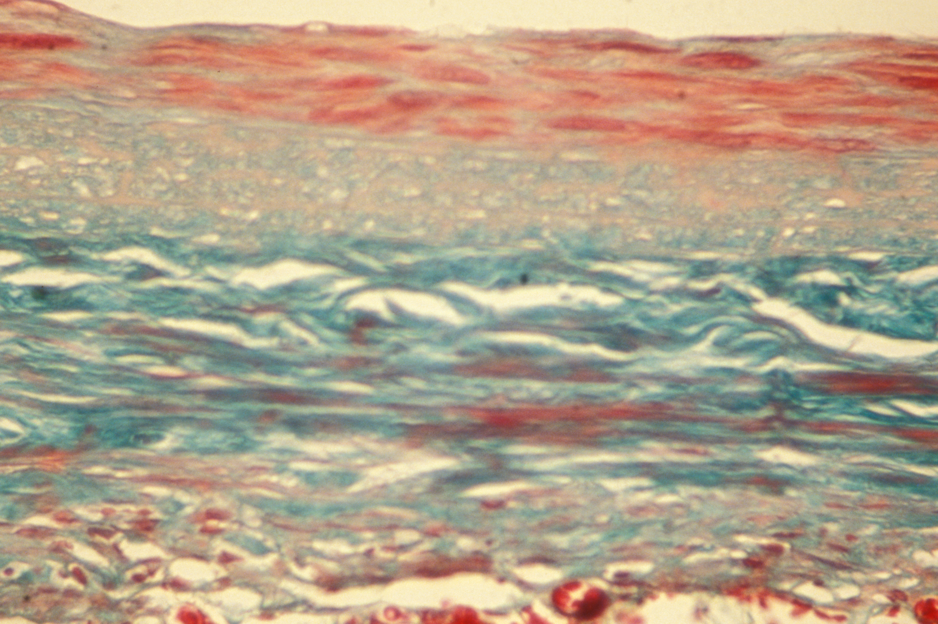
Prof. Alexander Loupy, Hospital Necker Children AP-HP and Prof. Carmen Lefaucheur, the Saint-Louis Hospital AP-HP and the University Paris Diderot in the Cardiovascular Research Center (Inserm / Paris Descartes University), showed, in an article published in the journal   New England Journal of Medicine September 20, 2018, the latest advances and applications of artificial intelligence carried out in the field of transplantation, including the diagnosis and the treatment of allograft rejection.

These interdisciplinary work focused on transplant patients of heart, kidney and lung. They have helped to change the past five years, three times, the International Classification of rejection. They contribute to improving the management of transplant patients on diagnosis and treatment plans.

The transplantation has become the treatment of choice at the onset of organ impairment. 120,000 new organ transplants are performed each year worldwide, but only one million people living with a functioning graft. This finding can be explained by a lack of improvement in graft survival in recent decades and a number of available organs sometimes limited.

The rejection of the organ caused by the production of antibodies by the recipient patient is recognized as one of the main causes of failure of a transplant. A better understanding of the mechanisms of this rejection now allows to diagnose accurately and offer a personalized therapeutic approach.

A multidisciplinary approach involving clinical specialists, pathologists, immunologists transplantation, epidemiologist and statisticians, has been developed in close collaboration with Professor Xavier Jouven, head of the cardiology department of the European Hospital Georges Pompidou AP-HP and the team “cardiovascular Epidemiology and sudden death” of the cardiovascular research Center Inserm and Université Paris Descartes, to evaluate this rejection to the population level. New diagnostic categories were established and patient groups likely to lose their accelerated graft were identified and defined.

The allograft rejection may for example be detected by

> An integrative analysis of multiple biomarkers (reactive antibodies directed against the donor, inflammatory markers); 

> A detailed study of the transplanted organ (identification of gene expression and characterization of cells infiltrating the graft that may cause rejection of the short or medium / long-term allograft).

Work by a team AP-HP / Inserm / Paris Descartes, and coordinated by Professor Alexander Loupy have thus demonstrated that the ultra-fine analysis of genes expressed by cells of the heart via a new technique called “molecular microscope “identifies precisely and patients with early beginnings of heart transplant rejection. (Read more:  >> Diagnosis of transplant rejection in heart: a French team shows the interest of a new method, molecular microscope (March 2017) ). Other more recent studies have demonstrated the usefulness of algorithms to improve efficiency and performance of clinical trials in transplantation *.

Finally, the interest in this approach to artificial intelligence “machine learning” applied to transplantation was realized by obtaining two funding within the hospital research future investment program (RHU ) and the European program for research and innovation 2020.

This research thus open the way to a medicine of the future in which the mathematical algorithms will be used for daily monitoring of patients and the medical decision making. A concrete example is the development of a predictive tool for the survival of kidney transplants.

* Complement-Activating Anti-HLA Antibodies in Kidney Transplantation: Allograft Gene Expression Profiling and Response to Treatment. J Am Soc Nephrol. 2018 Feb; 29 (2): 620-635. doi: 10.1681 / ASN.2017050589. Epub 2017 Oct. 17.

Prenatal exposure to cannabis impacts sociability of male rats

Crédits:  Matteo Paganelli – Unsplash

In a study performed in rats, researchers from Inserm and Aix-Marseille University reveal that prenatal exposure to cannabinoids has sex-specific effects on adult offspring. According to this study published in eLife, consuming cannabis during pregnancy can lead to behavioral and neuronal deficits in male descendants. The findings also point towards a potential pharmacological strategy to help reverse these effects in humans.

A study performed in rats by researchers from Inserm and Aix-Marseille University at the Mediterranean Institute of Neurobiology suggests that using cannabis during pregnancy can lead to less sociability and increased neuronal excitability in males.

According to Olivier Manzoni, Inserm Research Director in charge of the study at the Mediterranean Institute of Neurobiology and Director of the Inserm-Indiana University International Associated Laboratory CannaLab, says: “As cannabinoids can cross the placenta, they may interfere with fetal endocannabinoid signaling during neurodevelopment, which is involved in regulating a variety of processes (synaptic plasticity, appetite, pain sensation), and mediating the pharmacological effects of cannabis. This could in turn lead to some serious long-term deficits. But despite increasing reports of cannabis consumption during pregnancy, the long-term consequences of prenatal cannabinoid exposure remain incompletely understood.”

To fill this knowledge gap, the researchers in Marseille together with their counterparts from the University of Rome (Italy) and Indiana University (USA) examined how prenatal cannabinoid exposure influences the synaptic and behavioral functions of the medial prefrontal cortex – a brain region often implicated in neuropsychiatric disorders – in adult male and female rats.

Their results revealed that males exposed to cannabinoids while in the uterus were less sociable than normal animals, and spent less time interacting with others. Their social behaviors (interactions and play) were impaired, while the number of attacks among males remained unchanged.

Additionally, the researchers saw that the exposed males had a heightened excitability of pyramidal neurons in the prefrontal cortex and a loss of the synaptic plasticity normally mediated by the endocannabinoid system. None of these effects were seen in females.

The deleterious effects of prenatal exposure to cannabinoids on social behavior were specific to male offspring only,” explains co-first author and doctoral student Anissa Bara. “But while social interaction was specifically impaired in males, locomotion, anxiety and cognition remained unaffected in both sexes, suggesting sex-specific behavioral consequences.”

The results also revealed that expression of the mGlu5 gene – an effector of the endocannabinoid system in the prefrontal cortex – was reduced in the males exposed to cannabinoids in utero. The team discovered that amplifying mGlu5 signaling could normalize the synaptic and behavioral deficits induced by prenatal exposure to cannabinoids partly by activating the cannabinoid type 1 receptor (CB1R). Similarly, later tests also revealed that enhancing levels of anandamide (a type of endocannabinoid) in exposed males helped to restore normal social behaviors via the CB1R receptor.

However, prenatal exposure to cannabis does not leave females entirely unscathed. The researchers observed major modifications in the expression of synaptic protein genes in females exposed to cannabinoids in utero. The functional and behavioral consequences of these modifications remain to be identified.

“Altogether, these results provide compelling evidence for sex-specific effects of prenatal cannabinoid exposure,” concludes co-first author Antonia Manduca, also an Inserm Postdoctoral Researcher at the Mediterranean Institute of Neurobiology. “The fact that increasing mGlu5 signaling and enhancing anandamide levels helped to reverse the negative effects of early exposure in rats also hints at a new pharmacological strategy that could one day be trialled in humans.”

The FOReSIGHT project has been awarded University Hospital Institute status, reflecting the success of a long-term commitment by Inserm, Sorbonne Université, Quinze-Vingts National Ophthalmology Hospital and the Seeing and Hearing Foundation.

These founding partners of what is now the seventh center of excellence to receive the coveted University Hospital Institute (IHU) label are embarking on an exceptional project to address the challenges of vision disorders. Centered around the Vision Institute directed by Prof. José-Alain Sahel, the project combines fundamental and clinical research of excellence to improve the knowledge and treatment of age-related macular degeneration (AMD), glaucoma, diabetic retinopathy and degenerative disease of genetic origin. The focus of the IHU is not just to understand these diseases but to restore vision.

Visual impairment is one of the top fears of our fellow citizens and affects several million people in France (including over one million for AMD). The impact on education, employment, autonomy, cognitive functions and the emotional health of those affected is considerable. The human burden is incalculable, with the cost to society running into the tens of billions.

The major funding accompanying this new status will considerably intensify the momentum of diagnostic and therapeutic progress driven by the IHU teams and their partners. Their activities involve studying the mechanisms of visual perception from retina to cortex, and especially brain plasticity in a visual restoration context, characterizing aging and the pathological processes of the visual system to prevent vision loss, and finally developing and validating novel therapeutic approaches in regenerative and prosthetic medicine. This IHU is building on the many years of investment, by the founders having supported the Seeing and Hearing Foundation in the framework of the Labex LIFESENSES (Laboratory of Excellence) of the “Seeing and Hearing” Carnot Institute, by the Clinical Investigation Center and by the Inserm-CNRS-SU Joint Research Unit led by José-Alain Sahel.

The Vision Institute has enjoyed a number of successes in enterprise creation and in its industry partnerships. The IHU will have an additional leverage effect on the innovations generated directly by this new project and is at the heart of a large international partnership network with, among others, the University of Pittsburgh and Japan, with which it will work in synergy. The IHU will operate on the basis of flexible governance by public stakeholders and with the involvement of partners – particularly patient associations.

Project champion José-Alain Sahel thanks “the teams at the Vision Institute and Quinze-Vingts National Ophthalmology Hospital, as well as the public partners and stakeholders, associations and industry players which have made it possible, thanks to this State support, to embark on a new era of transformation serving patients and society”.

Director Jean-François Segovia and Medical Committee Chairman Christophe Baudouin declare: “Quinze-Vingts National Ophthalmology Hospital, the world’s oldest institution in the fight against blindness, together with its institutional partners, will be able to step up the innovation strategy conducted on the campus by the clinical teams and by the Vision Institute. The successes accomplished are also the measure of what separates us from our ultimate ideal: an end to blindness. There is no doubt that this IHU will be a potent accelerator of innovation, education and treatment”.

Jean Chambaz, for Sorbonne Université, declares: “This new success will enable the Vision Institute to pursue its integrated approach to vision, mobilizing numerous Sorbonne Université disciplines around a major public health issue. The IHU will enable it to extend its action via a personalized medicine network, bringing together patients and a broad network of healthcare players and structures”.

Yves Lévy, for Inserm, declares: “This success crowns the very lengthy investment of Inserm in an original research domain with a strong societal impact. We owe this success to the excellence of the fundamental and clinical research teams, and to the dynamism and ambition of project champion José-Alain Sahel at the service of patients, to whom I wish to extend my warmest congratulations.”

When Infection Strikes, Our Brain and Immune System Join Forces

©Adobestock

When infection strikes, what if our immune system was not alone in the fight? What if its major ally was in fact the brain? Researchers from Inserm, CNRS and Aix-Marseille University (AMU) have observed mechanisms of cooperation between the nervous system and the immune system in the response to pathogenic aggressions. This research, published in Nature Immunology, reveals the role of the brain in regulating the inflammatory reaction induced by the immune system in the event of infection and its protective effect against a potential self-destructive exacerbation of that inflammation.

In the event of infection with a virus or other pathogenic organism, the immune system springs into action to eliminate the infectious agent. The immune cells release inflammatory molecules called cytokines, which are responsible for the inflammation process necessary to fight the dissemination of pathogens in the body. There are times, however, when the inflammatory reaction is excessive and toxic to the body. It can provoke lesions in the infected organs which, when too severe, can lead to death.

Previous studies have shown that, in the event of infection, the brain is mobilized to regulate the inflammatory reaction. When it detects the cytokines produced by the immune cells, the brain induces the blood secretion of hormones known to be negative regulators of inflammation: glucocorticoids. The properties of these hormones are widely used in medicine in many pathological conditions, but little is known about their specific mode of action.

In this context, researchers from Inserm, CNRS and Aix Marseille University (AMU) at the Center of Immunology Marseille-Luminy (CNRS/Inserm/AMU) studied, in mice, the mechanism of action of the glucocorticoids produced following activation of the brain in controlling the intensity of the inflammatory reaction caused by viral infection.

Their findings show that glucocorticoids regulate the activity of a population of immune cells which produce inflammatory cytokines and exert a major antiviral and anti-tumor action: natural killer (NK) cells.

These cells possess a receptor which is activated by the glucocorticoids produced after the infection. This activation leads to the expression on the surface of the NK cells of a molecule called PD-1, which is attracting considerable interest within the medical community and is targeted in many cancer treatments due to its inhibitory action on the activity of the immune cells that express it.

The researchers observed that mutant mice which do not express the glucocorticoid receptor in their NK cells were more likely to develop a severe hyper-inflammation reaction and die in the event of an infection. This research demonstrates that expression of the glucocorticoid receptor by the NK cells is necessary to regulate the intensity of the inflammation so that the response against the virus does not become toxic to the body. In addition, the study shows that this regulation is governed thanks to the inhibitory effect of PD-1 which, in the infectious setting, limits the production of inflammatory cytokines by the NK cells.

According to Sophie Ugolini, Inserm researcher and study director: “The most unexpected part of our discovery was that this regulation prevents the immune system from going into overdrive and destroying healthy tissues while fully maintaining its antiviral properties necessary for the effective elimination of the virus. “

This study could enable the development of new therapeutic strategies which would target this regulation pathway. Aside from infections, the researchers especially hope to explore the potential role of this regulation pathway in some cancers.

“Together against rheumatism day” returns for a 5th edition in October 2018

Inserm/Aviesan, the French Society for Rheumatology, the French Arthritis Foundation and 17 partner associations will come together for the 5th Together Against Rheumatism day on October 12, 2018, under the high patronage of President Emmanuel Macron.

Its objective is twofold: fulfill the need for communication between patients, doctors and researchers and create a buzz around the need to support rheumatism and musculoskeletal disease research in France.

The event will take the form of two webinars for which you can register free online:


www.ensemblecontrelesrhumatismes.org


There will be two themes: the morning session (11:30 a.m. – 1 p.m.) will look at gene-environment interactions using the example of pre-rheumatoid arthritis, and the afternoon session (2 – 3:30 p.m.) will be devoted to the new cell and tissue therapies for rheumatism.

Each conference will be divided into three parts in order to promote dialog between patients and research professionals. A clinician/researcher will begin by describing the current research situation and give their clinical viewpoint. Then, representatives of the group of partner associations will put forward the questions and expectations of patients. The final part of the session will take the form of a discussion involving the various speakers, with the possibility for the webinar participants to ask questions.

Participants
In the morning, Prof. Alain Cantagrel, President of the French Society for Rheumatology at Toulouse university hospital, will speak followed by Nathalie de Benedittis (representing the association Pass Sapho), Gérard Thibaud (Andar) and Céline Danhiez (Kourir).

In the afternoon, Prof. Christian Jorgensen (Montpellier) will speak followed by Françoise Alliot-Launois (AFLAR) and Philippe Stévenin (ACS).

Find out more: www.ensemblecontrelesrhumatismes.org

One quarter of deaths and cases of serious after-effects related to meningitis in children can be avoided by applying the immunization schedule

© Chris Benson on Unsplash 

In a context of increasing vaccine hesitancy, researchers from Inserm and pediatricians from the Nantes and Grand-Ouest university hospitals together with the Paris public hospitals (AP-HP) sound the alarm concerning the consequences of severe bacterial infections in children. In a study published in Paediatric and Perinatal Epidemiology, the researchers showed, over a period of 5 years, that 25 % of deaths and 25 % of serious after-effects occurring in children with a severe bacterial infection (primarily meningitis) could have been avoided by simply applying the immunization schedule, notably against meningococcus and pneumococcus.

Bacterial infections are common and for the most part efficiently dealt with by our natural defenses, with an occasional helping hand from antibiotics. Nevertheless, children –and more particularly babies– form a vulnerable population in the face of some severe bacterial infections (meningitis, purpura fulminans, septic shock, etc.) which can lead to serious after-effects (paralysis, sensory deficit –particularly hearing loss–, hydrocephalus, epilepsy, amputation) and even death. While vaccines against the principal bacteria responsible for these infections exist (meningococcal C and pneumococcal conjugate vaccines), defiance towards vaccination, a growing phenomenon in France, is leading to a lack of complete vaccination in some children, putting them at risk of serious after-effects or death.

That is why researchers from Inserm and pediatricians wished to determine, in children having contracted a severe bacterial infection, the proportion of those which could have been avoided had immunization taken place according to the official recommended schedule.

In France’s Grand-Ouest administrative region, all children aged 1 month to 16 years admitted to pediatric intensive care for severe bacterial infection or who died prior to admission were prospectively included from 2009 to 2014 in a study coordinated by Inserm and funded by the Ministry of Health. An infection was considered theoretically preventable by vaccination if the child had either not been vaccinated or had been incompletely[1] vaccinated and that the bacterial strains identified in his or her body were targeted by the vaccines recommended at the time of infection onset.

According to the results of this study, meningococcus and pneumococcus were the principal bacteria causing severe infections in children (65 %), responsible for 71 % of the deaths and close to half of the cases of serious after-effects, despite the introduction of meningococcal C and pneumococcal conjugate vaccines to France’s vaccine schedule in 2002 and 2009.

Only 39 % of the children were correctly vaccinated against these bacteria, meaning that 61 % had either been incompletely vaccinated or not at all. More important still, according to the Inserm study, 25 % of the deaths and 25 % of the cases of after-effects could have been prevented by complete and timely immunization according to the recommended schedule.

In France, while vaccination against the two principal bacteria responsible for these severe infections is now mandatory for all children born after January 1, 2018 (meningococcal C and pneumococcal conjugate vaccines), the majority of the deaths related to meningococcus C occurred in children over 2 years of age who had not had their catch-up vaccination. These children are not currently concerned by the obligation to get vaccinated, which is why it is fundamental to apply the current catch-up recommendations.

As such, according to the researchers, “morbidity-mortality rates related to severe vaccine-preventable infections could be reduced by one quarter with better implementation of immunization programs in France. Such information could help enhance the perception of vaccine benefits and fight vaccine hesitancy”.

[1] “Incomplete vaccination” status was defined as the receipt of fewer injections than those recommended for the age of the child, with authorized delays of 15 days for the pneumococcal primary vaccination and 1 month for the pneumococcal booster and meningococcal C vaccination (according to Gras P, 2017)

fermer