Menu

Myeloid Leukemia: Understanding Treatment Resistance to Move Towards Personalized Medicine

mitochondries © Adobe Stock

The patients who best respond to the dual therapy in this study have a “Mitoscore signature” associated with strong mitochondrial activity. © Adobe Stock


While the care and treatment of acute myeloid leukemia (AML) have greatly improved in recent years, overall survival remains low. Resistance to the various treatments continues to present a major clinical challenge. Using animal models, and also by working with patients themselves, scientists from Inserm, CNRS and Université Toulouse III – Paul Sabatier at the Cancer Research Center of Toulouse have identified a new biomarker predictive of response to dual therapy (chemotherapy plus targeted therapy) used in AML, as well as resistance mechanisms behind relapses. The findings of this research have been published in Nature Cancer.

Leukemia groups several types of blood cancer that affect nearly 10,000 people each year in France. These include acute myeloid leukemia (AML), which affects the hematopoietic cells[1] in the bone marrow.

For a long time, intensive chemotherapy has been the treatment of choice for patients. Although the majority respond favorably and go into remission, overall survival in the longer term remains low, with certain resistant cancer cells persisting in the body following chemotherapy and leading to relapse.

In recent years, the development of targeted therapies has improved the treatment and response of patients, prolonging survival a little – particularly in elderly people ineligible for chemotherapy. However, even with these therapies, relapse remains a major issue. Understanding the mechanisms underlying resistance to leukemia treatments and finding a way to resolve them are a central focus of the work of Inserm researcher Jean-Emmanuel Sarry and his team at the Cancer Research Center of Toulouse (Inserm/CNRS/Université de Toulouse III – Paul Sabatier).

While most scientists working on the subject are more interested in the genetic mechanisms associated with resistance, Sarry’s team is studying the non-genetic mechanisms in order to understand why some patients are more likely to relapse.

Identification of a “Mitoscore signature”

In their new study, the researchers looked at a recently approved dual therapy (conventional chemotherapy combined with a new targeted therapy), which is increasingly used in the treatment of AML.

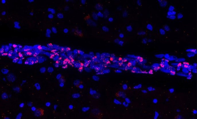
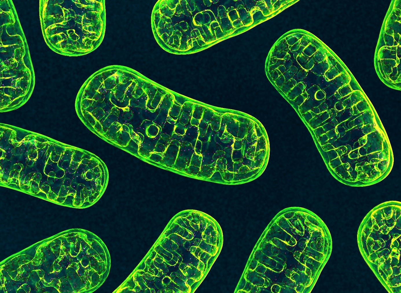
Using patient transcriptomes (i.e. all messenger RNA derived from genome expression), they show that people who respond best to the dual therapy and who have prolonged survival have a specific biomarker – a “Mitoscore signature” – that is associated with a high level of mitochondrial activity[2]. “In other words, this strong Mitoscore signature, which reflects a high level of mitochondrial activity, is predictive of an improved response to these treatments,” explains Sarry.

Finally, thanks to single-cell sequencing[3]of residual disease[4] following this dual therapy, the researchers observed a particular remodeling of mitochondrial function allowing cancer cells to adapt to therapies and induce the patient’s relapse. In mice, the team also showed that treatment based on a molecule that inhibits the action of the mitochondria makes it possible to block this mitochondrial function remodeling, prevent relapse, and prolong the animals’ survival.

“The objective is now to test this Mitoscore signature on very large cohorts in order to validate its utility. Ultimately, the idea would be to use this biomarker to improve patient follow-up and offer more personalized therapies – by giving dual therapy, possibly also with the mitochondrial inhibitor, for those likely to benefit from it. This research could therefore have a real clinical impact in the years to come,” explains Sarry.


1 Hematopoietic stem cells are made by the bone marrow and develop into the various blood cells: red cells, white cells, and platelets. Source INCa

2 Mitochondria are intracellular organelles whose role is to provide the cells with the energy they need. They therefore play a central role in cellular energy metabolism.

3 Single-cell sequencing is a set of molecular biology techniques used to analyze genetic information at single-cell level, using next generation sequencing technologies.

4 Residual disease is the persistence in tissue of malignant cells below the detection limit of conventional techniques.

Using Mechanical Tools Improves Our Language Skills


aires cérébrales liées au langage

The brain regions associated with language have increased during periods of technological boom, when the use of tools became more widespread. © Adobe stock


Our ability to understand the syntax of complex sentences is one of the most difficult language skills to acquire. In 2019, research had revealed a correlation between being particularly proficient in tool use and having good syntactic ability. A new study, by researchers from Inserm, CNRS, Université Claude Bernard Lyon 1 and Université Lumière Lyon 2 in collaboration with Karolinska Institutet in Sweden, has now shown that both skills rely on the same neurological resources, which are located in the same brain region. Furthermore, motor training using a tool improves our ability to understand the syntax of complex sentences and – vice-versa – syntactic training improves our proficiency in using tools. These findings could be applied clinically to support the rehabilitation of patients having lost some of their language skills. This study is published in November 2021 in the journal Science.

Language has long been considered a very complex skill, mobilizing specific brain networks. However, in recent years, scientists have revisited this idea.

Research suggests that brain areas, which control certain linguistic functions, such as the processing of word meanings, are also involved in controlling fine motor skills. However, brain imaging had not provided evidence of such links between language and the use of tools. Paleo-neurobiology[1] has also shown that the brain regions associated with language had increased in our ancestors during periods of technological boom, when the use of tools became more widespread.

When considering this data, research teams couldn’t help wondering: what if the use of certain tools, which involves complex movements, relies on the same brain resources as those mobilized in complex linguistic functions such as syntax?


Syntax exercises and use of tongs

In 2019, Inserm researcher Claudio Brozzoli in collaboration with CNRS researcher Alice C. Roy and their team had shown that individuals who are particularly proficient in the use of tools were also generally better at handling the finer points of Swedish syntax.

In order to explore the subject in greater depth, the same team, in collaboration with CNRS researcher Véronique Boulenger[1], developed a series of experiments that relied on brain imaging techniques (functional magnetic resonance imaging or MRI) and behavioral measurements. The participants were asked to complete several tests consisting of motor training using 30 cm-long pliers and syntax exercises in French. This enabled the scientists to identify the brain networks specific to each task, but also common to both tasks.

They discovered for the first time that the handling of the tool and the syntax exercises produced brain activations in common areas, with the same spatial distribution, in a region called the “basal ganglia”.


ganglions de la base

The handling of the tongs and the syntax exercises proposed to the participants produced activations in a region called “basal ganglia”. © Claudio Brozzoli


Cognitive training

Given that these two skill types use the same brain resources, is it possible to train one in order to improve the other? Does motor training with the mechanical tongs improve the understanding of complex phrases? In the second part of their study, the scientists looked at these issues and showed that this is indeed the case.

This time, the participants were asked to perform a syntactic comprehension task before and after 30 minutes of motor training with the pliers (see box for details of the experiment). With this, the researchers demonstrated that motor training with the tool leads to improved performance in syntactic comprehension exercises.

In addition, the findings show that the reverse is also true: training of language faculties, with exercises to understand sentences with complex structure, improved motor performance with the tool.

Motor training and syntax exercises

The motor training involved using the pliers to insert small pegs into holes that matched their shape but with differing orientations.

The syntax exercises which were completed before and after this training consisted of reading sentences with a simple syntax, such as “The scientist who admires the poet writes an article” or with a more complex syntax, such as “The scientist whom the poet admires writes an article.” Then the participants had to decide whether statements such as “The poet admires the scientist” were true or false. Sentences with the French object relative pronoun “que” are more difficult to process and therefore performance was generally poorer.

These experiments show that after motor training, the participants did better with the sentences that were considered to be more difficult. The control groups, which performed the same linguistic task but after motor training using their bare hands or no training at all, did not show such an improvement.

The scientists are now thinking about how to best apply these findings in the clinical setting. “We are currently devising protocols that could be put in place to support the rehabilitation and recovery of language skills of patients with relatively preserved motor faculties, such as young people with developmental language disorders. Beyond these innovative applications, these findings also give us an insight into how language has evolved throughout history. When our ancestors began to develop and use tools, this proficiency profoundly changed the brain and imposed cognitive demands that may have led to the emergence of certain functions such as syntax,” concludes Brozzoli.


[1] A field in which scientists study the evolution of our ancestors’ brain anatomy.

[2] Involved in these findings are the Lyon Neuroscience Research Center (Inserm/CNRS/Université Claude Bernard Lyon 1) and the Dynamics of Language laboratory (CNRS/Université Lumière Lyon 2).

Covid-19 : Le lourd impact de l’épidémie sur la santé mentale des étudiants, notamment pendant les périodes de confinements

Cohort study identifies genetic cause for rare form of diet-induced Cushing syndrome

Coupe de rein humain grossie 400 fois par un microscope à immunofluorescence polychromatique

Human kidney section magnified 400 times by a polychromatic immunofluorescence microscope. © Inserm/Oriol, Rafael


The team made up of researchers from the endocrinology and reproductive diseases department of Bicêtre AP-HP hospital, Inserm and Paris-Saclay University, carried out work, coordinated by Professor Peter Kamenický, to study the genetic cause of bilateral macronodular adrenal hyperplasia with diet-induced Cushing’s syndrome. This rare disease affects the two adrenal glands located above the kidneys and causes an overproduction of cortisol, a steroid hormone whose excess has harmful consequences for the body . Researchers were able to determine the molecular explanation for the occurrence of this disease 30 years after its initial description. This work was published on October 13, 2021 in the journal The Lancet Diabetes & Endocrinology .

This rare form of adrenal Cushing syndrome, studied by these researchers, is due to the abnormal expression of the GIP receptor (Glucose-dependent insulinotropic peptide) in both adrenal glands of patients. GIP is a hormone produced by the small intestine in response to the ingestion of food. In patients with this particular form of Cushing’s syndrome, cortisol levels increase abnormally after each food intake. Patients with this disease develop the typical clinical signs of Cushing’s syndrome such as weight gain associated with muscle atrophy, high blood pressure, diabetes mellitus, osteoporosis, and depression. The pathology is associated with an increase in mortality, especially cardiovascular causes.

In this international study involving researchers from six countries, and based in particular on close Franco-Quebec collaboration, the team reports that GIP-dependent macronodular hyperplasia of the adrenals, in both familial and sporadic forms, is a genetic disease caused by germline mutations of Lysine Demethylase 1A (KDM1A) with secondary loss of the second KDM1A locus, comprising the second copy of the gene, in adrenal tissue. KDM1A acts mainly as a transcriptional repressor (ie a regulator which prevents a gene from being expressed), the loss of its function results in deregulation of the expression of various genes in the adrenal tissue, including the GIP receptor but also of other receptors coupled to G proteins.

This discovery will make it possible to offer genetic counseling and earlier detection of this rare disease to patients and their relatives. Rare diseases are generally underdiagnosed. This is all the more important as the pathogenic variations of KDM1A also predispose to myeloma and other types of cancer.

In addition, this new role of KDM1A as an epigenetic regulator of the expression of the GIP receptor and other receptors coupled to G proteins could have pharmacological implications.

COVID-19: Artificial Intelligence Identifies Gene Signature Specific to Patients Suffering from Critical Forms

SARS-CoV-2

Covid-19: Intracellular observation of reconstituted human respiratory epithelium MucilAir™ infected with SARS-CoV-2. © Manuel Rosa-Calatrava, Inserm ; Olivier Terrier, CNRS ; Andrés Pizzorno, Signia Therapeutics ; Elisabeth Errazuriz-Cerda  UCBL1 CIQLE. VirPath (Centre International de Recherche en Infectiologie U1111 Inserm – UMR 5308 CNRS – ENS Lyon – UCBL1). Colorisé par Noa Rosa C.


What are the molecular and genetic characteristics that distinguish patients with critical forms of COVID-19 – and particularly acute respiratory distress syndrome (ARDS)? To answer this question, researchers from Inserm and Université de Strasbourg at Unit U1109 Molecular Immunology and Rheumatology, in collaboration with clinician-researchers at the Strasbourg University Hospitals, have investigated the biological and genomic data of a targeted cohort of young patients. Patients hospitalized in intensive care with ARDS were compared with COVID-19 patients hospitalized in a non-critical care ward.

As part of a Franco-US collaboration with researchers from the company Genuity Science in Boston and the University of Southern California in Los Angeles and using the most advanced artificial intelligence techniques to interpret these data, the scientists have succeeded in identifying a gene signature that differentiates these critical patients from their non-critical counterparts. Some of the genes included in this signature could ultimately become therapeutic targets for severe forms of COVID-19 or ARDS. The findings from this research have been published in Science Translational Medicine.

COVID-19 varies broadly from one patient to another. While some are asymptomatic, others develop flu-like symptoms. Then there are others who progress towards severe forms of the disease, in some cases developing acute respiratory distress syndrome (ARDS) that requires mechanical ventilation in an intensive care unit. Although this group of patients accounts for only a small proportion of those infected with the virus, its mortality rate is high – reaching around 25%.

While age and comorbidities such as diabetes and cardiovascular diseases are the main risk factors for developing these severe, potentially fatal, forms of COVID-19, scientists do not yet know why some younger and previously healthy patients also develop these forms. In molecular and genetic terms, what is it that distinguishes these patients with severe respiratory symptoms from the others?

There has been research into the subject since the start of the pandemic and some avenues have been identified, but so far each study addressed the question from a single methodological angle, generally focusing only on one aspect – genetic and metabolic factors, immune response parameters, etc.

Young patients with no comorbidities

The scientists from Inserm and Université de Strasbourg, in conjunction with the Strasbourg University Hospitals, were in this case interested in a patient cohort with restrictive and strict inclusion criteria. The patients had been hospitalized during the first wave of the pandemic, were under 50 years of age and had no major comorbidities. A total of 72 patients were recruited into two groups, one consisting of intensive care patients with ARDS and the other of less severe COVID-19 patients hospitalized in a non-critical ward. A “control” group of 22 healthy individuals was also studied.

“We chose to focus on a restricted but very well-defined patient cohort, excluding confounding factors such as age and certain diseases so that we could really study the molecular and genetic mechanisms directly associated with the severe forms, which are exclusively linked to viral infection and not to other pre-existing risk factors,” emphasizes Seiamak Bahram [1], last author of the study.

The scientists collected various samples in order to perform a multi-omics analysis –that is say retrieve and analyze the various genomic, proteomic, transcriptomic (investigation of all messenger RNA) data and other virological, immunological, and serological data from these patients. This allowed them to confirm that ARDS is associated with a major inflammatory state and an immune system surge (the so-called “cytokine storm”).

Using artificial intelligence

However, given the considerable mass of data generated as part of this multi-omics analysis, it was impossible to take the interpretation further without the help of artificial intelligence (AI). Thus, in collaboration with the AI Research Institute of Genuity Science [2] , a biotech in Boston (USA), the team was able to identify a network of 600 genes involved in the progression towards the critical forms of COVID-19, thanks to the cross-application of several AI algorithms (including one having run on the quantum computer made available by the University of Southern California in Los Angeles).

As part of this transatlantic collaboration, these large quantities of data were modelled and analyzed with the help of AI, making it possible to more accurately identify five genes that are overexpressed in these patients.

One of them, ADAM9, is a particularly interesting “driver gene,” with previous studies having shown that it interacts with SARS-CoV-2 proteins. The findings obtained here are consistent with that, suggesting that ADAM9 overexpression would “drive” some patients towards severe forms of COVID-19 and ARDS.

The researchers then conducted in vitro experiments which showed that blocking ADAM9 in cell lines is associated with a reduction in the quantities of SARS-CoV-2 in these cells, as well as reduced replication of the virus, thereby confirming not just its importance in critical disease but also its potential as a therapeutic target.

Of course, further studies will have to be carried out to confirm this last point, but the scientists believe that these findings have opened up an interesting therapeutic avenue, especially given the current clinical trials in oncology that are testing monoclonal antibodies which inhibit ADAM9. Therapeutic repurposing strategies could therefore be considered in the longer term.


[1] Professor Seiamak Bahram, a university professor and hospital practitioner, is the director of Inserm Unit 1109, head of the Strasbourg Precision Medicine Interdisciplinary Thematic Institute, and head of the Biological Immunology Department at Strasbourg University Hospitals.

[2] It has since become the company HiberCell

The Utility of a Two-Dose Ebola Vaccine Regimen Confirmed

vacnni anti Ebola

© Adobe Stock


In response to the 2014-2016 Ebola epidemic, the clinical development of a two-dose regimen of vaccines Ad26.ZEBOV and MVA-BN-Filo was accelerated. Approved in 2020 by the European Commission for use in epidemic emergencies, this regimen continues to demonstrate its relevance. An Inserm study led by Rodolphe Thiébaut (Inserm, INRIA, Université de Bordeaux, Vaccine Research Institute) has played a role in evaluating its safety and immunogenicity in healthy adults and in those with HIV and compared different time intervals between the two doses. It has confirmed that this regimen is well tolerated, that the antibodies acquired persist for at least one year, and that they can be easily reactivated by a booster shot. The findings of this trial have been published in Plos Medicine.

Since its discovery in 1976, Ebola has been found in several countries in Equatorial Africa with increasingly frequent epidemics (more than 30 to date). Infection with the virus begins in the form of flu-like illness but is often complicated by life-threatening organ failure and hemorrhage. During the largest epidemic in West Africa between 2014 and 2016, a total of 28,616 cases were identified leading to 11,310 deaths. During this epidemic, an initial single-dose vaccine was approved and used in the field (Ervebo). At the same time, pharma company Janssen launched the accelerated development of a different vaccine regimen1. This involves the administration of two different vaccines: the first being Janssen’s Ad26.ZEBOV, composed of an adenoviral vector containing the envelope protein of the Ebola Zaire virus that triggers antibody production, and the second being Bavarian Nordic’s MVA-BN-Filo, which uses a different viral vector and contains four different antigens of the Ebola virus family, including the Ebola Zaire virus glycoprotein.

Using the viral vector makes it possible for the Ebola virus antigen (the glycoprotein) to penetrate the immune cells in the manner of a Trojan horse, triggering the immune response. With this dual-administration strategy, we assumed that the immune response would last longer than with a single-dose vaccine,” explains Rodolphe Thiébaut, Inserm team leader and Professor of Public Health at Université de Bordeaux, which is participating in this EBOVAC accelerated development program.

This hypothesis has recently been validated by the publication of the findings of a Phase II clinical trial (EBOVAC 2) intended to assess the safety and immunogenicity of this regimen in healthy people and in those with HIV living in Africa. In the meantime, it was approved in July 2020 by the European Medicines Agency in adults and children over one year of age, based on data already available.

Nevertheless, this regimen continues to be developed in different populations (adults, children, pregnant women, caregivers), in different parts of the world (to confirm the robust immune response and tolerability profile) and with different follow-up durations, to confirm the preliminary data and evaluate its utility in prevention in populations potentially exposed to the virus – particularly rural populations in Equatorial African countries. The aim being to prevent the occurrence of a new epidemic. “While the epidemics are generated by the virus passing from bats to humans, their progression is facilitated by mobility that is increasing thanks to the advances in infrastructure,” warns Rodolphe Thiébaut, and “having vaccines tested in Africa with the collaboration of research teams from African countries to protect populations is a major development challenge for our countries with their limited means and limited hospital resources,” adds Houreratou Barry, investigator responsible for the trial at the Muraz Center in Bobo-Dioulasso, Burkina Faso.

This Phase II trial is moving in that direction. It enrolled 668 healthy adults between the ages of 18 and 70 and 142 adults between the ages of 18 and 50 with HIV (a common condition in African populations liable to influence immune response to the vaccine) whose viral load was controlled by antiviral therapy. Participants were recruited in Kenya, Burkina Faso, Côte d’Ivoire, and Uganda. They received the vaccine or a placebo solution according to the following regimen: one dose of Ad26.ZEBOV or placebo, followed 28, 56, or 84 days later by MVA-BN-Filo or placebo. In the group of healthy subjects, 90 people also received an additional booster dose of Ad26.ZEBOV one year later.

The researchers analyzed adverse event reports over the follow-up year as well as changes in the levels of antibodies to the Ebola virus glycoprotein. No vaccine-related serious adverse events were observed, only mild to moderate events common with vaccination, such as pain at the injection site, fatigue, headache, and muscle pain.

The increase in the interval between the vaccinations from 28 to 56 days improved the immune response and the antibodies persisted for at least one year in both the healthy subjects and in those with HIV. These antibodies were found in 78 to 88% of the participants. Extending the interval to 84 days provided no additional benefit (and would only extend the vaccination schedule unnecessarily), which confirmed the 56-day interval as optimal for this vaccine regimen.

In addition, the booster one year later sufficiently stimulated the production of antibodies with levels multiplied by 55, indicating that the first vaccination triggered an easily reactivable memory immune response, “which is very important in the context of the recurring epidemics observed in Africa,” concludes Thiébaut.


1 Supported by the IMI EBOLA+ program (a joint initiative of the European Commission and the European Federation of Pharmaceutical Industries and Associations [EFPIA]) within the framework of a consortium with the academic teams from Inserm, the London School of Hygiene and Tropical Medicine, the University of Oxford, and the Muraz Center in Burkina Faso.  This project was funded by the joint Innovative Medicines Initiative 2 (www.imi.europa.eu) under EBOVAC2 grant agreement no. 115861. This joint initiative is supported by the European Union and EFPIA’s Horizon 2020 research and innovation program.

Chatbot for addressing COVID-19 vaccine hesitancy

ordinateurResearchers from the CNRS, INSERM, and ENS-PSL show that such an interface is indeed capable of swaying the vaccine-hesitant. © seth schwiet on Unsplash


  • A considerable fraction of the population is reluctant to get vaccinated against COVID-19.
  • French scientists have designed a chatbot that offers personalised responses to questions posed by the curious or hesitant—and have demonstrated its effectiveness.

What if a few minutes of interaction with a chatbot could effectively address vaccine concerns? In an article published in the Journal of Experimental Psychology: Applied (28 October 2021), researchers from the CNRS, INSERM, and ENS-PSL show that such an interface is indeed capable of swaying the vaccine-hesitant.

Vaccine hesitancy is one of the major challenges in containing the COVID-19 pandemic. Previous studies have revealed that mass communication—through short messages relayed by television or radio—is not a very effective means of persuading the hesitant. In contrast, discussing your particular concerns with an expert whom you trust can be more persuasive, but having a face-to-face talk with every vaccine-hesitant individual is impractical.

To overcome this problem, a team of cognitive scientists from the Institut Jean-Nicod (CNRS / ENS-PSL) and the Laboratoire de Neurosciences Cognitives et Computationnelles (INSERM / ENS-PSL) created a chatbot that provides users with answers to 51 common questions about COVID-19 vaccines.1

Chatbots have the advantage of offering quick, personalized Q and A sessions while reaching a large number of people.

The team tested their chatbot with 338 individuals and compared their reactions to those of a control group of 305 participants who only read a brief paragraph that gave information about COVID-19 vaccines. After a few minutes of interaction with the chatbot, the number of participants with positive views of vaccination increased by 37%. People were also more open to getting vaccinated after using the chatbot: declarations of vaccine refusal fell 20%. Such changes in attitude were negligible in the control group.

It remains to be shown whether the effects of chatbot interaction are lasting, and whether they are the same across age groups, and among those most resistant to vaccination.2

Nevertheless, this study has demonstrated that a chatbot can indirectly reach a very large audience: half of the experimental group later tried to persuade others to get vaccinated, with three-quarters of them stating they drew on information provided by the chatbot to do so.

These findings suggest that a chatbot regularly updated to reflect the latest vaccine science could be an effective tool to help reduce vaccine hesitancy.


Notes

1The questions were selected on the basis of surveys on reasons for vaccine hesitancy as well as articles about vaccine-related preconceptions. Their answers were prepared from scientific sources and approved by COVID-19 vaccine specialists.

2On average, the group of participants was younger and more educated than the overall population.

COVID-19: How Does SARS-CoV-2 Infection Affect Vascular Irrigation of the Brain?

tissu cérébral humain post-mortem

Fluorescent image of post-mortem human brain tissue showing cell nuclei (blue) that reveal a blood vessel in which the vascular endothelial cells express the genetic material of SARS-CoV-2 (red). © Vincent Prévot/Inserm


A large number of researchers are currently mobilized to increase knowledge of SARS-CoV-2 in order to improve the treatment of infected patients and try to predict the future health impacts of infection with the virus. As part of an international collaboration, researchers from Inserm, Université de Lille, Lille University Hospital, and Pasteur Institute Lille within the Lille Neuroscience & Cognition laboratory, along with their colleagues from the CNRS1, have been the first to identify a direct effect of SARS-CoV-2 on the brain’s blood vessels. Certain cells, namely the cerebral vascular endothelial cells – essential components of the blood-brain barrier that protects the brain – are affected by a phenomenon of cell death. These findings, published in the journal Nature Neuroscience, particularly question the long-term impacts of the disease.

The blood vessels are comprised of endothelial cells. These include the vascular endothelial cells in the brain that make up the blood-brain barrier (BBB). The primary function of the BBB is to isolate the central nervous system from the bloodstream, preventing foreign substances or potentially toxic molecules from entering the brain and spinal cord while allowing the transfer of nutrients essential to their activity. As part of this effort, the vascular endothelial cells in the brain therefore play a key role in the proper irrigation of the organ, with their survival being essential for it to function correctly.

Within the framework of an international collaboration funded by the European Research Council3, the authors of the study looked at the vascular endothelial cells of the brain and the consequences of SARS-CoV-2 infection on their functioning.

Using preclinical research models and also by studying the cortex of patients who died as a result of SARS-CoV-2 infection, the researchers have shown that infection leads to the death of endothelial cells in the brain, resulting in the appearance of “ghost vessels” in the brain (empty tubes with no endothelial cells).

As a result, these essential cells can no longer perform their function in the BBB.

How does this endothelial cell death occur? What are the mechanisms involved? Thanks to state-of-the-art techniques2, the team has discovered that SARS-CoV-2 generates the manufacturing, from its own genetic material, of molecular scissors by the endothelial cells it infects. These scissors cleave a protein called NEMO which, being necessary for the endothelial cells to survive, therefore leads to their death.


The impacts of endothelial cell death on brain function

According to the scientists, the death of vascular endothelial cells in the brain can have two major consequences:

  • A temporary rupture of the BBB causing microbleeds in regions where the blood is not meant to have free access.
  • Hypoperfusion of some brain regions (due to the presence of non-functional ghost vessels), which is a decrease in blood flow that in the most serious cases can be fatal.

However, the study shows that the situation is reversible.

Furthermore, the scientists are interested in the long-term impacts of this phase of vulnerability during which brain irrigation is decreased. According to them, even if this hypothesis remains to be verified, this window of time could predispose certain people with the disease to develop cognitive or neurodegenerative disorders, or even dementia.

“This awareness of the severity of SARS-CoV-2 infection and its impacts on proper brain function is vital to enable the best possible management of infected patients in the years to come,” concludes Vincent Prévot, Inserm Research Director.


1 At the Center for Infection and Immunity of Lille (CNRS/Inserm/Institut Pasteur Lille/Université de Lille/Lille University Hospital)

2 Such as transgenesis, single-cell RNA sequencing, mass spectrometry and super-resolution microscopy.

3 Program funded by the European Research Council (ERC Synergy), with the participation of Drs. Prévot (Inserm, France), Nogueiras (University of Santiago de Compostela, Spain), and Schwaninger (University of Lübeck, Germany).

Découverte de nouveaux marqueurs génétiques à l’origine d’une maladie des artères essentiellement féminine

Towards the elimination of cholera in Haiti

robinet eau

Favored by poor access to clean water, sanitation, and hygiene, disease transmission has persisted in epidemic waves © Unsplash

In 2013, a medical team from the AP-HP, Sorbonne University, Inserm, the European Hospital of Marseille, the IRD, Aix-Marseille University (SESSTIM) and the AP- HM proposed to the Haitian government and to Unicef ​​a coordinated strategy to fight against cholera, aimed at breaking the chains of transmission. 

This strategy and its results, which show the apparent cessation of cholera transmission in Haiti since 2019, have just been the subject of a publication in the journal Emerging Infectious Diseases.

Unfortunately imported into Haiti in 2010 during a movement of troops from Asia, cholera caused in this Caribbean country one of the most violent epidemics of recent decades. Favored by poor access to safe drinking water, sanitation and hygiene, the transmission of the disease has persisted in epidemic waves to such an extent that the elimination of cholera in the country has been considered impossible by many. .

In 2013, a medical team from the AP-HP, Sorbonne University, Inserm, the European Hospital of Marseille, the IRD, Aix-Marseille University (SESSTIM) and the AP- HM proposed to the Haitian government and to Unicef ​​a coordinated strategy to fight against cholera, aimed at breaking the chains of transmission.

Mobile rapid response teams have thus carried out more than 50,000 interventions throughout the country in order to raise awareness, distribute water and hygiene treatment kits, and often “minute” antibiotic prophylaxis to contact subjects of patients received in the hospital. health centers.

In collaboration with the Haitian Ministry of Health, this medical team has just shown, in an article entitled ”  Towards cholera elimination in Haiti  ” that there is no longer the slightest sign of activity of the epidemic since February 2019 despite intense microbiological research.

These dramatic results suggest that case area targeted interventions led by rapid response teams have played a key role. They also question the theory that the bacteria responsible for the disease would persist in the country’s aquatic environments, preventing its elimination.

Study on the role of “ceramides” in infection by SARS-CoV-2 which could constitute a biomarker of severity and a therapeutic target in the management of Covid-19


SARS-CoV-2

Electron microscopy of a cell infected with SARS-CoV-2 © Philippe Roingeard, Anne Bull-Maurer, Sonia Georgeault, unité Inserm U1259 MAVIVH & Université de Tours, France.


Teams from the psychiatry and addictology department of the Corentin-Celton AP-HP hospital, the University of Paris and Inserm in collaboration with the University of Erlangen-Nuremberg and the University of Duisburg-Essen , coordinated by Dr Nicolas Hoertel, Prof Jo hannes Kornhuber and Prof Erich Gulbins, published on October 4, 2021 in the journal Molecular Psychiatry (Nature Publishing Group) a summary of the results of the international literature on the central role that “ ceramides ”, a class of lipids, may play a role in SARS-CoV-2 infection.

Results from in vitro data [1, 2] indicate that an enzyme found in cell lysosomes, acid sphingomyelinase (ASM), is activated by the virus upon binding to its ACE-2 cell receptor, inducing synthesis of a specific class of lipids, the “ceramides”, in the membrane of cells. These studies [1, 2] demonstrate that these ceramides serve as a gateway for the virus to infect cells.

Indeed, the reduction in the quantity of ceramides by functional inhibitors of ASM (called FIASMA, comprising in particular certain antidepressants such as fluoxetine or else fluvoxamine) or the use of anti-ceramide antibodies make it possible to greatly reduce infection in vitro according to these same studies [1-2] .

In addition, clinical data [3, 4] indicate that elevated plasma levels of ceramides are significantly and strongly associated with the clinical severity of infection and the severity of inflammation in patients with Covid-19.

Finally, preclinical [1, 2, 5, 6] , observational [7-9] and three clinical trials [10-12] , including two randomized placebo-controlled trials [10, 12] , conclude on potentially strong efficacy of fluvoxamine and fluoxetine against Covid-19. Several clinical trials using fluvoxamine or fluoxetine, necessary to confirm these very encouraging results, are underway in several countries (United States, Canada, South Africa, Brazil and Croatia).

This publication concludes that the activity of the enzyme ASM and the plasma levels of ceramides could allow a better understanding of this infection and its risk factors for poor prognosis, as well as the antiviral, anti-inflammatory and clinical effects observed with drugs that are functional ASM inhibitors, including fluoxetine and fluvoxamine.


References

  1. Carpinteiro, A. et al. Pharmacological inhibition of acid sphingomyelinase prevents uptake of SARS-CoV-2 by epithelial cells. Cell Rep. Med. 100142 (2020).
  2. Carpinteiro, A. et al. Inhibition of acid sphingomyelinase by ambroxol prevents SARS-CoV-2 entry into epithelial cells. J. Biol. Chem. 100701 (2021). doi: 10.1016 / j.jbc.2021.100701
  3. Marín-Corral J, Rodríguez-Morató J, Gomez-Gomez A, Pascual-Guardia S, Muñoz-Bermúdez R, Salazar-Degracia A, et al. Metabolic signatures associated with severity in hospitalized COVID-19 patients. Int J Mol Sci. 2021; 22: 4794.
  4. Khodadoust, M. Ceramide levels and COVID-19 respiratory distress, a causal relationship. Research Square. 2021. https://doi.org/10.21203/rs.3.rs-443020/v3 .
  5. Schloer, S. et al. Targeting the endolysosomal host-SARS-CoV-2 interface by clinically licensed functional inhibitors of acid sphingomyelinase (FIASMA) including the antidepressant fluoxetine. Emerg. Microbes Infect. 9, 2245–2255 (2020).
  6. Zimniak M. et al. The serotonin reuptake inhibitor Fluoxetine inhibits SARS-CoV-2 in human lung tissue. Sci Rep. 2021 Mar 15; 11 (1): 5890.
  7. Hoertel, N. et al. Association between antidepressant use and reduced risk of intubation or death in hospitalized patients with COVID-19: results from an observational study. Mol. Psychiatry (2021) .doi: 10.1038 / s41380-021-01021-4
  8. Diez-Quevedo C et al. Mental disorders, psychopharmacological treatments, and mortality in 2150 COVID-19 Spanish inpatients. Acta Psychiatr Scand. 2021 Jun; 143 (6): 526-534.
  9. Hoertel, N. et al. Association between FIASMAs and reduced risk of intubation or death in individuals hospitalized for severe COVID-19: an observational multicenter study. Clin Pharmacol Ther. 2021. https://doi.org/10.1002/cpt.2317 .
  10. Lenze, EJ et al. Fluvoxamine vs Placebo and Clinical Deterioration in Outpatients With Symptomatic COVID-19: A Randomized Clinical Trial. JAMA 324, 2292–2300 (2020).
  11. Seftel, D. & Boulware, DR Prospective Cohort of Fluvoxamine for Early Treatment of Coronavirus Disease 19. Open Forum Infect. Say. 8, ofab050 (2021).
  12. Reis, G., et al. Effect of Early Treatment with Fluvoxamine on Risk of Emergency Care and Hospitalization Among Patients with COVID-19: The TOGETHER Randomized Platform Clinical Trial. medRxiv . 2021; https://doi.org/10.1101/2021.08.19.21262323 .
fermer