Menu

Metastases: Tumour Cell Dissemination

When tumour cells acquire the capacity to move around and invade other tissues, there is a risk of metastases and cancer treatment becomes more difficult. Carine Rossé, INSERM research fellow and Philippe Chavrier, Research Director at CNRS, working alongside Dr. Anne Vincent-Salomon, medical researcher at the Institut Curie, have recently discovered one of the mechanisms that allow triple negative breast cancer cells to exit the mammary gland. These results were published on-line by PNAS on 21st April 2014.

Understanding how primary tumours infiltrate tissues and how certain cells detach and migrate to form metastases, is a major challenge in current cancer research. As such, the Institut Curie launched the incentive and cooperative research programme (PIC) “Breast Cancer: Invasion and Motility” in 2011 to give teams battling in this field access to important resources. Both programme coordinators Philippe Chavrier, Research Director at CNRS1 and medical researcher2 Anne Vincent-Salomon have joined forces in order to better understand how breast cancer cells move away and invade other tissues.

Furthermore, their latest discovery focuses on one of the most aggressive forms of cancer today – triple negative breast cancer. Dr. Anne Vincent-Salomon explains “this type of breast cancer is devoid of oestrogen and progesterone receptors with no overexpression of HER2. Women who have this type of cancer can neither benefit from hormonal therapy nor specific anti-HER2 therapy such as Herceptin”

A Tunnel in the Basement Membrane

Carine Rossé3 and Philippe Chavrier have discovered how these breast cancer cells break away from their original tissue. According to Philippe Chavrier “the tumour cells escape by digging a tunnel in the basement membrane that acts as a barrier around the mammary gland”.

His team has shown that the PKCλ protein and protease MT1-MMP are driving this cell “invasion”.  If PKCλ is “depleted” in stem cells derived from aggressive breast cancer, the supply of MT1-MMP to the cell surface is inhibited and cell invasion is no longer possible.

Thanks to the Biological Research Centre4 at the Institut Curie, who keep close to 60,000 tumour samples, researchers have also directly studied these proteins in tumour samples. “We have seen expressive correlation between both proteins associated with unfavourable prognosis in breast cancer” explains a researcher. “We have also identified a mechanism that allows both proteins work together to promote breast tumour cell invasion.”

Researchers have found an essential step in the quest for the early identification of highly invasive tumours, or even blocking the formation of metastases.

“We have identified some interesting potential therapeutic targets, but they remain to be validated” clarified a biologist. Drugs capable of halting these mechanisms will follow in time with the help of chemists.

Chain Reaction

interaction proteines

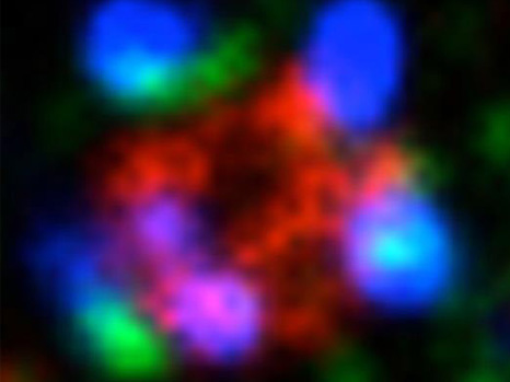
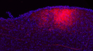
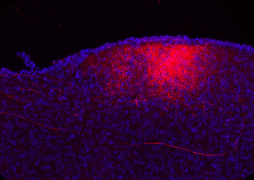
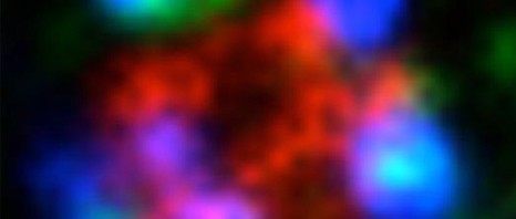
Zoom-in of the inside of a cell is used to see the reactions between various proteins triggered by PKCλ. This latest protease MT1-MMP traffic “control” (red) enables the association of cortactin (green) with dynamin 2 (blue). This reaction sequence is necessary to allow the cell separate from its neighbours and invade other tissues.
© Carine Rossé – Philippe Chavrier/ Institut Curie


1 1st class Research Director at CNRS, Philippe Chavrier is head of the “The Membrane and Cytoskeleton Dynamics” group “Subcellular Structure and Cellular Dynamics – Institut Curie/CNRS” laboratory, run by Bruno Goud.
2 Anne Vincent-Salomon is a medical pathologist in the Department of Biopathology at the Institut Curie Hospital Complex and has been a researcher in the “Genetics and Developmental Biology – Institut Curie/Inserm/CNRS” laboratory since January 2014, run by Prof. Edith Heard.
3 Carine Rossé is an INSERM research fellow within the “The Membrane and Cytoskeleton Dynamics” group, run by Philippe Chavrier.
4 Department of Biopathology at the Institut Curie Hospital Complex.

Discovery of two types of adrenal cancer

Adrenocortical carcinoma (also known as adrenal cortex cancer or ACC), is a generally aggressive tumour, with a mean survival rate of less than five years for those affected. Apart from metastasis, it exposes the patients to manifestations such as high blood pressure, diabetes, decreased potassium level, infections, etc. There is, however, some patient-dependent variation in tumour development. The team led by Prof. Bertherat at the Cochin Institute (Inserm – CNRS – Paris Descartes University) and the Expert Centre for Rare Adrenal Cancers at Cochin Hospital (AP-HP) has just published a molecular classification for this cancer in the journal Nature Genetics. The researchers identify many molecular abnormalities in these cancers that have not been well known until now, and thus reveal a new classification for these tumours.

This work involved 130 adrenocortical carcinomas, bringing together an initial cohort of some fifty tumour samples collected in the national research network COMETE (COrtico et MEdullosurrénale, Tumeurs Endocrines; COrtical and MEdullary adrenal Endocrine Tumours) and a second cohort of approximately 80 samples, collected within the European research network ENSAT (European Network for the Study of Adrenal Tumors). The complete genomes of these tumours were analysed by a combination of several high throughput genomic techniques, i.e. complete sequencing; study of the expression levels of all genes (transcriptomics) and of micro-RNAs (miRNAs); study of genetic variants (SNPs) and of gene methylation levels (epigenetics).

This study revealed the existence of two molecular types of adrenocortical carcinomas, one showing a relatively favourable prognosis for patients following complete surgery, and another for which the prognosis is unfavourable.

These two molecular types correspond to two different diseases. The type associated with a poor prognosis is characterised by a higher level of mutations, including recurrent alterations in a small group of genes already known to be involved in adrenocortical carcinoma (CTNNB1,TP53, CDKN2A, RB1, MEN1), or new genes (ZNRF3, DAXX, TERT, and MED12). In this study, ZNFR3 is specifically identified as a new tumour suppressor gene.

Moreover, specific profiles that distinguish these two cancer groups are shown by each of the following molecular analyses: gene and miRNA expression profiles, and pattern of methylation abnormalities.


This work opens up new clinical opportunities in the short term, especially in terms of predicting tumour prognosis following surgery on the lesion, and the possibility of conducting clinical studies according to tumour type. In the longer term, the research team suggests that results will allow identification of therapeutic targets specific for each of the subgroups. It is a further step in the development of a specific personalised medicine for rare cancers.


The researchers also anticipate new applications for these discoveries, especially applications based on the newly identified tumour suppressor gene ZNFR3.

Finally, the authors of the study emphasise the power of genomic methods and the importance of national and international multidisciplinary and multi-centre research networks, especially in the area of rare tumours.

This project has been developed in partnership with the French National Cancer League for several years, under the Cartes d’Identité des Tumeurs (Tumour Identity Card) programme.

Inserm and the Institut Pasteur identify a new variant of Ebola virus in Guinea

In an article which appeared in The New England journal of Medicine on 16 April, researchers from Inserm (Jean Mérieux-Inserm BSL-4 Laboratory, Lyon) and the Institut Pasteur have published their initial findings on the characteristics of the Ebola virus discovered in Guinea. Initial virological investigations enabled them to identify Zaire ebolavirus as the pathogen responsible for this epidemic. Performed in less than a month, sequencing of the complete genome and subsequent phylogenetic analysis show that the virus present in Guinea forms a clade (variant) that is distinct from strains previously identified in the Democratic Republic of Congo and in Gabon. Epidemiological investigations also linked the laboratory confirmed cases with the initial deaths recorded during the December 2013 outbreak.

Laboratoire P4 Jean Mérieux/Inserm.
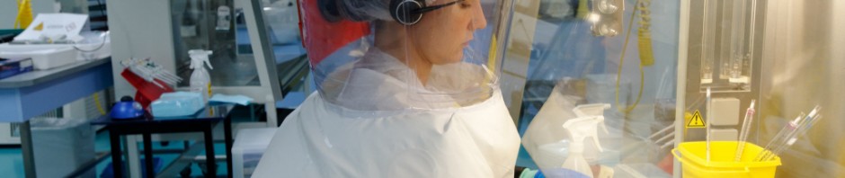
©Inserm/ Guénet François
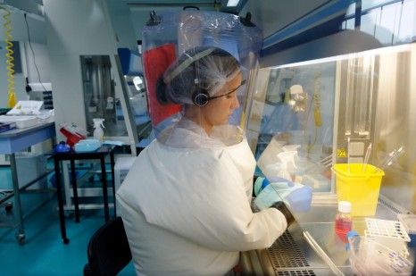
Ebola virus is a lethal, highly contagious virus for which there is presently no treatment. The symptoms are somewhat non-specific, and include fever, severe diarrhoea and vomiting. Between 30 and 90% of those infected with this organism die as a result.

On 2 April 2014, the World Health Organisation (WHO), in a communiqué published by the UN, reported that it had recorded 5 new cases of Ebola fever in Guinea. Since January, the total number of suspected and confirmed cases of Ebola fever in the present outbreak in Guinea is 127, with 83 deaths, according to WHO, which states that 35 cases were confirmed by laboratory testing.

The initial samples were analysed in Lyon in the Jean Mérieux-Inserm BSL-4 Laboratory directed by Hervé Raoul, Inserm Research Director, by the French National Reference Centre for Viral Haemorrhagic Fevers (attached to the Biology of Viral Emerging Infections Unit at the Institut Pasteur, directed by Sylvain Baize). A positive diagnosis was made.

A mobile BSL-4 laboratory was deployed in Guinea to provide assistance with diagnosis in the field. This mobile laboratory was developed as part of a European project, “EMP4,” coordinated by German researchers, and in which the Jean Mérieux-Inserm BSL-4 Laboratory is the French partner.

The researchers were able to analyse blood samples from 20 patients. Various tests were conducted by the scientists in order to establish a specific identity card for the virus.

Viral RNA was extracted from the blood samples, and then amplified and sequenced. These sequences were finally compared to 48 already known complete Ebola virus genomes. According to results, the analysis showed 97% identity with strains identified in the Democratic Republic of Congo in 1976 and 2007, and in Gabon in 1994 and 1996.

“These results demonstrate that we are facing the emergence of a new “form” of this virus in Guinea,” explains Hervé Raoul, Director of the BSL-4 Laboratory. This form is common to cases discovered since the month of December.

It would appear that the epidemic originated from a single introduction from animal to human.

Apart from the present epidemic, these results show that the endemic area for Ebola virus is greater than previously known, and that as a consequence, West Africa should henceforth be considered an area of risk for Ebola virus. Measures aimed at preventing transmission from wild fauna to humans and quickly identifying such events if there is a recurrence must be put in place in Guinea, as well as in neighbouring countries.

carte guinée


Further information on the BSL-4 laboratory

The Jean Mérieux BSL-4 Laboratory is a high-level containment laboratory dedicated to the study of Class 4 pathogens. The biological safety level applied is 4, the highest possible level. The researchers working there wear a full body, air-supplied, positive pressure suit to protect them from all contamination. The laboratory is itself maintained under negative pressure in order to protect the environment. Moreover, all wastes produced are completely inactivated, and the exhaust air is purified by a double absolute filtration system. This laboratory currently remains the structure offering the largest experimental capacity in Europe for this containment level.

Highly pathogenic agents:

Class 4 pathogens (or risk group 4) are highly pathogenic microorganisms characterised by a very high mortality rate, a lack of prophylactic or therapeutic measures to provide protection, and ready transmissibility. All class 4 pathogens currently known are viruses, and include viruses that cause haemorrhagic fevers or encephalitis. They include the Ebola, Marburg, Lassa, Junin, Machupo, Guanarito, Sabia, Crimean-Congo, Nipah and Hendra viruses.

To obtain photographs of the BSL-4 laboratory

Innovative strategy to facilitate organ repair

A significant breakthrough could revolutionize surgical practice and regenerative medicine. A team led by Ludwik Leibler from the Laboratoire Matière Molle et Chimie (CNRS/ESPCI Paris Tech) and Didier Letourneur from the Laboratoire Recherche Vasculaire Translationnelle (INSERM/Universités Paris Diderot and Paris 13), has just demonstrated that the principle of adhesion by aqueous solutions of nanoparticles can be used in vivo to repair soft-tissue organs and tissues. This easy-to-use gluing method has been tested on rats. When applied to skin, it closes deep wounds in a few seconds and provides a esthetic, high quality healing. It has also been shown to successfully repair organs that are difficult to suture, such as the liver. Finally, this solution has made it possible to attach a medical device to a beating heart, demonstrating the method’s potential for delivering drugs and strengthening tissues. This work has just been published on the website of the journal Angewandte Chemie.

In an issue of Nature published in December last year, a team led by Ludwik Leibler 1 presented a novel concept for gluing gels and biological tissues using nanoparticles 2. The principle is simple: nanoparticles contained in a solution spread out on surfaces to be glued bind to the gel’s (or tissue’s) molecular network. This phenomenon is called adsorption. At the same time the gel (or tissue) binds the particles together. Accordingly, myriad connections form between the two surfaces. This adhesion process, which involves no chemical reaction, only takes a few seconds. In their latest, newly published study, the researchers used experiments performed on rats to show that this method, applied in vivo , has the potential to revolutionize clinical practice.

In a first experiment, the researchers compared two methods for skin closure in a deep wound: traditional sutures, and the application of the aqueous nanoparticle solution with a brush. The latter is easy to use and closes skin rapidly until it heals completely, without inflammation or necrosis. The resulting scar is almost invisible.

Schéma plaie cutanée

Phase 1 Skin injury
Phase 2 Application of the solution
Phase 3 Using pressure to hold the edges together
Phase 4 Skin closure

Illustration of the first experiment conducted by the resear chers on rats: a deep wound is repaired by applying the aqueous nanoparticle solution. The wound closes in thirty seconds.
© “Matière Molle et Chimie” Laboratory (CNRS/ESPCI Paris Tech)

In a second experiment, still on rats, the researchers applied this solution to soft-tissue organs such as the liver, lungs or spleen that are difficult to suture because they tear when the needle passes through them. At present, no glue is sufficiently strong as well as harmless for the organism. Confronted with a deep gash in the liver with severe bleeding, the researchers closed the wound by spreading the aqueous nanoparticle solution and pressing the two edges of the wound toget her. The bleeding stopped. To repair a sectioned liver lobe, the researchers also used nanoparticles: they glued a film coated with nanoparticles onto the wound, and stopped the bleeding. In both situations, organ function was unaffected and the animals survived.

“Gluing a film to stop leakage” is only one example of the possibilities opened up by adhesion brought by nanoparticles. In an entirely different field, the researchers have succeeded in using anoparticles to attach a biodegradable membrane used for cardiac cell therapy, and to achieve this despite the substantial mechanical constraints due to its beating. They thus showed that it would be possible to attach various medical devices to organs and tissues for therapeutic, repair or mechanical strengthening purposes.

This adhesion method is exceptional because of its potential spectrum of clinical applications. It is simple, easy to use and the nanoparticles employed (silica, iron oxides) can be metabolized by the organism. It can easily be integrated into ongoing research on healing and tissue regeneration and contribute to the development of regenerative medicine.

1 Leibler was awarded the CNRS Medal for Innovation in 2013.
2 See the press release for this work: http://www2.cnrs.fr/presse/communique/3355.htm

Confirmation of the neurobiological origin of attention – deficit disorder

A study, carried out on mice, has just confirmed the neurobiological origin of attention – deficit disorder (ADD), a syndrome whose causes are poorly understood. Researchers from CNRS, the University of Strasbourg and INSERM1 have identified a cerebral structure, the superior colliculus, where hyperstimulation causes behavior modifications similar to those of some patients who suffer from ADD. Their work also shows noradrenaline accumulation in the affected area, shedding light on this chemical mediator having a role in attention disorders. These results are published in the journal Brain Structure and Function.

Attention – deficit disorder affects between 4 – 8% of children. It manifests mainly through disturbed attention and verbal and motor impulsiveness, sometimes accompanied by hyperactivity. About 60% of these children still show symptoms in adulthood. No cure exists at this time. The only effective treatment is to administer psychostimulants, but these have substantial side effects, such as dependence. Persistent controversy surrounding the neurobiological origin of this disorder has hindered the development of new treatments.

The study in Strasbourg investigated the behavior of transgenic mice having developmental defects in the superior colliculus. This structure, located in the midbrain, is a sensory hub involved in controlling attention and visual and spatial orientation. The mice studied were characterized by duplicated neuron projections between the superior colliculus and the retina. This anomaly causes visual hyperstimulation and excess noradrenaline in the superior colliculus. The effects of the neurotransmitter noradrenaline, which vary from species to species, are still poorly understood.

However, we do know that this noradrenaline imbalance is associated with signific ant behavioral changes in mice carrying the genetic mutation. By studying them, researchers have observed a loss of inhibition: for example mice hesitate less to penetrate a hostile environment. They have difficulties in understanding relevant information and demonstrate a form of impulsiveness. These symptoms remind us of adult patients suffering from one of the forms of ADD.

Currently, the fundamental work on ADD uses mainly animal models obtained by mutations that disturb dopamine production and transmission pathways. In mice with a malformed superior colliculus, these pathways are intact. The changes occur elsewhere in the neural networks of the midbrain. By broadening the classic boundary used to research its causes, using these new models would allow a more global approach to ADD to be developed. Characterizing the effects of noradrenaline on the superior colliculus more precisely could open the way to innovative therapeutic strategies.

Colliculus supérieur

In this image, marking shows the axons in retinal neurons (in red) that innervate the superior colliculus (in blue) in a “normal” mouse
© Michael Reber / Inserm Institut des neurosciences cellulaires et intégratives

(1) From the Laboratoire de Neurosciences Cognitives et Adaptatives (CNRS/Université de Strasbourg) and the Institut des Neurosciences Cellulaires et Intégratives (CNRS)

Caffeine and Alzheimer’s disease : a link with tau protein

Researchers at Inserm and University of Lille 2/University of Lille Nord de France directed by David Blum, Inserm Research Fellow, have provided experimental evidence of the beneficial effects of caffeine in an animal model of Alzheimer’s disease. This work, carried out on mice and published in Neurobiology of Aging, supports the idea that caffeine has a protective effect in some brain pathologies.

Post-it note with smiley face sticked on cup

©fotolia

Affecting more than 800,000 people in France, Alzheimer’s disease and related diseases are the leading cause of age-related loss of intellectual function. The cognitive impairment seen in Alzheimer’s disease mainly results from the accumulation of abnormal tau proteins in degenerating nerve cells. Regular caffeine consumption is known to reduce age-related cognitive decline, and the risk of developing dementia. However, the effects of caffeine on pathologies associated with tau protein, known as tauopathies, one of which is Alzheimer’s disease, had not been clearly elucidated.

Dr. David Blum, from the Alzheimer & Tauopathies laboratory at Joint Research Unit 837 (Inserm/University of Lille 2/University of Lille Nord de France), directed by Dr. Luc Buée, has just shown in mice that regular caffeine consumption prevents memory deficits and some of the modifications to tau protein. To arrive at this result, young transgenic mice that progressively develop age-related neurodegeneration associated with tau protein were given caffeine orally for 10 months.

“Mice treated with caffeine developed a less severe pathology from the point of view of memory, tau protein modifications, and neuroinflammation” explains David Blum, a research fellow at Inserm.

This study provides experimental evidence of a link between caffeine consumption and pathologies associated with tau protein in a neurodegeneration model of Alzheimer’s disease. This study also indicates that caffeine may act on different types of brain dysfunction involved in Alzheimer’s disease in order to exert its beneficial effects.

“This work indicates a significant role for environmental factors in the development of Alzheimer’s disease, emphasises the researcher. In the light of these results, we now hope to identify the molecular target responsible for these beneficial effects of caffeine on the one hand, and initiate a clinical trial to test the effects of caffeine on patients with Alzheimer’s disease on the other hand,” he adds.

This work received support from LabEx DISTALZ (Development of Innovative Strategies for a Transdisciplinary Approach to Alzheimer’s Disease) under the Investissements d’Avenir (Investment for the Future) programme, and from the France Alzheimer and LECMA/AFI associations.

A natural protein, Elafin against gluten intolerance?

Scientists from INRA and INSERM (France) in collaboration with scientists from McMaster University (Canada) and the Ecole polytechnique fédérale of Zurich (Switzerland) have shown that Elafin, a human protein, plays a key role against the inflammatory reaction typical of celiac disease (gluten intolerance). They have also developed a probiotic bacterium able to deliver Elafin in the gut of mice. This innovation, published online in the American Journal of Gastroenterology on 8 April 2014, paves the way to new strategies to treat gluten intolerance.

Celiac disease is an auto-immune pathology that occurs in individuals genetically predisposed to gluten intolerance. Affected people do not harbor the enzymes required to degrade gluten during digestion. Inflammatory reactions are induced by this abnormal digestion which can lead to the destruction of the gut barrier that is essential for nutrients absorption. Celiac disease causes chronic abdominal pain (diarrhea, cramps…) and predisposes to certain cancers (small intestine, lymphoma). Its prevalence is estimated between 1/500 and 1/300; no curative treatment currently exist and the only solution is a lifelong gluten-free diet.

Scientists from the French National Institute for Agricultural Research (INRA) and the French National Institute of Health and Medical Research (INSERM), along with Canadian and Swiss colleagues, have shown that Elafin, a protein with anti-inflammatory properties, is less abundant in patients with celiac disease than in healthy people. They identified that Elafin is capable of preventing the destruction of the gut barrier during inflammation, and that Elafin is able to interact with enzymes responsible for the abnormal breakdown of gluten: transglutaminase-2. Consequently, Elafin reduces gluten toxicity.

These observations led the scientists to propose a way to deliver the missing Elafin in celiac patients with help of a harmless bacterium that is often present in food: a lactic bacterium strain (Lactococcus lactis), that scientists transformed in order to express Elafin. The use of this strain, developed by the same teams from INRA and INSERM, enables a targeted and local production of Elafin, and represents a recent and innovative strategy. The first pre-clinical results pave the way to new therapies for Inflammatory Bowel Disease or IBD (no curative treatment exist).

In the present study, the scientists have administered this bacterium to gluten intolerant mice. They showed that the Elafin delivered by the probiotic decreases significantly the inflammatory reaction.

This strategy, patented by INRA in May 2013, opens promising prospects to treat celiac disease and gluten intolerance in general. The next step will consist in defining the mechanisms underlying the positive effects of elafin in celiac disease, and in the identification of bacteria that naturally produce proteins with anti-inflammatory properties similar to elafin.

Friedreich’s ataxia – an effective gene therapy in an animal model

The team led by Hélène Puccio, director of research for Inserm at the Institute of Genetics and Molecular and Cellular Biology (IGBMC) (Inserm / CNRS / University of Strasbourg) in close collaboration with Patrick Aubourg’s team (Inserm and Professor of Neuropaediatrics at Bicêtre Hospital) has demonstrated, in the mice, the efficacy of gene therapy for treating the heart disease associated with Friedreich’s ataxia, a rare hereditary neuro-degenerative disorder. The transfer, via a viral vector, of a normal copy of the gene deficient in patients, allowed to fully and very rapidly cure the heart disease in mice. These findings are published in Nature Medicine on 6 April, 2014.

Friedreich’s ataxia is a severe, rare hereditary disorder which combines progressive neuro-degeneration, impaired heart function and an increased risk of diabetes. The condition affects one in every 50,000 birth. There is currently no effective treatment for this disease. In most cases, Friedreich’s ataxia starts in adolescence with impaired balance and coordination (ataxia) of voluntary arm and leg movements, confining the majority of patients to a wheelchair after 10-20 years’ progression. However, complications affecting the heart are the major cause of death in 60% of patients, most often before the 35 years of age.

The disease is caused by a common mutation in the FXN gene which leads to a dramatic decrease in the production of the protein named ‘frataxin’. The reduced frataxin level disturbs the activity of mitochondria. These organelles are essential to cells and play a fundamental role in energy production. The nerve tissue (cerebellum, spinal cord etc.) and heart tissue are particularly vulnerable to this shortage of energy, which can lead to fatal heart failure.

The teams led by Hélène Puccio, director of research at Inserm and Patrick Aubourg have developed a therapeutic approach based on the use of an adeno-associated virus (AAV)[1], which is known to efficiently target and express a therapeutic gene in heart cells. The virus was modified to make it harmless but nevertheless capable of introducing a normal copy of the FXN gene in the heart cells, thus leading to the expression of frataxin.

Hélène Puccio’s team tested the efficacy of this treatment in a mouse model that faithfully reproduces the heart symptoms of patients suffering from Friedreich’s ataxia. The results show that a single intravenous injection of AAVrh10 expressing frataxin is not only capable of preventing the development of heart disease in animals before the appearance of symptoms, but also, more impressively, of fully and rapidly curing the hearts of animals at an advanced stage of heart disease. After three weeks of treatment, the heart become fully functional again; mitochondrial function and the appearance of heart tissue being very similar to those of healthy mice. “This is the first time that gene therapy has prompted full, lasting remission of heart disease so quickly in an animal model.” explains Hélène Puccio.
Figure 3 post review

The illustrations show a measurement of the activity of a mitochondrial protein (in blue) essential for cellular energy production, which is impaired when frataxin is absent (no staining in the untreated heart). Using gene therapy expressing frataxin, the activity of this essential protein can be corrected across the heart’s entire surface. © Inserm / H. Puccio

As the central nervous system is also a target of AAV vectors, Hélène Puccio and Patrick Aubourg’s teams are investigating whether a similar approach using gene therapy could be as effective for the spinal cord and cerebellum as it is for the heart.
Based on these promising results, work has begun on the necessary developments to propose to patients suffering from Friedreich’s ataxia and presenting a progressive cardiomyopathy a treatment by gene therapy. To this end, three of the paper’s authors have set up AAVLife, a French company specialising in gene therapy for rare diseases, to translate to clinic these important laboratory findings. A patent application has been submitted by Inserm Transfert for this gene therapy approach.

This study was backed by FARA[2], AFAF[3] and AFM[4].


[1] AAV: specifically the serotype AAVrh10.

[2] Friedreich’s Ataxia Research Alliance, an American association specialising in the treatment of Friedreich’s ataxia.

[3] French Friedreich’s ataxia association

[4] French muscular dystrophy association

Europeans and biomedical research

An Ipsos survey for Inserm

The French National Institute of Health and Medical Research (Inserm) is currently the leading biomedical research organisation in Europe, and plays a key role in developing European research. To mark its 50th anniversary, Inserm wanted to assess the perceptions of Europeans regarding biomedical research.

To create this European panorama, Ipsos carried out an Internet survey of over 4,000 Europeans (with 1,001 French, 1,004 German, 1,001 Italians and 1,005 British respondents) from 10 to 23 January 2014. Representative samples were obtained from each country involved using quota sampling.

According to results, health is still the area of research news that interests the greatest number of Europeans. This survey confirms the genuine confidence of Europeans regarding biomedical research and its players, at a time of moroseness and cynicism. Although they are aware that they do not understand all of its ramifications, they see it as a source of hope for themselves and their children, as well as a source of pride for their country.

The most memorable medical innovations for Europeans in the last 50 years have been organ transplants, medical imaging, and gene therapies.

Health, a theme of major interest for Europeans

  • Europeans show a particular interest in news items about health research, Inserm’s special area of interest. Indeed, nearly one out of two Europeans (45%) mentions health as the area of research news that most interests him/her, far ahead of information technology and the new technologies (21%), environment (14%), human sciences (9%), energy (6%) or space (5%).
  • Health-related research news stimulates particular interest among the French (53%) and the Germans (48%).
  • Health research also interests more women (58%, compared with 32% of men) and people aged 35 years or older (51%, compared with 33% of those under 35).

Biomedical research is above all a synonym for hope

  • For Europeans, research mainly evokes the word “hope” (34% put it in first place, 67% among the first three). The French are the most likely to consider that biomedical research represents hope for them more than anything else.
  • 82% of Europeans believe that biomedical research will enable their children to live better than they do today (22% say “much better”).

A high degree of confidence in researchers

  • Physicians and researchers in biomedical sciences remain trusted intermediaries in the eyes of Europeans: where a public health problem occurs, 45% of those surveyed named physicians among the three players that could be most trusted to tell them the truth, and 36% named researchers. 70% of Europeans also trust researchers to challenge opinion if they believe that their scientific research has important consequences for issues affecting society; 66% of Europeans trust researchers to refuse to let innovations arising from their work have negative consequences for public health, and 62% trust them to remain independent and to reject pressure regarding the results of their work.

A limited level of information and knowledge, but a questioning attitude nonetheless

  • Europeans who were surveyed predominantly admitted that they were not well informed about biomedical research, whether in terms of advances in this area (59% believed they were poorly informed), its consequences for their everyday lives (59%), or popular debates stimulated by some research projects (61%).
  • Their scientific literacy is also limited. On average, when Europeans were tested on approximately 20 scientific terms, they “really” understood only 4.5 terms. Only one of these was “really” understood by the majority—the term “animal experiments.” Finally, the terms “nanoscience,” “genome sequencing,” viral vector,” endocrine disrupters” or “epigenetics” were not understood by the majority of Europeans.
  • The result of this lack of scientific vocabulary is that Europeans are unable to understand the questions that may be raised by some areas of biomedical research. For these, they put all their faith in the experts and in researchers to “control” things. The results of the survey show that a subject may become part of public debate and generate intense discussion, even though most Europeans know very little about it.

The United States model, and development of European research funding

  • The USA seems to provide an ideal for Europeans regarding biomedical research: 84% of them name the United States as among the three most advanced countries in this area. Certainly, this figure primarily reflects the attractiveness of the USA to many Europeans. It also highlights the attraction of American laboratories for European students and researchers, explained by the large budgets and influence and its proven supremacy in terms of numbers of  publications and returns. Germany, Great Britain and France come next (mentioned by 41%, 39% and 29% of respondents, respectively).
  • In order for Europe and their countries to keep their positions in this ranking, Europeans seem to believe that developments in the modes of research funding are essential: 88% judge it necessary for the private sector to become more involved in funding scientific research. 94% of Europeans surveyed nonetheless strongly advocate that a substantial proportion of biomedical research be funded by the State.
  • Finally, even though American biomedical research remains the model for many Europeans, 82% of Europeans believe that biomedical research is an area in which their own country can be proud of its results. This is especially the case in France, since 90% of respondents think so.

Organ transplants, medical imaging and gene therapy considered the most important medical innovations in the last 50 years

  • For Europeans, the most important innovation of the last 50 years is the organ transplant (70% name it among the 5 most important), just ahead of medical imaging (65%) and gene therapy (51%).
  • Next among the innovations most often mentioned are: the recent development and implantation of the artificial heart, celebrated as an achievement “made in France” (47%), the potential offered by the decoding of the human genome (35%), triple therapy (34%) and reprogramming of stem cells (32%).
  • The ranking of the most often-mentioned innovations varies with the gender and age of respondents. Nonetheless, women (as well as those under 35 years) were found to be more likely to mention innovations related to reproduction. More women than men are likely to judge oral contraception as a major discovery (33% compared with 21% of men), together with in vitro fertilisation (21% compared with 15%) and epidural anaesthesia (15% compared with 11%). However, Viagra is more often mentioned by men than by women (7% compared with 5%), but nonetheless seems to be the least noteworthy innovation on the list they were offered.
fermer