Menu

A radiosensitivity test for predicting sequelae following radiotherapy

Researchers at Inserm Unit 1194, “Montpellier Cancer Research Institute” (Inserm/University of Montpellier/Montpellier Regional Cancer Institute) have confirmed the value of a new test to identify cancer patients who will be free of sequelae following radiotherapy. This test, conducted on a blood sample taken from 500 breast cancer patients, treated in 10 centres in France, and monitored for 3 years, showed that women with a high rate of radiation-induced lymphocyte apoptosis (RILA) had a very low rate of late breast fibrosis. These results, which are published in EBioMedicine, suggest that personalisation of curative intent radiotherapy could be considered, with tailoring of the radiation dose delivered to the patient and the radiotherapy technique employed.

Institut de Recherche en Cancerologie de Montpellier (IRCM)

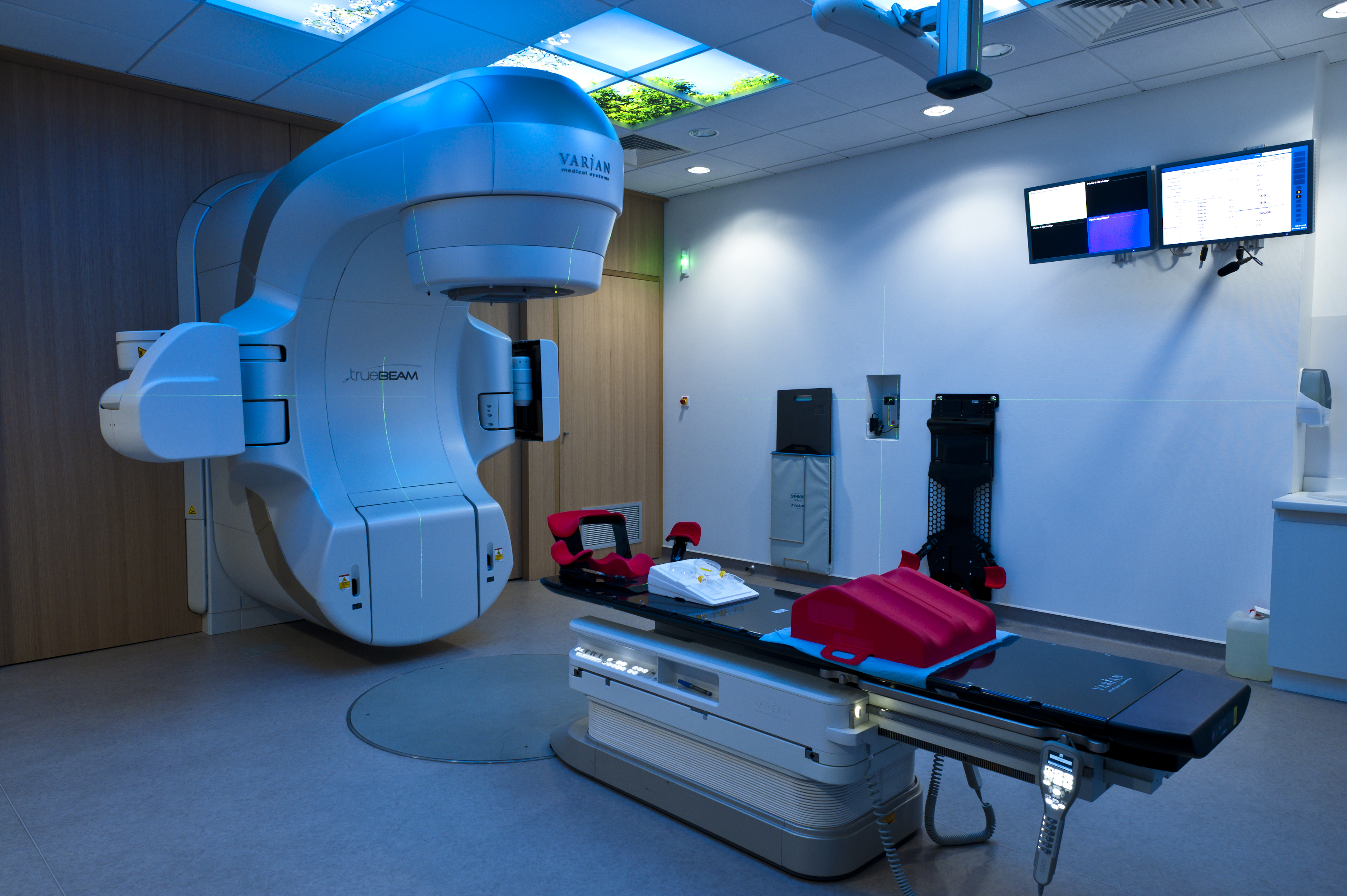
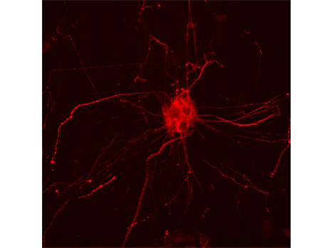
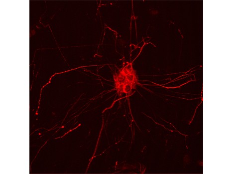
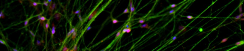
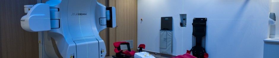
Radiotherapy is one of the treatments used to treat breast cancer. The irradiation destroys the malignant cells in a targeted manner. However, it also leads to the death of some healthy cells in the irradiation field. Using a single blood sample, the researchers analysed the rate of radiation-induced CD8 lymphocyte apoptosis (RILA) in the context of a prospective multicentre clinical trial which began in 2005. The objective of this trial is to develop a functional test based on RILA to predict radiosensitivity in tissues. The trial follows several pilot trials initiated in the last 15 years in relation to breast cancer, as well as other diseases.

In this context, 500 female patients with breast cancer and treated using radiotherapy were recruited in 10 French centres. The researchers at the Joint Research Unit “Montpellier Cancer Research Institute” (Inserm/University of Montpellier/Montpellier Regional Cancer Institute) performed RILA at 8 Gy for the patients before they underwent radiotherapy. The patients were then monitored for three years in order to assess late breast sequelae (fibrosis).

Results of the multicentre study provide large-scale confirmation of the preliminary data obtained by the researchers. They show that a high RILA value is correlated with a low incidence of late sequelae. A low rate of late breast fibrosis was observed, with a negative predictive value of over 90%. Conversely, almost all patients who showed a high level of fibrosis corresponded to the group with a low RILA value, predictive of more pronounced sequelae.

“This multicentre study provides a sufficient level of proof to allow the use of this test in routine clinical practice, and changes patient management. Given the results obtained, we can consider the possibility of increasing the total irradiation dose delivered locally, or of modifying the target volumes without compromising the oncological outcome,” explains David Azria, principal investigator in the study.

In practice, this test is carried out by taking a single blood sample, and results are obtained in 72 hours.

By providing the opportunity to identify patients who are not prone to sequelae and those at greatest risk, this test paves the way for the personalisation of curative intent radiotherapy.

It should not be used alone, but should be combined with other parameters in a predictive nomogram, a useful calculation tool, for which a patent application has been filed by the Montpellier team. “The results, when combined with the all the parameters, provide a reliable estimate of the risk of late sequelae following radiotherapy,” concludes David Azria.

Treating intestinal pain with bacteria

Faecalibacterium prausnitzii is an intestinal bacterium that is abundant in healthy adults but scarce in those suffering from inflammatory bowel disease. Using a mouse model, a team of scientists from INRA, INSERM, and the University of Auvergne has discovered that the bacterium possesses analgesic properties, a finding just published in Scientific Reports (January 18, 2016).Given that F. prausnitzii also exhibits anti-inflammatory effects, the bacterium shows great promise as a tool for improving human health and well-being.

PhotoCP web langella

Scanning electron micrograph of F. prausnitzii © MIMA 2 experimental facility, T. Meylheuc

Chronic abdominal pain is one of the most frequent symptoms experienced by patients suffering from intestinal diseases such as irritable bowel syndrome (IBS) or inflammatory bowel disease (IBD). In 2008, researchers at the INRA center in Jouy-en-Josas discovered that individuals with IBD had lower levels of the bacterium Faecalibacterium prausnitzii in their intestines and that F. prausnitzii exhibited anti-inflammatory effects.

The research team delved further into the phenomenon via a collaborative project carried out with other research teams from INRA, INSERM, and the University of Auvergne. Using a mouse model (an inbred line with colorectal hypersensitivity), they discovered that reduced F. prausnitzii abundance was associated with the occurrence of hypersensitivity. In humans, the latter can manifest itself via intestinal discomfort or abdominal pain. The researchers found that dosing the study animals with F. prausnitzii allowed them to recover normal levels of colonic sensitivity.

Based on these results and those of the 2008 study, it is reasonable to propose that F. prausnitzii could have both anti-inflammatory and analgesic properties in humans.

In individuals suffering from IBS or IBD, the reduction in F. prausnitzii abundance in the gut due to intestinal inflammation or chronic stress might act to exacerbate levels of inflammation and/or pain. To break this vicious circle, the researchers propose using nutritional supplements containing F. prausnitzii (i.e., probiotics) to restore “intestinal well-being.”
This research has not only added to our fundamental understanding of cellular microbiology but also has very practical implications for the medical industry.

Brand new intervertebral discs for back pain

Throughout life, extremely heavy demands are made on the spinal column. Spinal wear very soon affects the quality of life, and back pain is often described as the illness of the century. 40% of back pain may be due to irreversible deterioration of the intervertebral discs (which act as “cushions” between the vertebrae), which can no longer play their role as shock absorbers. Researchers from Inserm led by Jérôme Guicheux (Inserm Unit 791, “Laboratory of Osteoarticular and Dental Tissue Engineering,” Nantes) have successfully transformed adipose stem cells into cells that might be able to replace damaged discs. This work is published in the journal Stem cells.


Our spinal column is made up of a stack of vertebrae. Its articulation and flexibility are made possible by the presence of intervertebral discs that form “cushions” between the vertebrae. Degenerative diseases of the vertebral discs are related to the strong and repetitive demands made on the spinal column throughout life—carrying loads, sports, repetitive movements and twisting motions. With time, the discs become worn and deteriorate, and can no longer play their role as shock absorbers. Although these conditions appear slowly and progressively, they soon manifest as pain in the area where the discs are damaged. It is estimated that degeneration of the intervertebral discs is responsible for approximately 40% of lumbar pain. Current research is therefore aimed at developing treatments to slow or prevent the degeneration of the discs and the cells that constitute them.

From a physiological standpoint, the nucleus pulposus, the central part of the intervertebral disc, is the first to be affected. It is largely made up of water, which gives it its shock-absorbing properties. With age, the cells of the nucleus pulposus gradually become less proliferative, and are then subject to apoptosis and unable to produce this famous highly hydrated extracellular matrix.

How then can they be replaced with functional cells? The researchers focused on the adipose tissue, which constitutes a large reservoir of stem cells that can differentiate into a vast array of cell types. It was also necessary to find the right protocol to successfully enable stem cells from the adipose tissue to become transformed into nucleus pulposus cells.


A finely tuned recipe

The development of this protocol can be compared to a food recipe. The researchers were able to find the right ingredients and correct proportions for it to succeed. The winning strategy involved adding a combination of two growth factors, TGFβ and GDF5, to the cell medium. In 28 days the researchers obtained in vitro, from adipose tissue taken from nine patients, functional nucleus pulposus cells resembling those that exist naturally in the intervertebral discs.

“The protocol proved successful independent of patient age and weight,” says Jérôme Guicheux.

However, we had to go further, since these cells had no chance of survival if they were merely reimplanted into a damaged intervertebral disc, without all the nutrient substrate they need.”

The second trick was therefore to couple these cells to a synthetic biomaterial to recreate a favourable environment for their multiplication once they had been injected into the intervertebral disc. The researchers assessed the biological activity of these cells in vivo after transplanting them into mice. “This system most closely resembles intradiscal transplantation in humans. We have demonstrated that the protocol we apply to these cells was sufficient for them to retain their specific secretory activity and their specialised phenotype when reinjected in vivo.”

This work in regenerative medicine now allows the researchers to contemplate the next step before moving to the clinical setting: testing the therapeutic efficacy of these brand-new cells in a relevant animal model of degenerative vertebral disc disease.

schema 1 modif_161015-1

(c) S Renaudin/le Design de Solène for Inserm

Individual susceptibility to liver toxicity of alcohol depends on the intestinal microbiota

Despite excessive alcohol consumption, some individuals remain in good health while others develop liver disease. This inequality regarding the toxicity of alcohol for the liver depends on the intestinal microbiota. This has just been demonstrated by scientists and physicians from Paris-Sud University, Inserm, AP-HP (Antoine-Béclère Hospital), INRA (French National Institute for Agricultural Research), AgroParitech and Aix-Marseille University. These results are published online in the journal Gut.

PhotoCP web alcool

(c) Fotolia

Alcoholic liver disease is a major public health problem. The liver is affected to a variable degree, from a simple accumulation of fat to acute alcoholic hepatitis and cirrhosis. Nonetheless, people do not all react in the same way to the toxic effects of alcohol on the liver. For an equivalent intake of alcohol in terms of quantity and duration, only some excessive drinkers will develop liver disease. This unequal risk from the effects of alcohol toxicity indicates that other factors influence the initiation and progression of liver lesions. The intestinal microbiota is the complement of bacteria present in our digestive system. These bacteria perform many metabolic functions. Scientists and physicians from Paris-Sud University, Inserm, AP-HP (Antoine-Béclère Hospital, one of the Paris Public Hospitals), INRA, AgroParitech and Aix-Marseille University have studied the potential role played by the intestinal microbiota in the unequal toxicity of alcohol for the liver.

The scientists found an imbalance in the intestinal microbiota (dysbiosis) of patients with acute alcoholic hepatitis, which was not found in patients who consumed alcohol but did not have serious liver disease.

In order to ascertain whether this dysbiosis plays a causal role in the occurrence of lesions on the liver, the scientists humanised germ-free mice by transferring the microbiota of alcoholic patients to them. One group of mice received the microbiota of alcoholic patients suffering from acute alcoholic hepatitis, and another group of mice received the microbiota of alcoholic patients without serious liver disease. The mice were then fed alcohol. It was observed that the mice from the first group developed inflammation of the liver and adipose tissue, and a greater increase in intestinal permeability than those from the other group. Moreover, some harmful species of bacteria were specifically associated with sensitivity or insensitivity to alcohol. A study of metabolites showed that ursodeoxycholic acid levels were higher in alcohol-resistant mice.

When this type of experiment was repeated with conventional mice, which have the advantage of more closely mimicking the human disease, the work showed that it was possible to reduce the liver lesions in diseased mice by transferring the microbiota from alcoholic patients without liver lesions to them.

This work thus proves that individual susceptibility to alcoholic liver toxicity depends, at least to a substantial extent, on the intestinal microbiota. Moreover, the composition of the intestinal microbiota can be modified by diet, prebiotics, probiotics or by transfer of faecal microbiota. Furthermore, this type of experiment shows that it is possible to transfer alcohol susceptibility from human to mouse via the intestinal microbiota, and makes it possible to anticipate the development of treatments of this nature.

These results thus open up new possibilities, not only for identifying individuals likely to be sensitive to alcohol toxicity, but also for improving the treatment of alcohol-induced liver lesions by modulating the intestinal microbiota

An indicator for predicting emergence from coma

It is difficult to judge the progress of patients in a coma following head trauma or recovery from cardiac arrest. Researchers from Unit 825, “Brain imaging and neurological handicaps” (Inserm/Université Toulouse III – Paul-Sabatier), in collaboration with Toulouse University Hospital, show that the quality of communication between two structures in the brain predicts patient recovery at 3 months. This new indicator, obtained by conducting MRI analysis on the brain of patients at rest, may provide additional help in establishing a prognosis.

Results of this study are published in the 11 November issue of the journal Neurology.

See video on the discovery presented by Patrice Péran, Inserm Research Fellow

Consciousness appears to be a complex mental process, inseparable from our existence. In reality, this capacity is relatively unstable. It disappears in a cyclical manner during the day (wake-sleep), and can be modified by administering certain drugs (anaesthesia). Finally, it can be abolished more or less completely and permanently following an insult to the brain; this happens in a coma. It is very difficult to determine which patients will emerge from such a situation and regain a normal state of consciousness, and conversely, which will be left with major neurological sequelae leading to serious disability (vegetative state, state of minimal consciousness).

In this study, the researchers focused on the brain abnormalities responsible for the loss of consciousness observed during coma. They compared the activity of the brain at rest in 27 patients in this state, and of control subjects of the same age, using functional magnetic resonance imaging recordings.

Two regions of the brain no longer communicate with each other.

The scientists particularly analysed communications between the brain as a whole and one structure located at the back of the brain known as the posteromedial cortex (PMC). Activity in this key region is reduced during sleep or anaesthesia. This structure is made up of two areas that the researchers studied (the precuneus and the posterior cingulate cortex)

A major loss of communication between the PMC and the anterior part of the brain (medial frontal cortex, MFC), particularly at the level of the posterior cingulate cortex, is seen in all comatose patients. This bad connection is present regardless of the mechanism responsible (head trauma or cardiac arrest with subsequent recovery). This observation suggests a major role for the interaction between these two structures in the emergence of consciousness in humans.

Schéma mauvaire connexion en

3D representation of the brain and areas involved (posterior cingulate cortex in blue, medial frontal cortex in yellow) © Inserm Unit 825

The team went further, and evaluated the extent to which this connection becomes altered with time. The researchers at Inserm compared recordings a few days after the brain insult and onset of coma with the neurological profiles of the patients three months later. It turns out that patient recovery is closely associated with the degree of involvement of this connection.

“Patients who are going to recover consciousness show levels of connections comparable to those found in healthy subjects. Conversely, a reduction in communication between the two areas predicts an unfavourable progression towards a vegetative state, or minimally conscious state,”

explain Stein Silva and Patrice Péran, who made this discovery.

These results constitute an important step in understanding the emergence of conscious perception of the outside world. They are promising, since neurologists might use this parameter to form a prognosis and develop treatment plans for comatose patients. However, research must be continued in order to decipher the mechanisms underlying coma, which are still poorly known.

Medical diagnosis: will brain palpation soon be possible?

If there is one technique used by the physician to explore the human body during every medical examination in order to make a diagnosis or prescribe further tests, it is palpation. By its nature, however, the brain cannot be palpated without using a highly invasive procedure (craniotomy, or opening the skull), which is limited to rare cases. By drawing on seismology, Inserm researchers led by Stéfan Catheline (Inserm Unit 1032, “Therapeutic Applications of Ultrasound”) have just developed a noninvasive brain imaging method using MRI that provides the same information as physical palpation. Ultimately, it could be used in the early diagnosis of brain tumours or Alzheimer’s disease. This work is published in PNAS.

Many diseases involve structural changes in tissues, which are reflected in a change in their mechanical properties, such as elasticity. Using the sensitivity of their hands, and their detailed knowledge of the body, physicians, through an examination known as palpation, can assess the size and stiffness of a tumour, the presence of inflamed lymph nodes, or the size and position of the foetus in a pregnant woman, to mention a few examples.

This palpation has been supplemented or replaced by modern techniques that give the physician an indication of the elasticity of a biological tissue. They are based on the generation and detection of waves that propagate through the body at varying speeds depending on the stiffness of the organs (the stiffer the tissue, the slower the wave propagation, and vice versa). However, this method cannot be applied to the brain, which, doubly protected by the cranium and cerebrospinal fluid, is difficult for externally applied waves to access. It is therefore impossible to directly or indirectly palpate the brain, something that greatly complicates the work of neurosurgeons. On the other hand, the brain is the seat of natural vibrations created by the blood pulsating in the arteries and the circulating cerebrospinal fluid. There remained a significant unprecedented challenge: how to capture this complex field of natural shear waves, and represent it on a computer screen.

In this article, Inserm researchers, using MRI, have succeeded in detecting natural shear waves in the brain using computational techniques borrowed from seismologists and known as “noise correlation.” They were thus able to build images of the brain’s elasticity.

“If this method can be developed for clinical use, it will be a boon for both the patient and the physician, since making the brain vibrate is quite painful at the moment. Of course, this method will be complementary to those that already exist, and the future is in a multimodal medical diagnosis,” says Stéfan Catheline, Inserm Research Director and main author of this work.

 “Alzheimer’s disease, epilepsy, multiple sclerosis and hydrocephalus involve changes in the stiffness of the brain tissues. This new technique allows their detection, and could be used to avoid brain biopsies. ”

This method for palpating the brain could have other areas of application, such as for analysing the development of neurodegenerative processes, the impact of a lesion from a trauma or tumour, response to treatment, etc.Photorecadree

©Inserm/Stéfan Catheline

Antimicrobial Film for Future Implants

The implantation of medical devices is not without risks. Bacterial or fungal infections can occur and the body’s strong immune response may lead to the rejection of the implant. Researchers at Unit 1121 “Biomaterials and Bio-engineering” (Inserm/Strasbourg university) have succeeded in creating a film with antimicrobial, antifungal and anti-inflammatory properties. It may be used to cover titanium implants (orthopaedic prostheses, pacemakers…) prevent or control post-operative infections. Other frequently used medical devices that cause numerous infectious problems, such as catheters, may also benefit.

These results are published in the journal Advanced Healthcare Materials.

See video on the discovery presented by Philippe Lavalle, Research Director at Inserm (subtitles soon available)


Implantable medical devices (prosthesis/pacemakers) are an ideal interface for micro-organisms, which can easily colonize their surface. As such, bacterial infection may occur and lead to an inflammatory reaction. This may cause the implant to be rejected. These infections are mainly caused by bacteria such as Staphylococcus aureus, originating in the body, and Pseudomonas aeruginosa. These infections may also be fungal or caused by yeasts. The challenge presented by implanting medical devices in the body is preventing the occurrence of these infections, which lead to an immune response that compromises the success of the implant. Antibiotics are currently used during surgery or to coat certain implants. However, the emergence of multi-resistant bacteria now restricts their effectiveness.

A film invisible to the naked eye…

It is within this context that researchers at the “Bioengineering and Biomaterials” Unit 1121 (Inserm/Strasbourg University) with four laboratories[1] have developed a biofilm with antimicrobial and anti-inflammatory properties. Researchers have used a combination of two substances: polyarginine (PAR) and hyaluronic acid (HA), to develop and create a film invisible to the naked eye (between 400 and 600 nm thick) that is made of several layers. As arginine is metabolised by immune cells to fight pathogens, it has been used to communicate with the immune system to obtain the desired anti-inflammatory effect. Hyaluronic acid, a natural component of the body, was also chosen for its biocompatibility and inhibiting effect on bacterial growth.

illustration implant Inserm en

…with embedded antimicrobial peptides,

The film is also unique due to the fact that it embeds natural antimicrobial peptides, in particular catestatin, to prevent possible infection around the implant. This is an alternative to the antibiotics that are currently used. As well as having a significant antimicrobial role, these peptides are not toxic to the body that they are secreted into. They are capable of killing bacteria by creating holes in their cellular wall and preventing any counter-attack on their side.

…on a thin silver coating,

In this study researchers show that poly(arginine), associated with hyaluronic acid, possesses microbial activity against Staphylococcus aureus (S. aureus) for over 24 hours. “In order to prolong this activity, we have placed a silver-coated precursor before applying the film. Silver is an anti-infectious material currently used on catheters and dressings. This strategy allows us to extend antimicrobial activity in the long term” explains Philippe Lavalle, Research Director at Inserm.

…effectively reducing inflammation, preventing and controlling infection

The results from numerous tests performed on this new film shows that it reduces inflammation and prevents the most common bacterial and fungal infections.

On the one hand, researchers demonstrate, through contact with human blood, that the presence of the film on the implant suppresses the activation of inflammatory markers normally produced by immune cells in response to the implant. Moreover, “the film inhibits the growth and long-term proliferation of staphylococcal bacteria (Staphylococcus aureus), yeast strains (Candida albicans) or fungi (Aspegillus fumigatus) that frequently cause implant-related infection” emphasises Philippe Lavalle.

Researchers conclude that this film may be used in vivo on implants or medical devices within a few years to control the complex microenvironment surrounding implants and to protect the body from infection.

This work received the financial support from Institut Carnot MICA and from European Commission with the “Immodgel” project.

[1] The society Protip Medical, Heidelberg university, Institut de physique et chimie des matériaux de Strasbourg (CNRS/ Strasbourg university) and Institut Charles Sadron (CNRS)

Modulation of brain cholesterol: a new line of research in Alzheimer’s disease treatment?

We have known for some years that Alzheimer’s disease is characterised by two types of lesions, amyloid plaques and degenerated tau protein. Cholesterol plays an important role in the physiopathology of this disease. Two French research teams (Inserm/CEA/University of Lille/University of Paris-Sud[1]) have just shown, in a rodent model, that overexpressing an enzyme that can eliminate excess cholesterol from the brain may have a beneficial action on the tau component of the disease, and completely correct it. This is the first time that a direct relationship has been shown between the tau component of Alzheimer’s disease and cholesterol. This work is published in the 10 September 2015 issue of Human Molecular Genetics.
taualzheimer

Fluorescent labelling of the Tau protein in a hNT human cell © Inserm/U837

Excess brain cholesterol cannot freely cross the blood-brain barrier; to be eliminated it must be converted into 24-hydroxycholesterol (24-OHC) by the enzyme CYP46A1 (cholesterol-24-hydroxylase). At Inserm Unit 1169, Nathalie, Cartier, coordinator of this work, and Patrick Aubourg, director of the unit, proposed the hypothesis that increasing the efflux of cholesterol from the brain by overexpressing CYP46A1 might have a beneficial effect on the elements of Alzheimer pathology.

The first step in this work made it possible to show that injecting a viral vector, AAV-CYP46A1, effectively corrects a mouse model of amyloid pathology of the disease, the APP23 mouse. CYP46A1 thus appears to be a therapeutic target for Alzheimer’s disease.

Conversely, in vivo inhibition of CYP46A1 in the mice, using antisense RNA molecules delivered by an AAV vector administered to the hippocampus, induces an increase in the production of Aß peptides, abnormal tau protein, neuronal death and hippocampal atrophy, leading to memory problems. Together these elements reproduce a phenotype mimicking Alzheimer’s disease.

These results demonstrate the key role of cholesterol in the disease, and confirm the relevance of CYP46A1 as a potential therapeutic target (work published in Brain on 3 July 2015).

Taken together, this work now enables the research team coordinated by Nathalie Cartier, Inserm Research Director, to propose a gene therapy approach for Alzheimer’s disease: intracerebral administration of a vector, AAV-CYP46A1, in patients with early and severe forms (1% of patients, familial forms) for whom there is no available treatment.

“To achieve this objective, we are carrying out all the preclinical steps of development and validation of the tools (vector, neurosurgical protocol, elements of monitoring) for demonstrating the efficacy and tolerance of the strategy, in order to submit an application for authorisation of a clinical trial,” explains Nathalie Cartier.

This project is supported by the French Medical Research Foundation (FRM; Bioengineering for Health programme), and by Fondation France Alzheimer.

[1] Teams led by David Blum and Luc Buée (Jean-Pierre Aubert Research Center, Inserm Unit 1172/University of Lille/CHRU) and by Nathalie Cartier, Inserm Research Director (Inserm Unit 1169, “Gene Therapy, Genetics and Epigenetics in Child Neurology, Endocrinology and Development,” University of Paris-Sud, CEA, Paris), based at MIRCen (Molecular Imaging Research Center), Preclinical Research Facility, French Alternative Energies and Atomic Energy Commission (CEA) Centre, Fontenay-aux Roses

An electronic micropump to deliver treatments deep within the brain.

Many potentially efficient drugs have been created to treat neurological disorders, but they cannot be used in practice. Typically, for a condition such as epilepsy, it is essential to act at exactly the right time and place in the brain. For this reason, the team of researchers led by Christophe Bernard at Inserm Unit 1106, “Institute of Systems Neuroscience” (INS), with the help of scientists at the École des Mines de Saint-Étienne and Linköping University (Sweden) have developed an organic electronic micropump which, when combined with an anticonvulsant drug, enables localised inhibition of epileptic seizure in brain tissue in vitro. This research is published in the journal Advanced Materials.

Drugs constitute the most widely used approach for treating brain disorders. However, many promising drugs failed during clinical testing for several reasons:

  • they are diluted in potentially toxic solutions,
  • they may themselves be toxic when they reach organs to which they were not initially directed,
  • the blood-brain barrier, which separates the brain from the blood circulation, prevents most drugs from reaching their targets in the brain,
  • drugs that succeed in penetrating the brain will act in a non-specific manner, i.e. on healthy regions of the brain, altering their functions.

Epilepsy is a typical example of a condition for which many drugs could not be commercialised because of their harmful effects, when they might have been effective for treating patients resistant to conventional treatments [1].

During an epileptic seizure, the nerve cells in a specific area of the brain are suddenly activated in an excessive manner. How can this phenomenon be controlled without affecting healthy brain regions? To answer this question, Christophe Bernard’s team, in collaboration with a team led by George Malliaras at the Georges Charpak-Provence Campus of the École des Mines of Saint-Étienne and Swedish scientists led by Magnus Berggren from Linköping University, have developed a biocompatible micropump that makes it possible to deliver therapeutic substances directly to the relevant areas of the brain.

The micropump (20 times thinner than a hair) is composed of a membrane known as “cation exchange,” i.e., it has negative ions attached to its surface. It thus attracts small positively charged molecules, whether these are ions or drugs. When an electrical current is applied to it, the flow of electrons generated projects the molecules of interest toward the target area.

To enable validation of this new technique, the researchers reproduced the hyperexcitability of epileptic neurons in mouse brains in vitro. They then injected GABA, a compound naturally produced in the brain and that inhibits neurons, into this hyperactive region using the micropump. The scientists then observed that the compound not only stopped this abnormal activity in the target region, but, most importantly, did not interfere with the functioning of the neighbouring regions.

This technology may thus resolve all the above-mentioned problems, by allowing very localised action, directly in the brain and without peripheral toxicity.

“By combining electrodes, such as those used to treat Parkinson’s disease, with this micropump, it may be possible to use this technology to treat patients with epilepsy who are resistant to conventional treatments, and those for whom the side-effects are too great,” explains Christophe Bernard, Inserm Research Director.


Based on these initial results, the researchers are now working to move on to an in vivo animal model and the possibility of combining this high-technology system with the microchip they previously developed in 2013. The device could be embedded and autonomous. The chip would be used to detect the imminent occurrence of a seizure, in order to activate the pump to inject the drug at just the right moment. It may therefore be possible to control brain activity where and when it is needed.

In addition to epilepsy, this state-of-the-art technology, combined with existing drugs, offers new opportunities for many brain diseases that remain difficult to treat at this time.


PhotoCP-micropompe

The organic electronic micropump (represented by a purple cylinder) directly releases among the neurons, active molecules (spheres) that will control the activity of these neurons (here they will stop the epileptic activity).

© Adam Williamson, Christophe Bernard, ID Labs, Arab4D (Christophe Bernard: Controlling Epileptiform Activity with Organic Electronic Ion Pumps. DOI: 10.1002/adma.201500482. 2015. Copyright Wiley-VCH Verlag GmbH & Co. KGaA. Reproduced with permission)

[1]Epilepsy in brief

This disease, which affects nearly 50 million people in the world, is the most common neurological disorder after migraine.

The neuronal dysfunctions associated with epilepsy lead to attacks with variable symptoms, from loss of consciousness to disorders of movement, sensation or mood.

Despite advances in medicine, 30% of those affected are resistant to all treatments.

The biological basis of good health

In a biological sense, what does it mean to be “in good health”? This far-reaching question is the focus of the “Laboratoire d’Excellence” project Milieu Intérieur (“Environment Within”), coordinated at the Institut Pasteur by Professor Matthew Albert (Dendritic Cell Immunobiology Unit, Institut Pasteur/Inserm) and Dr. Lluis Quintana-Murci (Human Evolutionary Genetics Unit, Institut Pasteur/CNRS). The Milieu Intérieur cohort is the first French cohort of its kind, comprised of a thousand healthy donors. Studying this cohort will help define the parameters of a healthy immune system. The recent analysis of the initial results generated from the cohort represents an important milestone for the scientists in this consortium. In addition to having direct applications in the field of health, the results of this program are of direct interest to the scientific community, as they define a new control system for patient population studies. It should also improve our understanding of the variability between individuals, thereby providing reference tools for adapting treatments with a view to developing a personalized approach to patient care.

Despite the fact that immune responses are extremely complex and vary from one person to another, medical practices and public health policies are based on a single model of patient care and drug development. The Milieu Intérieur project was created specifically to address this paradox. This Laboratoire d’Excellence project involves more than thirty scientists from the top French research centers[1]. At the Institut Pasteur the project is coordinated by Professor Matthew Albert (Inserm research director) and Dr. Lluis Quintana-Murci (CNRS research director). Its primary objective is to define and increase understanding of the notion of a “healthy” donor, in order to give the research world an unprecedented opportunity to study the relationship between genetics, immunity and environment.

The scientists have just published the recruitment criteria for a cohort of healthy donors, comprising 500 French men and 500 French women aged between 20 and 69 and all in good health. The project has already led to the constitution of a biobank of different samples, including blood, nasal and stool samples as well as skin biopsies. The scientists have also collected medical, nutritional and sociodemographic data, as well as information about donors’ lifestyles.

The statistical approach initially adopted by the scientists enabled them to pinpoint the known correlation between certain biological profiles and the age and sex of donors. It confirmed in particular that LDL cholesterol levels increase with age and that creatinine – which measures renal function – is higher in men than in women. These initial results highlighted the integrity and significance of the data collected, and validated the Milieu Intérieur cohort as a benchmark group for the French population.

With the help of biological and epidemiological data, the scientists were also able to observe the impact of environmental and lifestyle factors on the immune system. For example they observed an increase in white cells circulating in the blood stream and a reduction in certain classes of antibodies (IgG.) that could identify smokers amongst the donors of the cohort. This work highlights a more general impact of tobacco on health, which goes beyond pulmonary toxicity. This and other environmental and lifestyle factors will be placed in the wider context once all additional phenotypic data have been analyzed.

In the long term, the Milieu Intérieur project aims to provide a new reference framework and data control system for patient studies. By providing a better understanding of the variability of immune responses between individuals, it should also constitute a first step towards personalized medicine, tailoring treatment to the individual and developing drugs and diagnostic tests that match the genetic and immune profile of each patient.

[1] Institut Pasteur, Institut Curie, Paris-Diderot University, Paris-13 University, INSERM and CNRS

Production of human motor neurons from stem cells is gaining speed

The motor neurons that innervate muscle fibres are essential for motor activity. Their degeneration in many diseases causes paralysis and often death among patients. Researchers at the Institute for Stem Cell Therapy and Exploration of Monogenic Diseases (I-Stem – Inserm/AFM/UEVE), in collaboration with CNRS and Paris Descartes University, have recently developed a new approach to better control the differentiation of human pluripotent stem cells, and thus produce different populations of motor neurons from these cells in only 14 days. This discovery, published in Nature Biotechnology, will make it possible to expand the production process for these neurons, leading to more rapid progress in understanding diseases of the motor system, such as infantile spinal amyotrophy or amyotrophic lateral sclerosis (ALS).


Human pluripotent stem cells have the ability to give rise to every cell in the body. To understand and control their potential for differentiation in vitro is to offer unprecedented opportunities for regenerative medicine and for advancing the study of physiopathological mechanisms and the quest for therapeutic strategies. However, the development and realisation of these clinical applications is often limited by the inability to obtain specialised cells such as motor neurons from human pluripotent stem cells in an efficient and targeted manner. This inefficiency is partly due to a poor understanding of the molecular mechanisms controlling the differentiation of these cells.

Inserm researchers at the Institute for Stem Cell Therapy and Exploration of Monogenic Diseases (I-Stem – Inserm/French Muscular Dystrophy Association [AFM]/University of Évry Val d’Essonne [UEVE]), in collaboration with CNRS and Paris-Descartes University, have developed an innovative approach to study the differentiation of human stem cells and thus produce many types of cells in an optimal manner.

“The targeted differentiation of human pluripotent stem cells is often a long and rather inefficient process. This is the case when obtaining motor neurons, although these are affected in many diseases. Today, we obtain these neurons with our approach in only 14 days, nearly twice as fast as before, and with a homogeneity rarely achieved,” explains Cécile Martinat, an Inserm Research Fellow at I-Stem.

To achieve this result, the researchers studied the interactions between some molecules that control embryonic development. These studies have made it possible to both better understand the mechanisms governing the generation of these neurons during development, and develop an optimal “recipe” for producing them efficiently and rapidly.

“We are now able to produce and hence study different populations of neurons affected to various degrees in diseases that cause the degeneration of motor neurons. We plan to study why some neurons are affected and why others are preserved,” adds Stéphane Nedelec, an Inserm researcher in Cécile Martinat’s team.

In the medium term, the approach should contribute to the development of treatments for paralytic diseases such as infantile spinal muscular amyotrophy or amyotrophic lateral sclerosis. “Rapid access to large quantities of neurons will be useful for testing a significant number of pharmacological drugs in order to identify those capable of preventing the death of motor neurons,” concludes Cécile Martinat.

These results are the subject of a patent application with Inserm Transfert.

fermer