Menu

Working toward improved management of cranial trauma

Under normal conditions and because it cannot store oxygen, the brain cannot withstand being deprived of oxygen for more than a few minutes without risking serious consequences. After an accident (cranial trauma or stroke), emergency teams therefore try to restore cerebral oxygenation as quickly as possible. The faster and more precisely physicians work, the greater the chances of recovery. A multi-disciplinary team at the Grenoble Institute of Neuroscience (GIN, Inserm/ Grenoble-Alps University/Grenoble teaching hospital) comprising physicists, biologists and physicians (neurologists and anaesthetists) has developed a new method for measuring cerebral oxygenation using MRI. Besides being non-invasive, this technique identifies the least oxygenated areas of the brain with precision. Ultimately, it could be used to guide therapeutic interventions and make them more precise, less risky and more effective.
These results are published in the Journal of Cerebral Blood Flow and Metabolism.

Although the brain accounts for just 2% of bodyweight, it uses 20% of the total oxygen consumed by the body. It is unable to create ‘reserves’ and the slightest shortage of oxygen can result in serious consequences (e.g. loss of language functions and motor skills) and even death if not restored very quickly. Ideally in a trauma situation, it should be possible to immediately identify the areas of the brain most affected by a lack of oxygen and where possible avoid physically operating on already weakened bodies.

After an accident, it is vital to monitor oxygenation of the brain. Nowadays, standardized international methods, notably the installation of a probe in the brain, allow brain oxygenation to be estimated locally. This operation entails a neurosurgical procedure and only allows a highly localized measurement of cerebral oxygenation. The only means of mapping the brain’s oxygen levels is by a radioactive measurement of oxygen using functional imaging (a technique developed in the 80s). However, this method also has its limits as it is expensive and requires PET (positron emission tomography) equipment which is scarce in France. Finally, while the brain’s total oxygen consumption remains constant, it varies between the various regions of the brain.

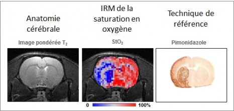
The new MRI method developed by Inserm researchers measures brain tissue oxygen saturation or the quantity of oxygen present in the tissue microvascularization (expressed as StO2).

Thanks to this technique, researchers have observed a major reduction in cerebral oxygenation in an animal model ischemic stroke and, to a lesser extent, following cranial trauma. By mapping cerebral oxygenation, it has been possible to identify regional heterogeneity of StO2 in strokes, cranial trauma and a model brain tumour.

This innovative approach offers a number of benefits including the possibility of immediate use with humans. Unlike positron emission imaging (or PET imaging), which is the gold standard technique, no radioactive tracer needs to be injected when performing an MRI examination. StO2 maps with enhanced spatial resolution can also be produced by MRI. Moreover, MRI examinations are less expensive than PET examinations and MRI imaging devices are more widespread in France than PET imaging devices.

The results of this preclinical trial match those achieved with the two gold standard methods (measurement of blood gas levels and histological mapping of oxygen deprived areas) for different oxygenation levels.

CP_E_Barbier_StO2

From a clinical perspective, several applications may be foreseen, which are illustrated in this project by 3 situations:

  • In patients suffering from cranial trauma, this map could help identify the least oxygenated areas of the brain. Follow-up and therapeutic strategy could therefore be adapted to optimal effect.
  • In stroke patients, this technique should enable more effective identification of the area of ischemic brain tissue which is likely to recover after treatment.
  • Finally, certain malignant brain tumours contain poorly oxygenated areas which are known to be resistant to treatments such as chemotherapy and radiotherapy. It is strongly suspected that these poorly oxygenated areas contain tumour stem cells which cause tumour recurrence. By mapping them more effectively, it should be possible to adapt and customize treatments.


Although the reliability of this system has now been established, it remains to be seen to what extent it is capable of guiding the work of neurologists and anaesthetists. “In several years’ time, it is possible that a precise map of brain oxygenation will enable us to supply drugs to the right place or more effectively configure surgical operations to reduce intracranial hypertension” explains Emmanuel Barbier, Inserm research director.

This work was carried out using the IRMaGe platform, which is a platform of the ‘France Life Imaging’ national life imaging infrastructure. It was funded notably by the Agence Nationale pour la Recherche [French National Research Agency] (IMOXY project).

Laser bioprinting in Bordeaux : an innovative approach

In recent years, 3D printing has developed in the area of health. Custom medical devices and prostheses were the first applications for this new technology. In 2011, for example, the first prosthesis (a titanium jaw) made with the help of 3D printing was implanted. Two years later, a 3D printed cranium (the largest ever placed) was implanted into a 22-year-old woman in the Netherlands.


In addition to these medical devices made of inert materials, researchers took on a challenge of an entirely different nature : bioprinting involves printing living cellular materials!

In contrast to traditional 3D printing, bioprinting involves consideration of a 4th dimension: the time dimension, in which the printed cells assemble themselves, migrate and differentiate autonomously to form functional tissues.


ModuLAB 1

Bioprinting 3D © Inserm/ Guillemot, Fabien – Alphanov / Lescieux, Ludovic


Laser bioprinting in Bordeaux, an innovative approach

One of the goals of the laboratory is to develop laser and microfabrication technologies with the aim of printing tissues in vitro and in vivo. The researchers in the laboratory were pioneers in Europe, developing laser-assisted bioprinting from 2005. This Inserm/University of Bordeaux joint research unit is one of a very few worldwide to use this process. The objective of Fabien Guillemot’s team is therefore not only to position cells in 3D, but to define and model the self-assembly dynamic of the printed cells.

What are the results using the laser approach?

In the laboratory, bioprinting employs the principles of 3D printing to assemble the components of biological tissues (such as the cells and the extracellular matrix) layer by layer in digitally designed predefined patterns.

Laser-assisted bioprinting enables the manufacture of complex tissues with the help of printing using bioinks with cell concentrations close to physiological conditions, with a high degree of resolution (micron scale, pL volume) and a high operating speed (.> 10,000 droplets per second).

“In the area of bioprinting, laser technology offers the highest resolution,” explains Fabien Guillemot, Inserm Research Fellow.


Since 2005, the research team has succeeded in printing different structures and cell types with multiple layers of keratinocytes (cells of the superficial layer of the skin and appendages—nails and body and head hair) and collagen.

Impression de peau

Impression de peau réalisée en juin 2014
© Ludovic Lescieux Alphanov / Fabien Guillemot Inserm.


Printed cells are viable (97% viability after 6 h), and the researchers confirmed that bioprinting did not affect cell differentiation in the case of human adult stem cells.

At the moment, researchers are working on printing corneal and skin tissues in order to meet the needs of regenerative medicine, pharmacology, cosmetics, etc.

At the same time, the research team also conducts in vivo experiments in mice. In 2010 it succeeded in printing mesenchymal stem cells in the bone of live mice. The next step will involve testing computer-aided surgery which would allow in vivo printing of tissues directly where required.

What is the outlook for tomorrow?

The challenge of bioprinting remains the production of functional tissues with the aim of creating:

  • Here and now….Predictive models that reproduce the physiology of healthy human tissues or diseased tissues, enabling the more predictive testing of drugs, components and candidate drugs. These physiological models will be used in the pharmaceutical field. (For cosmetic applications, the overall market for alternative methods has been estimated at €1 billion in 2015 (Source: Transparency Market Research), with an annual growth of 13.1%.)

  • In the next 3-5 years…Individualised tissues, made using patient cells, that allow in vitro selection of treatment based on these tissues, and development of personalised treatment solutions. Fabien Guillemot’s team hopes to include bioprinting in the developments of the new Cancer Plan concerning individualised medicine.

  • In the next 7-10 years…Implantable tissues for regenerative medicine. The development and manufacture of biological tissues represent major socioeconomic challenges. The market for tissue engineering was valued at $15 billion in 2014, and should double by 2018 (source: MedMarket Diligence, LLC.). Moreover, because of the increase in life expectancy, and in the incidence of major diseases such as cancer and diabetes, the number of people waiting for an organ transplant is constantly increasing (51,000 people in Europe in 2013).

Despite advances in research, it is not presently possible to print functioning organs. 

“Once researchers are able to create functional tissues, they will then be able to modify these tissues to improve them. Ethical debate will be needed to determine the extent to which tissue modification is possible, and for what purposes,” emphasises the researcher.


Innovative strategy to facilitate organ repair

A significant breakthrough could revolutionize surgical practice and regenerative medicine. A team led by Ludwik Leibler from the Laboratoire Matière Molle et Chimie (CNRS/ESPCI Paris Tech) and Didier Letourneur from the Laboratoire Recherche Vasculaire Translationnelle (INSERM/Universités Paris Diderot and Paris 13), has just demonstrated that the principle of adhesion by aqueous solutions of nanoparticles can be used in vivo to repair soft-tissue organs and tissues. This easy-to-use gluing method has been tested on rats. When applied to skin, it closes deep wounds in a few seconds and provides a esthetic, high quality healing. It has also been shown to successfully repair organs that are difficult to suture, such as the liver. Finally, this solution has made it possible to attach a medical device to a beating heart, demonstrating the method’s potential for delivering drugs and strengthening tissues. This work has just been published on the website of the journal Angewandte Chemie.

In an issue of Nature published in December last year, a team led by Ludwik Leibler 1 presented a novel concept for gluing gels and biological tissues using nanoparticles 2. The principle is simple: nanoparticles contained in a solution spread out on surfaces to be glued bind to the gel’s (or tissue’s) molecular network. This phenomenon is called adsorption. At the same time the gel (or tissue) binds the particles together. Accordingly, myriad connections form between the two surfaces. This adhesion process, which involves no chemical reaction, only takes a few seconds. In their latest, newly published study, the researchers used experiments performed on rats to show that this method, applied in vivo , has the potential to revolutionize clinical practice.

In a first experiment, the researchers compared two methods for skin closure in a deep wound: traditional sutures, and the application of the aqueous nanoparticle solution with a brush. The latter is easy to use and closes skin rapidly until it heals completely, without inflammation or necrosis. The resulting scar is almost invisible.

Schéma plaie cutanée

Phase 1 Skin injury
Phase 2 Application of the solution
Phase 3 Using pressure to hold the edges together
Phase 4 Skin closure

Illustration of the first experiment conducted by the resear chers on rats: a deep wound is repaired by applying the aqueous nanoparticle solution. The wound closes in thirty seconds.
© “Matière Molle et Chimie” Laboratory (CNRS/ESPCI Paris Tech)

In a second experiment, still on rats, the researchers applied this solution to soft-tissue organs such as the liver, lungs or spleen that are difficult to suture because they tear when the needle passes through them. At present, no glue is sufficiently strong as well as harmless for the organism. Confronted with a deep gash in the liver with severe bleeding, the researchers closed the wound by spreading the aqueous nanoparticle solution and pressing the two edges of the wound toget her. The bleeding stopped. To repair a sectioned liver lobe, the researchers also used nanoparticles: they glued a film coated with nanoparticles onto the wound, and stopped the bleeding. In both situations, organ function was unaffected and the animals survived.

“Gluing a film to stop leakage” is only one example of the possibilities opened up by adhesion brought by nanoparticles. In an entirely different field, the researchers have succeeded in using anoparticles to attach a biodegradable membrane used for cardiac cell therapy, and to achieve this despite the substantial mechanical constraints due to its beating. They thus showed that it would be possible to attach various medical devices to organs and tissues for therapeutic, repair or mechanical strengthening purposes.

This adhesion method is exceptional because of its potential spectrum of clinical applications. It is simple, easy to use and the nanoparticles employed (silica, iron oxides) can be metabolized by the organism. It can easily be integrated into ongoing research on healing and tissue regeneration and contribute to the development of regenerative medicine.

1 Leibler was awarded the CNRS Medal for Innovation in 2013.
2 See the press release for this work: https://www2.cnrs.fr/presse/communique/3355.htm

Efficacy of gene therapy demonstrated in canine and murine models of myotubular myopathy

A team of French researchers, led by Dr. Anna Buj-Bello (Genethon/Inserm) and teams at the University of Washington and Harvard Medical School in the United States, have demonstrated the efficacy of gene therapy in models of myotubular myopathy, an extremely severe neuromuscular disease in children. Transfer of the MTM1 gene, which is deficient in the disease, corrected the affected muscles in mice and dogs and prolonged the survival of treated animals. This work, published today in Science Translational Medicine, has been achieved thanks to donations from the French Telethon and the support of the Myotubular Trust.

Discover the images of treated dogs.

ADN

©fotolia

Myotubular myopathy is an X-linked genetic disease affecting 1 in every 50,000 newborn boys. It is caused by mutations in the gene MTM1 encoding myotubularin, a protein involved in the functioning of muscle cells. In its most serious form, it causes hypotonia, generalized muscle weakness and death in the first years of life. There is currently no effective treatment for this severe rare disease.

The study by the French team at Genethon, and the U.S. team at the University of Washington, aimed at evaluating the efficacy of a single intravenous injection of an adeno-associated virus (AAV) expressing myotubularin in the muscles of mice and dogs which carry an MTM1 mutation.

In 2009, the group directed by Dr. Anna Buj-Bello performed the first study of gene therapy on mice with this disease at Genethon. Their success led to the development of a study in dogs which naturally carry this genetic abnormality, in collaboration with U.S. teams from Boston and Seattle. The vectors used for gene therapy have been developed and manufactured at Genethon.

Exceptional results: normalization of muscle strength and respiratory function and prolonged survival

The results of the study indicate an increase in muscle strength and improved respiratory function as well as improved mobility, and prolonged survival.

This normalization is the first demonstration of persistent correction by a single injection of AAV intravenously in a large animal model of neuromuscular disease. A single dose of drug-vector permitted the long-term expression of myotubularin in muscles.

For Dr. Anna Buj Bello, principal investigator at Genethon: “These results are the culmination of four years of research and show how gene therapy is effective for this genetic muscle disease. We finally can envision a clinical trial in patients. These are very promising results. ”

For Dr. Martin Childers from University of Washington: « The implications of the pre-clinical findings are extraordinary for inherited muscular diseases. Two of our dogs treated with AAV-mediated gene therapy appear almost normal with little, if any, evidence, even microscopically, of disease caused by XLMTM. »

For Dr. Alan Beggs, director of the Manton Center for Orphan Disease Research at Boston Children’s Hospital: “Demonstrating that gene therapy is effective in prolonging the lives of these dogs is extremely exciting, providing us with the necessary information to start planning clinical trials in humans.

Fulvio Mavilio, Chief Scientific Officer Genethon and co-author of the study: “These results have a significant impact on the prospect of developing treatments neuromuscular diseases. They are indeed very promising.

Frédéric Revah, CEO Genethon: “For the first time, researchers have obtained a systemic therapeutic effect on neuromuscular disease in dogs with a single intravenous injection: the treatment does not act locally but throughout the body. Genethon is proud to have worked with the best teams in the world and our next goal is working on the implementation of a clinical trial in humans.

Laurence Tiennot-Herment, President of the AFM-Téléthon and Genethon : “This result achieved by our laboratory Genethon, in association with the best American teams, is a major step forward for families who constantly fight the disease. Our determination to defeat the disease is stronger than ever and, thanks to the support of donors from Téléthon, we move step by step toward new victories.

The AFM-Telethon in France, Muscular Dystrophy Association in the United States, Myotubular Trust in Britain, Anderson Family Foundation and Joshua Frase Foundation participated in the financing of this study.

Parkinson’s disease: an immense step forward thanks to gene therapy

A French and English team (AP-HP, Inserm, UPEC, CEA/Mircen, Oxford Biomedica, Cambridge University) has conducted a clinical phase 1/2 gene therapy study among patients suffering from an evolved form of Parkinson’s disease. Fifteen patients were able to benefit from this new treatment, which involves injecting a vector expressing the genes of three enzymes that are essential for the biosynthesis of dopamine, which is lacking in Parkinson’s disease. Thanks to this therapy certain cells in the brain begin to produce and secrete dopamine again. In all the patients, the motor symptoms of the disease were improved for up to 12 months after administration of the treatment. After a period of four years, this study is at this stage demonstrating innocuousness and tolerance of the lentiviral vector used for the first time in human beings. This study was coordinated by Prof. Stéphane Palfi, head of neurosurgery at Henri-Mondor Hospital (AP-HP) within the framework of the neurolocomotor research cluster directed by Prof. Césaro.
It is the subject of a publication in The Lancet

Palfi opération © APHP

Parkinson’s: a common neurodegenerative disease

With about 120,000 patients in France, Parkinson’s disease is the most common neurodegenerative disorder after Alzheimer’s disease. It essentially manifests itself through motor symptoms that steadily grow and become more severe such as trembling, rigidity of the limbs and diminished movement of the body. This pathology is due to the degeneration of neurons that produce dopamine, a neurotransmitter that participates in motor control .

Currently, the treatment of people affected by this disease consists of taking medication that mimics the action of the dopamine missing in the brains of these patients. While this treatment makes it possible to improve motor activity considerably during the first stages of the disease, severe undesirable effects appear at the end of this time such as fluctuations in the effect of the treatment and abnormal involuntary movements, called dyskinaesia.

Developing a new treatment that permits the physiological restoral of missing dopamine

For several years, experts on Parkinson’s disease, researchers and doctors, have held the hypothesis that the intermittent intake of medication during the day alters the functioning of the brain by stimulating neurons in an excessively irregular manner. This phenomenon would constitute the origin of the complications connected with dopaminergic treatment.

The currently most pressing issues in the treatment of Parkinson’s disease thus concern the development of a technology that would make it possible to induce:

  • sustained dopaminergic stimulation;
  • local dopaminergic stimulation in order to induce beneficial motor effects while avoiding the complications that follow stimulation in other regions of the brain not affected by Parkinson’s disease .

This is why researchers today are turning to gene therapy, which consists of causing a therapeutic gene to be expressed directly by brain cells.

Gene therapy consists of introducing therapeutic genes in vivo so that they express directly in the targeted cells.

It rests on the use of viral vectors such as lentiviruses, adenoviruses and AAVs (adeno-associated viruses), which have the ability to introduce their genetic material into the nucleus of host cells.

Some requirements must be absolutely satisfied for a wild virus to be able to be transformed into a vector with the ability to ensure the transfer of genes of therapeutic interest in complete security. These viral envelopes are stripped of their properties for multiplication and rendered non-pathogenic.

Increasing the synthesis of dopamine through gene therapy

In the majority of cases, Parkinson’s disease does not have a genetic origin. However, the biochemical modifications responsible for the symptoms can be corrected by using a gene therapy strategy of the ‘replacement or restoral of function’ type in order to increase the synthesis of dopamine (by expressing genes involved in the biosynthesis of dopamine) and restore the function of dopaminergic cells partially.

It is this approach that was adopted in the phase I/II biomedical study coordinated by Prof. Stéphane Palfi (Henri-Mondor Hospital, AP-HP), the results of which have just been published.

Fifteen patients were operated on by Prof. Palfi, coordinating investigator, in two centres of excellence in neurosurgery – Henri Mondor Hospital (AP-HP) in France and Addenbrookes Hospital in Cambridge, UK.

For the first time in human beings, the team used a lentiviral vector which expresses the genes of three enzymes – AADC (decarboxylase of aromatic amino acids), TH (tyrosine hydroxylase) and CH1 (GTP-cyclohydrolase 1) – essential in the biosynthesis of dopamine. The product was administered in the area of the brain called the striatum during a heavy surgical operation.

Once in the right place, the genes contained in the lentivirus can express themselves and reprogramme cells, which begin to produce and secrete dopamine in the extracellular environment.

Three increasing dosage levels (1×, 2× and 5×) were tested.

‘This biomedical gene therapy study shows innocuousness over the long-term transfer of genes by the lentiviral vector when it is injected directly into the brain of patients suffering from Parkinson’s disease’, explains Prof. Stéphane Palfi. ‘The clinical analysis suggests that the vector used enables a reduction in motor symptoms depending on the vector dose administered, with the strongest dose being the most effective .

The objective of future clinical developments of the vector will be to confirm an improved viral construction that would make it possible to induce an increased release of dopamine (phase 2a). This phase will be followed by a study of the therapeutic effect of ProSavin® by comparing a group of patients receiving the treatment and another group not receiving the treatment (phase 2b). This study, which is pioneering the use in gene therapy of a lentivirus injected in situ, will definitely open up new therapeutic perspectives for diseases of the nervous system.’

Architecture of phase I/II clinical trial

The local and sustained production of dopamine in vivo was restored in 15 patients suffering from an evolved form of this disease. The long-term monitoring of these patients (4 years) evidenced undeniable innocuousness, tolerance and signs of the therapeutic effectiveness of the viral vector depending on the administered dose, with the strongest dose of the vector inducing the most substantial therapeutic effects.

Key figures

15 patients treated

1 lentiviral vector used for the first time in humans

3 dosage levels tested

Research initiated in 2009

This clinical trial follows on from a preclinical study published in 2009, which showed for the first time the effectiveness and innocuousness of the medication in an animal model. Carried out within the framework of the MIRCen translational platform of the CEA, it has opened the door to the clinical study of ProSavin®.

Cancer treatment: a step towards personalized chronotherapy

Cancer chronotherapy consists in administering treatment at an optimal time. Because the body is governed by precise biological rhythms, the efficacy of anti-cancer drugs can be doubled and their toxicity reduced five-fold depending on the exact timing of their administration. However, important differences in biorhythms exist between individuals, which chronotherapy has not been able to take into account until now. An international study conducted on mice and coordinated by researchers from Inserm, CNRS and Université Paris-Sud[1] has paved the way towards personalized chronotherapy treatments. In an article published in the journal Cancer Research, the team has shown that the timing of optimal tolerance to irinotecan, a widely used anti-cancer drug, varies by 8 hours depending on the sex and genetic background of mice. They then developed a mathematical model that makes it possible to predict, for each animal, the optimal timing for administering the drug. They now hope to test this model on other drugs used in chemotherapy.

The body’s metabolism follows a 24 hour rhythm, driven by the circadian clock. Consequently, at certain precise times of the day or night, a given drug may prove to be more toxic to cancer cells and less aggressive to healthy cells. Cancer chronotherapy, discovered some twenty years ago by Francis Lévi, seeks to improve the efficacy of chemotherapy treatments. His research has shown that this efficacy can be doubled, depending on the time at which they are administered. Furthermore, it is precisely at this optimal time that the drugs prove to be five times less toxic to the body.

However, research points to the need for personalizing chronotherapy. Indeed, biorhythms can change from one person to the next. For example, although the optimal timing is the same for 50% of patients, the remaining 50% are either ahead of or behind this time. The team headed by Lévi wanted to elucidate the factors that affect these differences in biorhythms.

To do this, the researchers studied the toxicity of irinotecan, an anti-cancer drug widely used in the treatment of cancer of the colon and pancreas, as a function of the timing of its administration in four strains of male and female mice. For the first time, they were thus able to observe that the time of best tolerance to treatment varied by up to eight hours from one group of rodents to the next, depending on their sex and genetic background.

The researchers then worked on developing a method able to predict this optimal drug timing independently of sex and genetic background. To do this, they measured the expression of 27 genes in the liver and colon over 24 hours and then analyzed these measurements using a methodology derived from systems biology. In this way, the researchers were able to construct and validate a mathematical model to precisely predict the timing at which irinotecan is less toxic to the body using the expression curve of two genes, known as Rev-erbα and Bmal1, which regulate the metabolism and proliferation of cells.

The researchers are now aiming to validate this model on other drugs used in chemotherapy. In addition to gene expression, they would also like to find other physiological parameters related to the biological clock that could help predict the optimal timing of treatments for each patient. This work should make it possible to enhance the efficacy and tolerance of such treatments as well as considerably improve the quality of life of patients.

This project was funded in particular by the European Union (7th Framework Programme for Research and Technological Development) and ERASYSBIO+, the European consortium of funding bodies, ministries and project management agencies


[1] Coordinated by the Unité Rythmes Biologiques et Cancers (Inserm/Université Paris-Sud), this work also involved the Institut de Biologie de Valrose (CNRS/Inserm/Université de Nice Sophia Antipolis), the Laboratoire des Signaux et Systèmes (CNRS/Supélec/Université Paris-Sud) and the Milan Institute of Pharmacology.

Pandoravirus : missing link discovered between viruses and cells

Researchers at IGS, the genomic and structural information laboratory (CNRS/Aix-Marseille University), working in association with the large-scale biology laboratory (CEA/Inserm/Grenoble Alpes University) have just discovered two giant viruses which, in terms of number of genes, are comparable to certain eukaryotes, microorganisms with nucleated cells. The two viruses – called “Pandoravirus” to reflect their amphora shape and mysterious genetic content – are unlike any virus discovered before. This research appeared on the front page of Science on July 19, 2013.

With the discovery of Mimivirus ten years ago and, more recently, Megavirus chilensis[1], researchers thought they had reached the farthest corners of the viral world in terms of size and genetic complexity. With a diameter in the region of a micrometer and a genome incorporating more than 1,100 genes, these giant viruses, which infect amoebas of the Acanthamoeba genus, had already largely encroached on areas previously thought to be the exclusive domain of bacteria. For the sake of comparison, common viruses such as the influenza or AIDS viruses only contain around ten genes each.

In the article published in Science, the researchers announced they had discovered two new giant viruses:
Pandoravirus salinus, on the coast of Chile;
Pandoravirus dulcis, in a freshwater pond in Melbourne, Australia.

Detailed analysis has shown that these first two Pandoraviruses have virtually nothing in common with previously characterized giant viruses. What’s more, only a very small percentage (6%) of proteins encoded by Pandoravirus salinus are similar to those already identified in other viruses or cellular organisms. With a genome of this size, Pandoravirus salinus has just demonstrated that viruses can be more complex than some eukaryotic cells[2]. Another unusual feature of Pandoraviruses is that they have no gene allowing them to build a protein like the capsid protein, which is the basic building block of traditional viruses.

Despite all these novel properties, Pandoraviruses display the essential characteristics of other viruses in that they contain no ribosome, produce no energy and do not divide.

This groundbreaking research included an analysis of the Pandoravirus salinus proteome, which proved that the proteins making it up are consistent with those predicted by the virus’ genome sequence. Pandoraviruses thus use the universal genetic code shared by all living organisms on the planet.

This shows just how much more there is to learn regarding microscopic biodiversity as soon as new environments are considered. The simultaneous discovery of two specimens of this new virus family in sediments located 15,000 km apart indicates that Pandoraviruses, which were completely unknown until now, are very likely not rare. It definitively bridges the gap between viruses and cells — a gap that was proclaimed as dogma at the very outset of modern virology back in the 1950s. It also suggests that cell life could have emerged with a far greater variety of pre-cellular forms than those conventionally considered, as the new giant virus has almost no equivalent among the three recognized domains of cellular life, namely eukaryota (or eukaryotes), eubacteria, and archaea.

Notes
[1] Arslan D, Legendre M, Seltzer V, Abergel C, Claverie JM (2011) “Distant Mimivirus relative with a larger genome highlights the fundamental features of Megaviridae.” PNAS. 108:17486-91
[2] Parasitic microsporidia of the Encephalitozoon genus in particular.

Discovery of a molecule derived from cholesterol that has anti-cancer properties

Although excessive quantities of cholesterol in the body are known to have adverse effects on health, researchers might polish up its reputation via one of its derivatives. The research team from Inserm and the CNRS led by Marc Poirot and Sandrine Silvente-Poirot at the “Centre de recherche en cancérologie de Toulouse” (Inserm / CNRS / Université Toulouse III – Paul Sabatier), has not only discovered a new molecule derived from cholesterol (known as dendrogenine A), but has also provided proof in mice that this molecule has anti-cancer properties.

This work has been published in the journal Nature Communications.

Cholesterol is involved in different chronic pathologies, such as cardiovascular diseases and cancer. Current knowledge led us to suppose that cholesterol had a negative effect on cancers, mainly for two reasons. Firstly, cholesterol is a precursor of androgens and estrogens, both of which are involved in the development of cancers known as “hormone-dependent” cancers. Secondly, the synthesis process of cholesterol (that contains over 20 different stages) activates tumour-related genes.

Blocking the upstream biosynthesis pathway using inhibitors such as statins should have provided protection against cancer or some measure of anti-cancer efficiency, but this was not confirmed by clinical studies involving very large patient cohort studies; This suggests that its metabolism is much more complicated[1].

So the team of researchers led by Marc Poirot and Sandrine Silvente-Poirot concentrated particularly on the metabolism of cholesterol. Using screening and chemical synthesis techniques, the researchers were able to show that the product of the chemical reaction between a cholesterol derivative and a histamine generates a new class of sterols known as Dendrogenine A (DDA). This molecule, obtained in laboratory conditions, shows remarkable properties for inducing differentiation and death of cancer cells.

These observations led the team to search for this molecule in the tissues of mammals. And they discovered that DDA is present in healthy tissue and cells in humans, whereas it is not detectable in tumour cells. In women, its level is 5 times lower in breast tumour tissue than in normal tissue.

“Our results suggest that DDA protects the cells from carcinogenic processes” points out Marc Poirot.

In order to test this hypothesis, the researchers then attempted to repair the DDA deficiency in tumours implanted in animals. And in these animals, the administration of DDA controlled the proliferation of the tumour and extended the life expectancy.

For the researchers, this is a major discovery, because it provides the proof of the existence of a new metabolic pathway in humans, at the interface between the cholesterol metabolism and the histamine metabolism, and also because, thanks to its anti-cancer properties, DDA could be used to treat different cancers.

 Dendrogine_ENG

© Sandrine Silvente-Poirot / Marc Poirot


[1] A group of processes than involve both synthesis and breakdown of cholesterol into products that can be used by the body.


A chip that is 100% biocompatible to measure brain activity

Interpreting the signals emitted by the brain and translating them into commands usable by humans is a goal that researchers have been pursuing in the development of what is known as brain-machine interfaces. With respect to health, these interfaces could be used by people suffering from paralysis. Up to now, researchers have encountered technological difficulties because the sensors used to record brain activity do not yet do so with enough accuracy.

With the support of the Bioelectronics Department of the St Etienne Ecole des Mines, a research team headed by Christophe Bernard of INSERM Unit 1106 “Institut de neurosciences des systèmes” have designed a system for sensing brain activity that is 100% biocompatible and made of organic matter. The medium, which is only a few microns thick, is as thin and flexible as cellophane, and yet very tough. The model was tested on an animal suffering from epilepsy. The quality of the brain signal recorded was 10 times better than the traditional brain activity recording systems. The research was published in the Nature Communications journal.

Des nouvelles techniques au service de la santé, OpenVibe. Une interface cerveau-ordinateur ou ICO (en anglais Brain-Computer Interface ou BCI) permet à son utilisateur d'envoyer des commandes à un ordinateur ou à une machine uniquement par la pensée. © P Hirsch/Inserm

Man-machine interfaces have been playing a central role for several years in the diagnosis and treatment of certain conditions, in the movements of artificial limbs (exoskeletons) and even in the design of artificial sensory organs. In the case of brain-machine interfaces, the problem consists in detecting the signals emitted by the brain and translating them into commands that are usable by humans. These signals are used for diagnostic purposes (such as, for example, to determine whether a person is epileptic and which regions of the brain are responsible for the attacks), to link an artificial eye to the regions of the brain that process visual information or to control the movement of exoskeletons in people who are paralysed by recording the neurons in the regions of the brain that control the motor function of limbs.

For the capture of the maximum number of signals emitted by the brain, there is a need for direct contact with the central nervous system. This is very hard to achieve when using non-invasive measurement systems (i.e. electrodes placed on the head). Another disadvantage is that most of sensors used today are not biocompatible, thus triggering a defence reaction in the tissues, resulting in loss of signal after a certain amount of time. Finally, and this is the most important aspect, the signals detected are pre-amplified at a distance from the source, and this results in the presence of considerable interference on the recordings, thus preventing them being used to their best advantage.

One solution: organic transistors

The Bioelectronics Department of the St Etienne Ecole des Mines in Gardanne, the Institut de Neuroscience des Systèmes (INSERM Unit 1106) and the Microvitae SME at Gardanne have contributed a technological solution to these problems.

The researchers designed a system of brain activity sensors made of an organic material (based on carbon composites) that is 100% biocompatible. The medium is only a few microns thick, as thin and flexible as cellophane, and very tough.

But the revolutionary technology lies in enabling the recording site to work as an organic transistor that amplifies the signal locally. The system was tested in an animal model suffering from epilepsy. The quality of the signal was amplified tenfold in comparison with the traditional systems an amount that the researchers claim to be considerable.

A technological solution such as this makes it possible to record numerous neurons and interface with brain structures in the long term. The immediate clinical applications could include assistance with diagnosing epilepsy and functional mapping in the context of neurosurgery for brain tumours. Naturally, these transistors could also be used for non-invasive recordings in direct contact with the head.

In addition to disease control, the technology will enable major advances to be made in basic research, and especially in the context of the Human Brain Project which has received one billion euros of finance from the European Union. Recording systems based on organic transistors are precursors to the Man-machine interfaces of tomorrow.

By the sixth month of pregnancy, the human brain is capable of processing speech

A discovery by INSERM Unit U1105 “Groupe de Recherche sur l’Analyse Multimodale de la Fonction Cérébrale” [Research Group for Analysis of the Multimodal Cerebral Function] of the University of Picardy Jules Verne (UPJV) and INSERM Research Unit U992 “Neuroimagerie cognitive” [Cognitive neuroimagery], NeuroSpin/CEA

The immature brain of a premature infant is capable, at the age of three months pre-term, of distinguishing syllables uttered by male and female voices. These results obtained by INSERM researchers at the University of Picardy Jules Verne and the CEA’s NeuroSpin Imaging Centre, were published in the PNAS journal dated 25 February 2013. They highlight the very early sophisticated organisation of the regions of the brain involved in language-processing and social communication in humans.

At birth, infants are capable of distinguishing between similar syllables, of recognising their mother’s voice and of differentiating between several human languages. Are these abilities in the human infant due to the presence of innate mechanisms specific to the human race for processing speech or do they indicate swift learning of the characteristics of the mother’s voice during the final weeks of pregnancy?

To discover the truth, Fabrice Wallois, Director of the mixed UPJV/INSERM research unit known as the “Research Group for Analysis of the Multimodal Cerebral Function” (GRAMFC), and Ghislaine Dehaene-Lambertz, (INSERM, NeuroSpin/Atomic Energy and Alternative Energies Commission (CEA), University of Paris-Sud) in collaboration with hospital doctors at the Amiens Picardy University Hospital, tested the auditory discrimination abilities of twelve premature babies, born at 28 to 32 weeks of amenhorræa, i.e. born two to three months’ pre-term.

At this stage of development, the brain is immature since the neurones are still in the process of migrating to their final destination. Yet the first connections between the brain and the outside world are being set up, especially those that enable the fœtus to hear sounds, thus enabling it to record the first brain responses to external stimulation.

The authors of this study stimulated premature infants by using audio stimulation, exposing them to the sounds of two similar syllables (“ga” and “ba”) pronounced by either a man or a woman. They recorded the brain responses by using functional optical imaging (near infrared spectroscopy (NIS)). The researchers were thus able to show that despite the fact that their brains were still immature, the premature babies were receptive to changes in the voice (male or female) and changes in phonemes ( “ba” or “ga”) (figure 1).

Furthermore the sets or networks of neurones involved in premature babies are very similar to those described in the adult when performing the same type of task. They are asymmetrical and especially involve the frontal areas. As in adults, the right frontal area responds to newness, regardless of the change involved, whereas the left frontal area or Broca’s region, only responds to a change of phoneme.

Figure 1: Projection of brain activation in a premature baby born at 30 weeks of amenorrhæa. The change of phoneme produces an increase in brain activity in the temporal and frontal areas, especially the left. The response to a change in the voice is more limited and only involves the change of voice is more limited, and only involves the lower right frontal area.

©Wallois

These results show very early, as soon as the first cerebral connections are established (three months before term) and above all before any learning process has occurred, that the human brain is capable of processing special characteristics of human speech thanks to the sophisticated organisation of certain language areas of the brain (the right and left perisylvian regions). Since the organisation of the regions of the brain being governed by genetic expression during the development of the fœtus, the authors suggest that the emergence of language is largely influenced by genetics and thus by innate mechanisms.

This study received support from the Picard Regional Council and the ERDF (European Regional Development Fund). 


Inauguration of TherA-Image, imaging technology-assisted treatment platform

When research, medicine and high tech work together in Rennes

Imagine, in this hybrid operating theatre, surgeons, physicians and engineers surrounded by control screens, using enhanced and roboticised reality systems, enabling interventions that are even more accurate and safe. At the Signal and Image Processing Laboratory (University of Rennes 1 / Inserm), researchers, engineers and doctors at the Rennes CHU Cardio-pneumological Centre have been working together to bring the TherA-Image platform into existence.

They have designed and implemented interventional cardiology and mini-invasive surgery techniques, using image-guidance and computer-assistance through the TherA-Image platform. These procedures are designed to reduce to a minimum the time taken for the intervention and trauma from the operation. This means that the frailest patients will be able to have access to these innovative medical techniques that are designed to improve post-operative and the prognosis thanks to instruments and skills that are unique in Europe.

TherA-Image is a hybrid operating theatre, used for both patient care and de research in the field of health technology. It is a medico-technical platform located at the interface between the Rennes University Hospital, the University of Rennes 1, INSERM and the medical industry. It is here that computerised approaches are devised and deployed for planning interventions, assistance with operating procedures and assessment of these procedures.

TherA-Image possesses state-od-the-art imaging equipment (3D intra-operating observation, enhanced reality, cardiac electrophysiology), operating assistance (endovascular navigation and a catheterisation robot) as well as video broadcasting (to obtain remote expertise, provide training, etc.)


Treating cardiac insufficiency through resynchronisation therapy /©L. Després

This combination of equipment and skills in the same operating theatre in Rennes is unique in Europe. It is the result of a convergence of views that originated long ago in the LTSI multidisciplinary teams, that included doctors, researchers and engineers, and a solid partnership has been formed for the long-term, including industry leaders in their own field of interest.


©Replacing the thoracic aorta

Today, TherA-Image makes it possible to explore new approaches to cardiovascular treatment, in order to:

  • Treat cardiac insufficiency, especially through the type of treatment known as cardiac resynchronisation. Patients sometimes present with defects in the synchronisation of the heart ventricles. This means that the chambers of the heart do not all contract at the same time, so the heart no longer works efficiently and the patient becomes out of breath when making the slightest effort. Thanks to the TherA-Image platform, doctors and researchers can optimise the techniques and the implantable devices for electrically stimulating the heart.
  • Eliminate the sources of electrical disturbances inside the heart muscle that are the cause of cardiac dysrhythmia. Techniques and intra-corporeal navigation models have been developed for this purpose, as well as diagrams of the electrical current inside the heart muscle. The aim is to identify the sources of electrical disturbance and check that they have been eliminated after treatment, through localised heating of the tissue.
  • Promote the development of less invasive surgical techniques. For example, it should become possible to reliably replace heart valves percutaneously (passing through an artery), without having to open up the chest cavity, relying on location and algorithms and guiding the surgical instruments through the body.
  • Treating aneurisms (abnormal dilatation of the walls of a blood vessel) and stenoses (shrinking or narrowing of a major blood vessel). Current mini-invasive techniques are becoming increasingly complex, but thanks to TherA-Image, the surgeon can guide his/her instruments through the blood vessels using tools for planning the route (as for GPS) and effective assisted-imaging methods (enhanced reality) to reach the lesion and install a prosthesis at the site in complete safety.


Simplified dynamic 3D modelling of the Théra-Image room/©LTSI

TherA-Image is an instrument that has caused a major development in medical research and the occupational culture associated therewith, making it possible to design, deploy and assess the surgical interventions of tomorrow, to the benefit of the patient.

TherA-Image is financed through the State-Region Project Contract 2007-2013 in an amount of €5.2 million and has received support from the European Union (FEDER: €1.7 million), and the State (€2 million), the Brittany Region (€370,000), the General Council of Ille-et-Vilaine (€640,000) and from Rennes Métropole (€526,000).

fermer