Menu

Auto-immune disease: the viral route is confirmed

Why would our immune system turn against our own cells? This is the question that the combined Inserm/CNRS/ Pierre and Marie Curie University/Association Institut de Myologie  have strived to answer in their “Therapies for diseases of striated muscle”, concentrating in particular on the auto-immune disease known as myasthenia gravis. Through the project known as FIGHT-MG (Fight Myasthenia Gravis), financed by the European Commission and coordinated by Inserm, Sonia Berrih-Aknin and Rozen Le Panse have contributed proof of the concept that a molecule imitating a virus may trigger an inappropriate immune response, causing muscular function to deteriorate. These results have been published in Annals of Neurology, accessible on line.

Myasthenia, a rare auto-immune disease

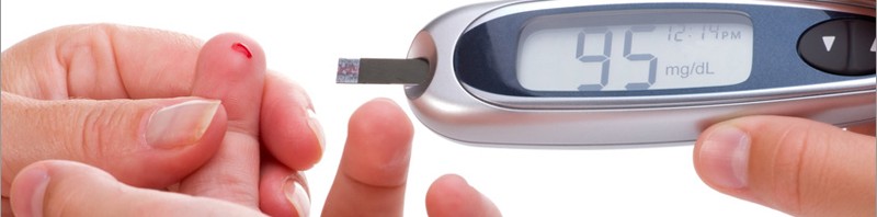
Myasthenia gravis is a rare auto-immune disease (5,000 to 6,000 patients in France) that produces muscular weakness and exhaustion. It generally affects the facial muscles first, and may then become generalised through the muscles of the limbs or the respiratory muscles, causing respiratory distress.

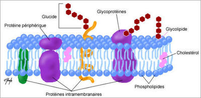
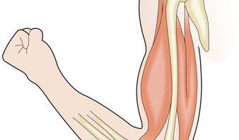
This is due to the production of circulating auto-antibodies that block the acetylcholine receptors (RACh), these neurotransmitters being necessary for transmitting the motor nerve signal to the neuro-muscular junction.

Could a viral infection be the origin of myasthenia?

Myasthenia is a multi-factorial disease in which environmental factors seem to play a key triggering role. Viral infections are suspected but it is hard to prove the role of a virus in triggering the condition. In fact, diagnosis of myasthenia is often made months, or even years, after the actual start of the illness when the virus is no longer detectable, even though the signature left by the virus is visible long after the infection.

Proof of the concept of a viral origin contributed by researchers

Under the European FIGHT-MG project, the team of researchers managed to decode the trigger for the illness by using a molecule that mimics the RNA double viral strand (Poly(I:C)).

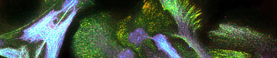
To do this, they concentrated on the organ that plays a central role in the disease – the thymus. It is in this gland located in the thorax that the T-lymphocytes mature, these being the key players in immune response that are normally programmed to avoid the development of any auto-immunity.

The researchers were thus able to show in vitro that the Poly(I:C) was capable of specifically inducing an over-expression of RACh through thymal epithelial cells, while activating three proteins (the “toll-like” receptor 3 (TLR3), the protein kinase R (PKR) and interferon-beta (IFN-â)); it is this last that produces inflammation in the thymus.

At the same time, they analysed pathological thymus glands of myasthenia sufferers in whom they observed over-expression of these same three proteins in the immune system, characteristic of a viral infection.

Finally, the researchers also managed to identify the same molecular changes in the thymus glands of mice, after they had been injected with Poly(I:C). After a prolonged injection period, they also observed a proliferation in the mice of B anti-RACh cells, the presence of auto-antibodies blocking the RACh receptors and clinical signs synonymous with the muscular weakness found in myasthenia.

These original results show that molecules that mimic a viral infection are capable of inducing myasthenia in the mouse, something that had never been demonstrated before.

This set of papers published in the Annals of Neurology provides proof of the concept that a viral infection can cause inflammation of the thymus and lead to the development of auto-immune myasthenia.

The next stages of the research will consist in determining which exogenous virus this may be or whether it is a case of the abnormal activation of an anti-viral response by endogenous molecules.

© Inserm / R. Le Panse 

The introduction of a double strand of RNA (Poly(I:C) into the thymal epithelial cell induces the over-expression of the acetylcholine receptors (RACh), via the activation of the “toll-like” receptor 3 (TLR3) and the protein kinase R (PKR),  as well as the production of interferon-beta (IFN-β)). These changes in the thymus gland cause the formation of B anti-RACh cells and the production of circulating auto-antibodies that block the acetylcholine receptors present in the neuromuscular junction.

FIGHT-MG (Fighting Myasthenia Gravis) – a European collaboration making giant leaps forward

The FIGHT-MG project seeks to determine the genetic and environmental risk factors associated with the occurrence of the illness and its development. The project aims also to identify the key immunological molecules associated with its appearance, and to study the pathogenic mechanisms at the neuromuscular junction, establish new diagnostic tests, as well as new treatments (cellular treatments, immuno-regulatory treatments, immuno-absorption of pathogenic auto-antibodies and other pharmacological treatments).

“When one is working on a rare disease, it is essential to work through networking, so as to be able to share our facilities and resources to promote fundamental and clinical research. It is also crucial to communicate permanently with patient associations. It is this combination that enables us to take giant steps in the treatment of rare conditions,” explains Sonia Berrih-Aknin.

FIGHT-MG : https://www.fight-mg.eu/ 

FIGHT-MG started in December 2009 and will last for four years, with a total budget of about six million euros funded by the European Union (FP7). The project involves 12 partners based in seven European countries:

The 12 partners:

Inserm (coordinator), France
Hellenic Pasteur Institute (HPI), Greece
Open University of Israel (OUI), Israel
Fondazione Istituto Neurologico “Carlo Besta” (INNCB), Italy
Oslo University Hospital (OUS), Norway
Hadassah Hebrew University Medical Center (HMO), Israel
Israel Institute of Technology (TECHNION), Israel
University of Paris 6 Pierre and Marie Curie (UPMC), France
University of Basel (UNIBAS), Switzerland
ProteoSys (PSY), Germany
Genopolis Consortium for Functional Genomics (GENOPOLIS), Italy
INSERM TRANSFERT SA (IT), France

The “Myasthenia” team

The “Myasthenia” team, headed by Sonia Berrih-Aknin joined the Institute of Myology directed by Professor T. Voit, just over a year ago in order to get closer to the reference centre for neuromuscular diseases run by Prof B. Eymard, at the Pitié-Salpêtrière Hospital in Paris. The Institute of Myology is an international center of expertise on the muscle and its diseases, a member of the Institute of Biotherapy of rare diseases created by the AFM-Telethon. The Sonia Berrih-Aknin’s team is interested in the etiological and physio-pathological mechanisms of myasthenia and innovative treatments that could improve patients’ quality of life.

Even though winning a European project is very competitive, this team has exceptionally been granted three other projects since 2001, and was responsible for their coordination. Sonia Berrih-Aknin was the coordinator of the “Mechanisms of Myasthenia” project (2001-2005) under FP5, the MYASTAID (2006-2010) project under  le cadre du FP6, as well as the Euromyasthenia Project (2006-2009) through the European Public Health  Directorate. These projects brought a total of more than fifty teams of clinicians, researchers and associations of sufferers in Europe.


Un diagnostic prénatal des maladies génétiques sur simple prise de sang

The radical restructuring of brain networks in comatose patients

Researchers from Inserm, CNRS and the Université Joseph Fourier in Grenoble, in collaboration with Cambridge university, Strasbourg university and clinical practitioners from the Strasbourg University Hospital Centre, have analysed data from 17 comatose patients using functional MRI data. Their research reveals that the brain networks of these patients have been restructured. The results, published in PNAS on 26 November 2012, could help clinical practitioners diagnose comatose patients.

The researchers are focusing on analysing brain networks of brain-damaged comatose (non-traumatised) patients, a state where the individual is considered to be unconscious.

The authors of the study used an original graph theory-based methodology, where images were constructed using functional MRI data at rest and using robust statistical signal-processing methods. Local and overall effectiveness indices of functional brain networks were obtained for 17 brain-damaged patients and 20 healthy volunteers. Correlations in  417 brain regions were extracted to produce brain connection graphs using the statistically significant correlations.

Inserm unit 836 “Grenoble Institut des neurosciences”, CNRS researchers from the “GIPSA lab” and from the Behavioural and Clinical Neuroscience Institute in Cambridge, in collaboration with clinical practitioners from the Strasbourg University Hospital Centre, have been able to highlight restructured brain networks in brain-damaged (non-traumatized) comatose patients.

Through comparisons with the healthy subjects, the results demonstrate that the overall cerebral connectivity is preserved in comatose patients. By analysing the connectivity at a local level, the authors of the study have observed that some brain regions (“hubs”), which are highly connected in healthy volunteers, are less well connected in comatose patients. Conversely, the less densely connected regions in the network in healthy subjects become “hubs” in comatose patients.

Brain imaging obtained from connectivity graphs

Photos: © Sophie Achard – Petra Vertes

The connectivity graph method is used to summarize in a single image data acquired through MRI scanning. It translates the effectiveness of connections in a single region compared to all the others. By grouping the most interconnected regions, modules are revealed (each represented by a different colour). Patients and healthy volunteers both have different models in their spatial location, representing radical alterations to the brain connections.

According to current hypotheses, consciousness disorders in persistently comatose patients could be linked to disconnection phenomena between specific cortical regions, particularly the precuneus. The results of this study also point in this direction. “From an overall perspective, the topology of brain connections resists well to traumatism by reorganising the most interconnected regions in the network. It therefore seems that comas may be linked to changes in the location of “hubs” among the brain networks” suggests Chantal Delon Martin, an Inserm researcher.

An assessment of brain injury and comas

Patients with brain injury may go through various clinically-defined states: vegetative state that is characterized by the preserved sleep-wake cycle (eyes opening spontaneously, autonomous breathing, etc.); minimally conscious state where patients have partially preserved environmental consciousness (eye movement capacity, reaction to stimulation); locked in syndrome where the patient is paralysed but conscious (communication using eyes); brain death when the coma is irreversible flat line EEG, no blood flow).

Coma (from the Greek κῶμα kôma meaning “deep sleep”) is one of the different states where self awareness and consciousness of the outside world is eradicated further to an accident (cerebral, cardiac, etc.). There are two coma phases: the “acute” coma phase (a few days after the accident) and the “chronic” phase (one month or more). Brain restructuring was observed by researchers during the “acute” phase, when it is not known which coma type the patient will develop.

Assessments of brain injuries in comatose patients are currently conducted through clinical examination, morphological MRI, evoked potentials and by SPECT (Single-photon emission computed tomography) or TEP (Positron emission tomography (PET). “The results of this study could help clinical practitioners in the difficult diagnosis process for comatose patients, since this method makes it possible to characterize each patient individually”, conclude the researchers.

Towards the development of bisphenol A substitutes

Bisphenol A (BPA), which is present to a great extent in our domestic environments, is suspected of inducing hormonal effects in humans. Researchers from Inserm and CNRS in Montpellier have studied the interaction of BPA and its derivatives with the oestrogen receptor, one of its main targets, at molecular level. In this study published in PNAS, the researchers describe for the first time how this compound acts at molecular level and present a bioinformatic tool that can predict in 3D its interaction with the receptor and can evaluate whether potential substitutes bind to this receptor. These results will ultimately guide the synthesis of new compounds that keep their industrial characteristics but do not have hormonal properties.

Bisphenol A (BPA) is a chemical compound used in the composition of plastics and resins. For example, it is used in the manufacture of food containers such as plastic bottles and babies’ feeding bottles. It can also be found in the protective film inside beverage cans and in till receipts where it is used as a developing agent. Recent studies have shown that this industrial compound has adverse effects on the reproduction, development and metabolism of laboratory animals. BPA is currently strongly suspected of having the same effects on humans. The manufacture and sale of babies’ bottles produced using bisphenol A has been banned in Europe since January 2011. The studies are continuing and some have already revealed significant levels of BPA in blood, urine and the human placenta.

Although considered to be an endocrine disruptor that can upset certain cellular receptors by mimicking the action of natural hormones, the molecular action that causes these adverse effects remained obscure. By combining complementary approaches from cellular and structural biology, two teams from Montpellier  (Inserm/UM1/CRLC Unit 896 ‘Montpellier Cancer Research Institute’ and Inserm/CNRS/UM1 Unit 1054 ‘Centre for Structural Biochemistry’) have shown how BPA and its derivatives interact with the oestrogen receptor and modulate its activity.

The researchers first showed using biological tests that the regions of the receptor activated by the binding of bisphenols A, AF and C differ from those activated by estradiol, the hormone that naturally binds with it.

These results suggest that bisphenols might not reproduce all the effects of estradiol in the various target tissues and underline the importance of choosing the right tests when assessing whether bisphenols are ‘endocrine disruptors’,”

explain Patrick Balaguer and William Bourguet, the two Inserm researchers who are the main authors of the study.

To find out how bisphenols bind to the oestrogen receptor, they then characterised the interaction at atomic level using X-ray crystallography. This technique, which uses state-of-the-art instruments like the ESRF Synchrotron in Grenoble, consists of obtaining a crystal made of proteins (in this case measuring 1/10 mm) from the compounds to be analysed and lighting it up with an X-ray beam to find out its atomic structure.

Unlike the theoretical models designed on the basis of the structure of the receptor when estradiol is present, the results of the X-ray crystallography analysis give a very precise picture in 3D of the actual structures present when bisphenol binds to the receptor. From these results, the researchers developed a bioinformatic tool that can predict the interaction between bisphenols and their various target receptors (oestrogen receptors, androgen receptors and the oestrogen-related receptor gamma).

Cette structure cristallographique montre comment le BPA (atomes de carbone gris et atomes d’oxygène rouge) se lie au récepteur des estrogènes (surface moléculaire bleue clair). Certains acides aminés importants pour l’interaction sont représentés en orange.

Figure 1: This crystallography structure shows how BPA (grey carbon atoms and red oxygen atoms) binds to the oestrogen receptor (light blue molecule surface). Certain amino acids that are important for the interaction are shown in orange. 

©Inserm CNRS / V. Delfosse et W.Bourguet

“The results of our study and the cellular, biophysical and bioinformatic tools we have developed will make it possible to guide the synthesis of new compounds so they keep their industrial characteristics but do not have hormonal properties,” explain Patrick Balaguer and William Bourguet, research directors at Inserm.

“We are currently continuing our work to identify the crystallographic structures formed with other endocrine disruptors such as alkylphenols, pesticides, parabens and benzophenones, and to extend our IT programme to these environmental pollutants. The availability of these results should also make it easier to evaluate all the molecules referred to by the REACH Regulation (140,000 compounds) to find out whether they are ‘endocrine disruptors’,” conclude the researchers.


A novel eye writing device

A novel device that makes it possible to draw and write using just eye movements has been developed by a CNRS researcher at the Centre de Recherche de l’Institut du Cerveau et de la Moelle Epinière (CNRS/UPMC/Inserm). Subjects can use it like a pen to write numbers, letters, figures, their signature and even use their eyes to draw using a very simple technique comprising an oculometer[1] and a computer screen. This remarkable feat is based on a visual illusion that enables the eyes to follow smooth and clear trajectories after only several hours of training. This system could improve the living conditions of patients suffering from paralysis of the limbs. This work is published on the 26 July 2012 in the journal Current Biology.

©J Lorenceau, CNRS

Current writing devices using eye movements only allow users to choose from words or letters that are displayed on a screen but do not give them the freedom to draw their own figures. Until now, this was thought to be impossible. In fact, although the eye can follow a moving object very efficiently, it is not capable of performing smooth, regular movements in front of a static background. Any attempt in this sense results in a succession of quite irregular jerks.

To obtain smooth trajectories of the eye, Jean Lorenceau, CNRS researcher at the Centre de Recherche de l’Institut du Cerveau et de la Moelle Epinière (CNRS/UPMC/Inserm) came up with the idea of using a visual illusion called reverse-phi, which has been known since the 1970s but which has not until now found any applications. The illusion occurs when several hundred disks whose luminance[2] varies over time at a frequency of around 10-15 Hertz (Hz) are displayed on a screen. When the user’s eyes move over this flickering background, the subject has the clear impression that the disks move with the displacement of the eyes. Since the human eye is capable of following with precision moving objects, the illusory movement of the disks induced by the movement of the eyes gives them a sort of moving support, allowing them to realize regular and non-jerky trajectories. An oculometer1 records the movements of the user’s eye and very simple software enables these movements to be visualized on a screen. Two to four training sessions lasting around 30 minutes are all that are needed to be able to manage eye movements and draw letters. In tests carried out, subjects firstly learned to perceive the reverse-phi movement then to “cling” onto this movement somewhat like a surfer who “clings” onto a wave. Then, the subjects progressively learned to “surf” on this visual illusion of movement to guide their eye movements at will. Thanks to this system, well-trained individuals can write with their eyes at more or less the same speed as with their hand. Although the concentration required to draw figures can become tiring in the beginning, training enables automatisms to be created that facilitate writing.

©J Lorenceau/CNRS

This device could give persons suffering from limb paralysis the means to personalize their writing, write their own signature and, more generally, to express themselves and communicate in a more creative and free manner.

The next step in this research will consist in proposing to persons suffering from amyotrophic lateral sclerosis to use the invention. However, Lorenceau believes that this system opens the way to other applications: it could be used to train pilots, surgeons, sportsmen, artists[3] and other persons whose activities require precise oculomotor control. It could also make it possible to design security systems based on the recognition of eye movements.


[1] An oculometer enables eye movements to be recorded by analyzing images of the human eye captured by a camera to calculate the direction in which the subject is looking.

[2] Luminance is the intensity of a broad source in a given direction, divided by the apparent area of said source in the same direction. It is a photometric quantity, in other words it depends on the sensitivity of the human eye.

[3] A collaboration is currently underway with the contemporary artist Michel Paysant, who creates eye drawings, the objective of which is to test the new possibilities of this device => https://www.michelpaysant.fr/onlab/

(French) Au cœur de l’infiniment petit

Sorry, this press release is only available in French.

(French) Progéria – Découverte du mécanisme moléculaire qui préserve les cellules neurales du vieillissement accéléré

Sorry, this press release is only available in French.

Asthma: a vaccination that works using intramuscular injection

Asthma is a chronic inflammatory and respiratory disease caused by an abnormal reactivity to allergens in the environment. Of the several avenues of exploration that are currently being developed, vaccination appears to be the most promising approach. In a publication soon to appear in the review Human Gene Therapy, the research scientists at Inserm and CNRS ( Institut du thorax, CNRS/Inserm/University of Nantes) reveal an innovatory vaccine against one of the allergens most frequently encountered in asthma patients. After vaccine was directly injected into the muscle of an asthmatic mouse, a nanovector significantly reduced both the hypersensitivity to the allergen and the associated inflammatory response.

Allergic asthma is a chronic respiratory disease that affects 300 million people throughout the world. The number of people suffering from asthma has doubled over the last ten years and almost 250, 000 people die prematurely from this problem each year. In most cases, asthma is caused by an abnormal reaction to substances in the environment known as allergens. From a physiological point of view, this hypersensitivity results in serious inflammation of the bronchial tubes and the bronchioles in sensitive persons. This alters their ability to breathe correctly.

Current treatment consists in administering corticoids that treat the symptoms and temporarily relieve the disorder, but without curing it. An alternative, long-lasting treatment for allergic asthma is based on a specific immunotherapeutic protocol commonly known as desensitization. Repeated, increased doses of the allergen are administered in order to decrease the hypersensitivity and reduce the symptoms in the event of subsequent exposure. However, the efficiency of this protocol is limited and varies greatly from one patient to another.

Then the research scientists came up with the idea of a vaccination technique using the DNA of the allergenic substance. Rather than administering repeated doses of allergen extract in order to reduce sensitivity, we worked on specific DNA sequences of the allergen responsible for the allergy. “Several studies demonstrated the therapeutic potential of this strategy, but we still had to find techniques that were reliable in human beings”, explains Bruno Pitard, Director of the Biotherapy Innovations team at the Institut du thorax (CNRS/Inserm/University of Nantes). Using these techniques on human subjects meant that the treatment had to be efficient when only a small dose of DNA was injected.

The researchers first tried proving the efficiency of this DNA-based vaccination against the specific allergen Derf1, using a relevant animal model developed by the Bronchial and Allergic Pathologies team led by Antoine Magnan. In Europe,Dermatophagoides farinae 1 (Derf1) is a very common allergen carried by the dust mite Dermatophagoides farinae. More than half of patients presenting allergies to dust mites produce specific IgE type antibodies (Derf1) against this substance that are characteristic of asthma.

In practice, the researchers associated useful genetic sequences of the allergen Derf1 with a nanovector consisting of a synthetic polymer. This DNA sequence, transported by a sort of “molecular taxi” into the muscle cells that ensure protein synthesis of the allergen, modulated the allergic response in asthmatic animals (1).

The vaccine developed in a healthy mouse model was then optimized in a model composed of asthmatic mice. In the asthmatic mice, the vaccine triggers the production of specific anti Derf1 antibodies and a specific cellular response to Derf1, so that the immune system reacts with a protective non-allergizing response when the body comes into contact with the allergen. Two injections were administered at 3 weekly intervals. They significantly reduced the hypersensitivity of the airways and the levels of inflammatory cytokines, that were found in the lungs of asthmatic mice that had not been vaccinated.

These new results validate the whole potential for the use of this new nanovector in DNA vaccination. It is currently undergoing regulatory pre-clinical development with a view to future clinical trials in humans.

Footnote:

(1) Recently, this new class of vector was also used to treat hepatocellular carcinoma (see press release of September 9, 2010Combine a first-rate target with a first-rate vector to build an effective immunotherapy strategy against cancer!”

How sleep reduces our awareness of the outside world

While we sleep, our self-awareness and our awareness of the outside world are reduced. Why? Habib Benali, Director of Research at the Inserm functional imaging laboratory (Unit 678 Inserm/UPMC) and his team have been investigating this phenomenon. The research scientists have shown that during nonrapid eye movement sleep, the brain’s activity is re-organized into networks that communicate less with each other than during wakefulness. The results of this study on brain activity have been published in the PNAS review.

Deep slow sleep is a sleep phase in which our self-awareness and our consciousness of the outside world are considerably reduced. Yet the neurons in our brain are still highly active during this phase. So consciousness is not just associated to brain activity, but rather to its ability to process information. This is the conclusion of the study carried out by the research scientists from the functional imaging laboratory (Inserm/Université Pierre et Marie Curie) of Pitié Salpêtrière, and the Cyclotron research center of Liège.

© H. Benali / Inserm

Graphical representation showing the flow of information inside the brain during

wakefulness (on the left) and during nonrapid eye movement sleep

By measuring the quantity of information exchanged between different areas of the brain, the research scientists were able to show that the flow of information in the brain during sleep was different from that observed during wakefulness.

During sleep, the activity of the brain seems to be organized into a multitude of smaller networks. Inside each network, data exchange is just as active, even more active than during wakefulness, but communication between these networks decreases“, explains Habib Benali, Director of Research at Inserm.

The figure shows that the brain (B) contains different networks (colored circles) that group together several brain areas (small peripheral black dots). The longer the black lines connecting these structures (colored circles and black dots), the more able they are to process information independently. During sleep (situation on the right), the distance between these structures increases, meaning that the information in these small clusters of brain areas is processed on a more local scale.

The research team believes that this is due to a hierarchical reorganization of the flow of information during NREM sleep, since this has been observed repeatedly at several levels in the brain. “We suggest that the modification of the information flows between areas of the brain reduces the brain’s ability to generate a unified representation of the person and the outside World” states Habib Benali.

Biological time-keeper linked to diabetes

Sleeping disorders have been known for some years to increase the risk of diabetes. A French-British team coordinated by Philippe Froguel from the Genomics and Metabolic Diseases Laboratory (CNRS/Université Lille 2/Institut Pasteur, Lille, EGID Research Federation) (1) working with Ralf Jockers’ team (Institut Cochin, CNRS/Inserm/Université Paris Descartes, Paris),) has just linked a gene that plays a key role in synchronising biological rhythms to type 2 diabetes. Researchers in Lille and Paris demonstrated that mutations in the melatonin receptor gene (melatonin or the “hormone of darkness” induces sleep) lead to an almost sevenfold increase in the risk of developing diabetes. This research, which was published in Nature Genetics on 29 January 2012, could contributed to the development of new drugs for the treatment or prevention of this metabolic disease.

Type 2 diabetes is characterised by excess blood glucose and increased resistance to insulin. It is the most common form of the disease and affects 300 million people in the world, including 3 million in France. This figure should double in the next few years, driven by the obesity epidemic and the disappearance of ancestral lifestyles. It is known that genetic factors, combined with a high-fat, high-sugar diet and lack of exercise, can also contribute to the onset of the disease. Furthermore, several studies have shown that sleeping disorders that affect the duration and quality of sleep are also high risk factors. Shift workers, for example, are at greater risk of developing the disease. No previous research has described any mechanism linking the biological clock to diabetes.

The researchers focused their attention on the receptor of a hormone called melatonin, which is produced by the pineal gland (2) as light fades. Melatonin, also known as the hormone of darkness, can be seen as a biological “time-keeper”, synchronising biological rhythms with nightfall. The teams sequenced the MT2 gene, which encodes its receptor, in 7600 diabetics and persons with normal glycaemia. They found 40 rare mutations that modify the protein structure of the melatonin receptor, 14 of which made the receptor in question non-functional. The team went on to demonstrate that the risk of developing diabetes is nearly seven times higher in people affected by such mutations, which make them melatonin-insensitive.

It is known that the production of insulin, the hormone responsible for controlling blood glucose levels, drops at night to prevent any risk of hypoglycaemia. Insulin production starts up again, however, to avoid excess blood glucose during the day, which is when most people eat.

This study could lead to new drugs aimed at preventing or treating diabetes. Researchers could, for example, adjust MT2 receptor activity to control the metabolic pathways associated with it (3). The work also highlights the importance of genome sequencing as a means of personalising treatment for diabetic patients. There are many genetic causes for diabetes and the therapeutic approach needs to be adapted to the metabolic pathways concerned by each patient’s particular disorder.

Footnotes:

(1) Research conducted in collaboration with Imperial College London and the Sanger Institute in Cambridge.

(2) A small endocrine gland, part of the epithalamus in the vertebrate brain.

(3) Drugs that mimic melatonin already exist . They are used to treat jet lag and seasonal depression due to the fewer daylight hours in winter.

Identification of a recurrent chromosomal anomaly in neural cells derived from pluripotent stem cells (ES and iPS)

At a time when the first regenerative medicine clinical trials are being performed using pluripotent stem cells, teams from the I-Stem Institute, directed by Marc Peschanski (Inserm Research Director), are continuing to explore the quality criteria that must be adopted to best ensure patient safety.

Three years ago, an I-Stem team identified a genomic anomaly that very frequently appeared in undifferentiated cell lines when the latter were forced to perform too great a number of proliferation cycles[1]. The same team, directed by Nathalie Lefort (Inserm Research Engineer), has today demonstrated the systematic occurrence of a genomic anomaly in differentiated neural stem cells, beginning at these lines, after several dozen replication cycles. The details of this research are published in the Journal of Clinical Investigation, dated 24 January. This research received backing from Inserm and the AFM thanks to donations from a Telethon.

Pluripotent stem cells can differentiate into any other cell in the body if they are subjected to a suitable environment. As such, they represent a major beacon of hope for the treatment of several degenerative diseases, since they can conceivably be used to replace sick or lost cells. Last year, the American Regulations Agency (FDA) authorized the launch of the first cell therapy clinical trials (based on differentiated cells using pluripotent stem cells). All the trials currently in progress use progenitor cells from the nervous system (central or retinal).

Nathalie Lefort’s team has been focussing on neural progenitors of the same type, resulting from the differentiation of pluripotent stem cells of an “iPS” embryonic or lined origin (induced pluripotent through the genetic reprogramming of adult cells). The researchers were surprised to observe that they could be cultivated over very long periods – well over one hundred replication cycles – without ever reaching senescence. This is surprising since all cells in the body are programmed to fulfil a limited number of divisions before reaching senescence (generally to the order of a few dozen).

The occurrence of some chromosomal anomalies may lend mutated cells the capacity to divide up an infinite number of times. Nathalie Lefort and her collaborators thus concentrated their research on these anomalies. They found them in the neural progenitors that they cultivated. Interestingly, they were not random disorders. A single type of chromosomal rearrangement was observed: duplication of long arm chromosome 1 (arm 1q), accompanied by a translocation of this supernumerary arm to another chromosome (random). This type of chromosomal anomaly has already been described in haematological malignancies under the name of “jumping translocation”, and sometimes in solid tumours (breast cancer, hepatocellular carcinoma, retinoblastoma, paediatric brain tumours). The presence of this chromosomal rearrangement is still associated with a poor prognosis for patients. Therefore, this new data demonstrates that, during the long-term cultivation of neural progenitors derived from pluripotent stem cells, the duplication of arm 1q provides a massive advantage, resulting in the selection of abnormal cells. An additional interesting result of this research: it is not one of the chromosomal anomalies identified in the undifferentiated pluripotent stem cells, which means that it did not predate the differentiation of neural progenitors, it appeared afterwards.

This discovery provides researchers and clinical practitioners with the opportunity to identify this recurrent anomaly at each stage of cell therapy; thus systematically eliminating the preparations that would be likely to present a risk for the patient.


[1] Lefort N et al., Human embryonic stem cells reveal recurrent genomic instability at 20q11.21. Nature Biotechnology 2008 ; 26 : 1364-6

fermer