
Inserm is wishing you a Happy Holiday!

Researchers analyzed media coverage of WWII in order to identify the shared collective representations associated with that period. Credits: Adobe Stock
For sociologists, our individual memories are shaped by the collective memory of our community. Until now, this phenomenon had never been studied at the neurobiological level. Inserm researchers Pierre Gagnepain and Francis Eustache (Inserm/Université de Caen-Normandie/Ecole Pratique des Hautes Etudes/Caen University Hospital/Cyceron Public Interest Group), in association with their colleagues from EQUIPEX MATRICE led by CNRS researcher Denis Peschanski, studied the collective representations of WWII in France, using brain imaging to show how collective memory shapes individual memory. Their findings have been published in Nature Human Behaviour.
In the last century, French sociologist Maurice Halbwachs declared that personal memories are influenced by their social contexts. From this perspective, the memory function of individuals cannot be understood without taking into account the group to which they belong and the social contexts related to collective memory.
Until now, these theories had never been tested by neuroscientists. Inserm researchers Pierre Gagnepain and Francis Eustache (Inserm/Université de Caen-Normandie/Ecole Pratique des Hautes Etudes/Caen University Hospital/Cyceron Public Interest Group), in association with their colleagues from EQUIPEX MATRICE (involving several French teams), led by CNRS historian Denis Peschanski, decided to take a closer look, using brain imaging techniques. They have for the first time revealed in the brain the link between collective memory and personal memories – innovative research which has been published in Nature Human Behaviour.
Collective memory is comprised of symbols, accounts, narratives and images that help to construct a community identity. For a better grasp of this concept, the researchers began by analyzing the media coverage of WWII in order to identify the shared collective representations associated with it. They studied the content of 30 years of WWII reports and documentaries, broadcast on French television between 1980 and 2010, and transcribed.
Using an algorithm, they analyzed this unprecedented corpus and identified groups of words regularly used when discussing major themes associated with our collective memory of WWII, such as the D-Day Landings. “Our algorithm automatically identified the central themes and the words repeatedly associated with them, thereby revealing our collective representations of this crucial period in our history”, states Gagnepain.
Visiting the Caen Memorial Museum
But what is the link between these collective representations of the war and individual memory? To answer that question, the researchers recruited 24 volunteers to visit the Caen Memorial Museum and asked them to view photos from that period, which were accompanied by captions.
Based on the words contained in the captions, the team was able to define the degree of association between the photos and the various collective memory themes identified previously. If words which had previously been associated with the theme of the D-Day Landings were found in the caption, for example, the photo was then considered to be linked to this theme in the collective memory. In this way, the researchers were able to establish proximity between each of the images: when two photos were linked to the same themes, they were considered to be “close” in the collective memory.
Gagnepain and his colleagues then turned their attention to the perception of these photos in the memory of the individuals. They tried to find out whether there was the same degree of proximity in the memories of the individuals as between the photos. The volunteers underwent an MRI examination during which they recalled the images seen at the Memorial Museum the day before. The researchers were especially interested in the activity of their median prefrontal cortex, a brain region linked to social cognition.
As such, the researchers compared the level of proximity between the photos by looking at the collective representations of WWII in the media and, via brain imaging, by looking at the individual memories that people had of these images following a visit to the Memorial Museum. The team showed that when photo A was considered close to photo B – because it was linked in the same way to the same collective theme – it also had a higher probability of triggering brain activity similar to photo B in the brains of the subjects.
This novel approach enabled indirect comparison between collective memory and individual memory. “Our data demonstrate that collective memory, which exists beyond the individual level, organizes and shapes personal memory. It constitutes a shared mental model making it possible to link the memories of individuals across time and space”, emphasizes Gagnepain.
Other research is ongoing in order to deepen our understanding of the interaction between collective and individual memory. Nevertheless, one lesson can already be learned from this study: the functioning of our memories cannot be researched without taking into account the social and cultural context within which we evolve as individuals.

©AdobeStock_301035832
Could atmospheric pollutants have an impact on proper menstrual cycle function? This was the question asked by a research group led by Inserm researcher Rémy Slama at the Institute for Advanced Biosciences (Inserm/CNRS /Université Grenoble Alpes). They measured levels of hormones in the urine of 184 women throughout a complete menstrual cycle and compared them to the levels of pollution to which these women were exposed during the 30 days preceding the cycle. The researchers found an association between the concentration of fine particles in the air and the length of the follicular phase of the cycle (the phase preceding ovulation), which tended to increase in line with the levels of pollution.
These original findings, which have been published in Environmental Pollution, point to the need for more extensive studies to confirm these results.
Atmospheric pollution contains thousands of gas, liquid, and solid elements. Many previous studies have demonstrated the toxic effects of several of these elements, particularly fine particles, with a fraction of these inhaled pollutants able to reach not only the lungs, but also the bloodstream, heart, brain, and reproductive organs. The effects on mortality and cardiovascular function have been well characterized, while in relation to reproductive function, there is a probable impact on fetal growth and the risk of preeclampsia. Until now, however, very few studies have looked at the impact of pollution on ovarian activity and the various phases of the menstrual cycle.
The menstrual cycle is divided into two main phases separated by ovulation: the follicular phase, during which the egg grows prior to its release, and the luteal phase, which takes place after ovulation. Proper regulation of these phases is handled by the hypothalamic-pituitary-ovarian axis, a chain of hormonal information between the hypothalamus in the brain, the pituitary gland (the gland located under the hypothalamus), and the ovaries, which is itself affected by other hormonal regulatory chains. Some studies have suggested that this axis may be altered by exposure to fine particles.
As part of the French Observatory for Fertility study (Obseff),[2] the researchers recruited and monitored 184 women who were not on hormonal contraception. The women agreed to collect a urine sample every one to two days throughout a complete cycle. Their hormone levels were then measured to assess the day corresponding to ovulation and the length of the follicular phase and luteal phase.
The levels of pollution (fine particles or PM10 and nitrogen dioxide) at the home address of these women were estimated and averaged across the 30 days preceding the cycle,[3] using information provided by the network of permanent monitoring stations and a national model.
The researchers found that each 10 µg/m3 increase in the concentration of fine particles (PM10) in the air during the 30 days preceding the cycle in question was associated with an increase in the length of the follicular phase by around 0.7 days. However, no clear difference was observed in the luteal phase or total length of the cycle.
According to Rémy Slama, “These findings are consistent with more fundamental data suggesting that atmospheric pollution may disrupt the axis that controls the menstrual cycle, and stress hormones such as cortisol, which can affect it.” He concludes that “This is original research that has generated a new hypothesis. It will likely take some time to disprove or confirm it on the basis of larger population samples, given the cost and effort such studies require.”
[1] The other research group involved in the study was Applied Mathematics at Paris 5 (CNRS/Université de Paris) [2] The French Epidemiology Observatory for Fertility was a very large representative study carried out by Inserm in 2007-09 with support from Santé Publique France (French Public Health Agency), ANR (French National Research Agency), and ANSES (French Agency for Food, Environmental and Occupational Health & Safety), which produced data to characterize the frequency of subfertility across the country. Around 50,000 households were contacted to identify about a thousand women of reproductive age not using any contraception, a subgroup of whom agreed to take part in this study. [3] For reasons related to the confusion bias that can arise when looking at the long-term effects of exposure, and the existence of an experimental animal study suggesting a short-term effect, the researchers limited the study to a window corresponding to the menstrual cycle preceding the one in which hormone levels were measured.

Adobe Stock
Using cerebral imaging, connections between the various brain regions can be visualized. These connections form a veritable “map” of its structure, specific to each individual. A team led by Christophe Bernard, Inserm researcher, and Viktor Jirsa at the Institute of Systems Neuroscience (Inserm/Aix-Marseille Université), has shown that having the knowledge of these “maps” is enough to predict not just brain function but also the potential development and treatment of neurological diseases. Their findings have been published in PNAS.
Over the previous three decades, rapid progress in brain imaging has enabled major advances in the field of neurosciences. Magnetic resonance imaging (MRI), has paved the way for a deeper understanding of the brain and of the mechanisms of certain diseases.
Using MRI, researchers can access the general organization of the brain and more particularly the map of the neural connections between its different regions – a bit like a map of the roads that link various towns. “This map is unique to each individual and is more accurate than even a fingerprint”, highlights Inserm researcher Christophe Bernard. To pursue this analogy, neurological diseases such as Alzheimer’s and epilepsy are associated with a reorganization of the “maps”: the connections between brain regions are modified and some “roads” disappear.
While it is possible to accurately visualize the brain of each individual after using MRI to obtain a map of its connections, is it also possible, just from this map, to accurately predict its function and the potential development of diseases, as well as their treatment? Is it enough just to have a knowledge of this “map” to make these types of personalized predictions for patients?
The “Virtual Brain”
These are questions that Bernard, researcher at the Institute of Systems Neuroscience (Inserm/Aix-Marseille Université) and his colleagues have tried to answer in a new study published in PNAS. They began their research by using brain imaging techniques to visualize very precisely the connections between the brain regions of several mice.

Mapping the brain connections of a mouse makes it possible to predict its brain activity. Results obtained following virtualization of mouse brains using The Virtual Brain platform. Credits: Christophe Bernard
Based on these “maps”, they then created virtual models of the brain of each mouse using a technology called The Virtual Brain[1], in collaboration with researchers from Technion in Israel. In each of these virtual brains, the researchers generated electrical activity, mimicking what happens in a real brain.
This enabled them to study which brain regions communicate together – findings that were compared with the experimental data obtained using functional imaging from each mouse in a resting state. The researchers were thus able to show that having a knowledge of the “map” of each mouse is sufficient to explain the brain activity of that same mouse as seen using functional imaging. They were also able to show which connections make each brain unique.
Although these findings remain to be validated in humans, they are already paving the way for the personalized medicine of the future. “Our research validates the strategy of patient brain virtualization in order to explore, using a computer, the optimal therapeutic strategies prior to their personalized transfer. We can imagine that the ability to predict the development of certain diseases in an individual based on the unique map of their brain will enable us to envisage prevention strategies and personalized therapeutic options”, Bernard highlights.
[1] The Virtual Brain, a neuroinformatics platform developed by Viktor Jirsa at the Institute of Systems Neuroscience (Inserm/Aix-Marseille Université) in collaboration with Randy McIntosh (Baycrest Centre, Toronto) and Petra Ritter (Charité, Berlin), is used to create individual brain models. Currently being evaluated in patients with drug-resistant epilepsy, it can, for example – following virtualization of the patient’s brain – explore and predict the best form of neurosurgery to cure such epilepsy.

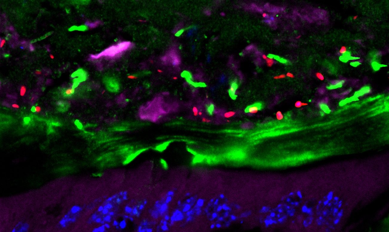
The colon viewed using a confocal microscope, showing the microbiota bacteria (in red), the intestinal mucous (green), the intestinal cells (purple) and their DNA (blue). Credits: Benoît Chassaing
In animals, a vaccine modifying the composition and function of the gut microbiota provides protection against the onset of chronic inflammatory bowel diseases and certain metabolic disorders, such as diabetes and obesity. This research was conducted by the team of Benoît Chassaing, Inserm researcher at Institut Cochin (Inserm/CNRS/Université de Paris), whose initial findings have been published in Nature Communications.
Chronic inflammatory bowel diseases, such as Crohn’s and ulcerative colitis, are linked to abnormalities of the gut microbiota in humans and in animals. The subjects concerned generally present reduced bacterial diversity in their intestinal flora along with excessive levels of bacteria that express a protein called flagellin, which favors their mobility. This enables them to penetrate the layer of mucous that covers the intestinal wall and is usually sterile. The purpose of this layer is to form a bacteria-resistant wall between the internal digestive tract and the rest of the body, thereby protecting it from the risk of inflammation linked to the presence of the billions of bacteria of the intestinal flora.
Previous research had already shown that antibodies are naturally found within this mucous layer, some of which are directed against flagellin. This means that the body spontaneously develops immune protection against flagellin, making it possible to control the presence of the bacteria that express it. With the aim of reducing the risk of chronic inflammation, Inserm researcher Benoit Chassaing and his colleagues had the idea of stimulating this anti-flagellin antibody production in order to reduce the presence of the bacteria that express flagellin in the gut microbiota.
As described in their study published in Nature Communications, the researchers administered flagellin to mice intraperitoneally, thereby inducing a marked increase in the anti-flagellin antibodies, particularly in the intestinal mucosa. The researchers then applied a protocol in order to induce chronic intestinal inflammation, whereupon they observed that immunizing against flagellin gave the animals significant protection from intestinal inflammation. In addition, detailed analysis of their microbiota and intestines revealed not just a reduction in the levels of bacteria that strongly express flagellin but also their absence in the intestinal mucosa, as opposed to the unvaccinated group.
Given that excess flagellin in the gut microbiota has also been linked to metabolic disorders such as diabetes and obesity, the researchers tested their vaccine strategy in mice exposed to a high-fat diet. Whereas the unvaccinated animals developed obesity, the vaccinated animals were protected.
“This vaccine strategy can be envisaged in humans, because such abnormalities of the microbiota have been observed in patients with inflammatory and metabolic diseases. With this in mind, we are currently working on a means of locally administering flagellin to the intestinal mucosa“, explains Chassaing. The researchers are considering, for example, the possibility of developing ingestible flagellin-filled nanoparticles. Finally, in addition to the preventive aspect, they now wish to test this vaccination in curative mode, in animals already presenting chronic inflammatory disease or metabolic disorders.

©Frédérique Koulikoff/Inserm
Developmental coordination disorder (DCD), also known as dyspraxia, is a commonly occurring condition that affects 5% of children on average. When a certain level of motor coordination is required, children with DCD do not perform as well as their peers in daily life activities (washing, dressing, eating, etc.) and at school (handwriting).
Inserm was commissioned by the French National Solidarity Fund for Autonomy (CNSA) to produce a Collective Expert Review of the scientific knowledge of DCD. Over a two-year period, Inserm’s Collective Expert Reviews Unit coordinated a dozen researchers and consulted ten specialists in order to review a scientific corpus of over 1400 international articles and issue recommendations on improving the diagnosis and management of children with DCD.
DCD varies markedly in its intensity and expression and is often associated with other neurodevelopmental disorders (of language, attention and learning), as well as a high risk of developing emotional, behavioral or anxiety disorders. These impact the child’s quality of life and participation in activities, particularly schoolwork. One of the key obstacles to their academic integration concerns handwriting.
To limit these impacts, the Expert Review states that spotting the signs is paramount when it comes to establishing rapid monitoring and individualized management for the child according to the severity of their condition, verbal competency, age, and any concomitant disorders.
The recommendations put forward by this Collective Expert Review can be summarized according to three main areas of focus.
The first consists of guaranteeing access to diagnosis for everyone, and as soon as possible following identification of the initial signs. In this respect, the Expert Review highlights the need to train professionals, emphasizing the importance of deepening the criteria and standardizing the tools needed to establish a diagnosis according to international standards.
Establishing such a diagnosis involves at the very least the participation of a doctor trained in developmental disorders, as well as that of a psychomotricity therapist or occupational therapist.
The second area of focus concerns what happens post-diagnosis, where there is no one standard intervention of unanimously recognized efficacy. Once diagnosed, it is therefore important to establish appropriate interventions that take into account the child’s profile, quality of life, and that of their family. The experts advise prescribing group sessions for the least affected children and one-on-one sessions for the others. They also recommend preferring interventions that focus on learning the skills needed for school and day-to-day life. Finally, these interventions must increase the involvement of the child’s family, teachers and anyone else interacting with them (sports coaches, etc.).
The objective of the third and final area of focus is to enable each child to get the right support at school. This requires that the school and its staff make the necessary additional arrangements for the child during their exams, in accordance with the French Disability Act (2005). This also involves raising the awareness of and training those involved in supervising and interacting with the child in everyday life, whether at home, school or during leisure

Adobe/Stock
Teams from Dijon-Bourgogne University Hospital, Inserm and CEA have recently established the results of the whole-genome analysis of severely ill neonates, hospitalized in neonatal ICUs – the time of which was slashed from the current 18-month average to just 38 days. Thanks to this rapid analysis, the resulting diagnosis of two-thirds of the infants enrolled in the project enabled one third of them to receive faster and more appropriate treatment. The deployment of this process over the coming years will make it possible to optimize the management of these patients.
Although a number of countries use whole-genome sequencing for diagnostic purposes and while France has recently launched its 2025 Genomic Medicine Plan (PFMG2025)1, its urgent use in a neonatal setting is not very widespread at present. Yet rapid genetic examination is a crucial factor when a diagnosis is required urgently – a common situation when it comes to rare diseases with early pediatric onset or rapid progression. Teams from Dijon-Bourgogne University Hospital, Inserm and CEA conducted a feasibility study of fast high-throughput genome sequencing before envisaging such a process for the future, in the framework of the PFMG2025.
In this pilot study, called Fastgenomics2, some thirty children hospitalized in neonatal ICUs across eight university hospitals belonging to the AnDDI-rares network3 underwent fast genome analysis in the previous nine months. High-throughput sequencing of the genomes of the children and their parents and a primary bioinformatics analysis were carried out on the sequencing platform of the French National Research Center for Human Genomics (CEA-CNRGH), in collaboration with the Very Large Computing Center (TGCC) of the CEA and the Computing Center of Université de Bourgogne (CCuB). The genomics data were interpreted by the TRANSLAD University Hospital Federation (FHU TRANSLAD), in close collaboration with the Inserm U1231 GAD (Genetics of Developmental Disorders) research team.
Mobilizing the teams meant that it was possible to obtain the analysis results within 49 days, with the most rapid turnaround being 38 days. This is particularly fast for a genetic analysis, given that despite the considerable advances made, the average time to obtain a genetic diagnosis in France continues to remain long: 1.5 years on average, and up to 5 years for 25% of patients. Rapid analysis of the genomes of these neonates made it possible to diagnose two thirds of them, with one third able to receive quicker and more appropriate treatment.
Such analysis has been made possible thanks to major advances in the high-throughput sequencing of the gene set. The new-generation high-throughput DNA sequencing technologies – which analyze a person’s entire genome – have emerged in recent years as a tool of choice in the study of rare diseases. These cutting-edge technologies are used at CNRGH and have already implicated numerous genes in numerous diseases. The FHU TRANSLAD team from Dijon-Bourgogne University Hospital was one of the first in France to demonstrate the value of exome sequencing (with the exome representing 1% of the total size of the genome) in the diagnosis of severe diseases with early pediatric onset, as well as developmental abnormalities and intellectual disability.
Diagnosing rare diseases in the neonatal period
Rare diseases (affecting fewer than 1 in 2,000 people) are a major public health problem because they represent around 8,000 conditions and affect more than 3 million people in France. Given that the majority of these diseases are of pediatric onset, they are responsible for 10% of deaths before 5 years of age. Up to 80% of these diseases are considered to have a genetic origin. Establishing a diagnosis brings multiple benefits to the patients and their families: it clarifies the cause and the prognosis, enables access to treatment or clinical trials, determines risk of recurrence, cuts out needless diagnostic tests, enables the management of known complications, facilitates the acquisition of specific financial and practical support, and gives them the possibility to forge links with other families dealing with the same challenges.
Obtaining a diagnosis is a major challenge in diseases with early pediatric onset and rapid progression, and whose genetic causes are highly heterogenous, such as epilepsy, metabolic diseases, cardiac diseases, musculoskeletal diseases and other polymalformative syndromes. The 3rd French National Plan for Rare Diseases (PNMR3) envisages reducing diagnostic delay to one year, given that it is responsible for “the potential worsening of the condition of patients, the delayed possibility for genetic counseling and the wastage of medical resources (due to multiple diagnostic consultations)“.
When it comes to severe neonatal diseases, obtaining rapid diagnosis is particularly important. Diagnosis, when accurate, makes it possible to modify how the patient is managed, whether in terms of treatment adaptation (such as in metabolic diseases or epilepsy), referral to specialists, dietary adjustments, additional examinations, reassessment of any need for surgery, or the consideration of these results when discussing the continuation of care.
1 In 2016, France launched its 2025 Plan for Genomic Medicine (PFMG2025). It aims to implement mass whole-genome sequencing for the diagnosis of rare diseases through the establishment of very high-throughput platforms, as well as pilot studies to define the prescribing conditions for such investigations.
2 Fastgenomics: French national pilot study prepared by the AnDDI-rares national healthcare network, FHU TRANSLAD and CEA-CNRGH, and supported by a financial donation from SANOFI-GENZYME.
3 AnDDI-rares: National rare diseases network dedicated to diseases with somatic and cognitive developmental abnormalities. http://anddi-rares.org

Des billes fluorescentes sont filmées à très haute vitesse, et analysées pour suivre leur trajectoire, pour comprendre l’influence des forces fluidiques sur l’arrêt d’une cellule tumorale sur un tapis de cellules endothéliales. ©Harlepp S. /Goetz J
Circulating tumor cells use the blood and lymphatic networks to disseminate within the body, forming metastases distant from the primary tumor. Inserm researcher Jacky Goetz and his Tumor Biomechanics team at the Molecular ImmunoRheumatology laboratory (Inserm/Université de Strasbourg) have helped to show that the flow properties of these biological fluids have a huge influence on the risk of developing metastases. They observed that the slowdown of blood flow where the arteries branch out gives cancer cells the opportunity to attach to and pass through the vessel wall, and then colonize the tissues. The team describes its work in a review article published in Nature Reviews Cancer.
Cancer cells largely exploit the biological fluids in order to disseminate within the body and form metastases in locations remote from the primary tumor. While some access the bloodstream directly, others do so first by escaping from the tumor via the interstitial fluid and lymphatic network to then colonize the lymph nodes before joining the bloodstream.
Jacky Goetz, Inserm Research Director of the Tumor Biomechanics team at the Molecular ImmunoRheumatology laboratory (Inserm/Université de Strasbourg), has been studying these mechanisms for a number of years. His research has been showcased in Nature Reviews Cancer.
The researchers were able to observe that the cancer cells circulate rapidly in the large arteries without the ability to stop. However, when the arterial diameter narrows and the blood network branches out, the blood flow slows markedly, giving the tumor cells the opportunity to attach to the vessel wall, pass through it — a phenomenon known as extravasation —, and then colonize the tissues. The team identified a certain number of “hotspots” for this extravasation, corresponding exactly to the most common metastatic sites in humans, there where the blood network is made up of many small capillaries: the brain, lungs and liver. This research was confirmed in collaboration with teams from Germany, in a cohort of 100 patients with brain metastases. Goetz’ team also discovered that it is possible in the zebrafish to change the location of the hotspots, and therefore that of the metastases, by changing the speed of the blood flow.
Also with the help of the zebrafish, they observed that metastatic formation was preceded by the release of tumor‑derived extracellular vesicles. These contain proteins, DNA, RNA, and appear to act as veritable scouts for the tumor cells, possibly to prepare their implantation. The research team showed that the behavior of these vesicles in the blood is similar to that of the tumor cells, and also depends on the force of the blood flow.
Finally, the researchers correlated the force of the flow with the action of two proteins located on the tumor cell surface, which can only act when the blood flow is slowed down. The first, CD44, acts like a brake by attaching to the vessel wall. The second, α5ß1 integrin, enables the cell to stop and to pass through the vessel wall and out of the blood circulation. In zebrafish and mice, the absence of α5ß1 integrin strongly slows metastatic growth.
“Overall, this research shows that if we are to prevent the development of metastases, we cannot just focus on the inherent properties of the tumor, its microenvironment or that of the metastases, we must also consider the role played by the biological fluids. Preventing the circulating cells from stopping or from attaching to the vessel wall could, for example, reduce this risk”, concludes Goetz.
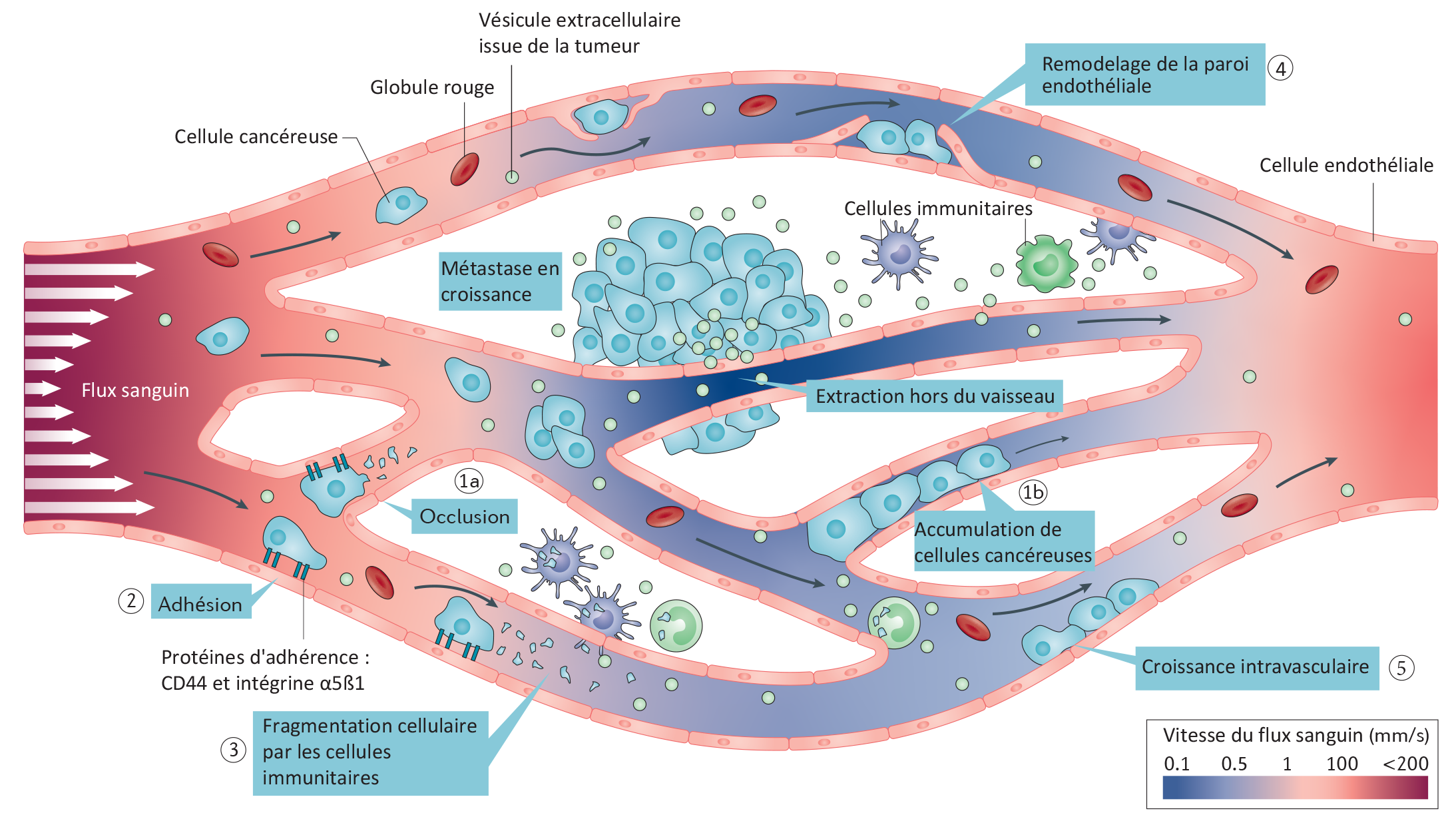
Circulation, adherence and extravasation of tumor cells and vesicles to form a metastasis according to changes in blood flow speed. ©Jacky Goetz / Nature Reviews Cancer, 2019
French | English |
Cellule cancéreuse | Cancer cell |
Globule rouge | Red blood cell |
Vésicule extracellulaire issue de la tumeur | Tumor‑derived extracellular vesicle |
Remodelage de la paroi endothéliale | Endothelial wall remodeling |
Cellule endothéliale | Endothelial cell |
Cellules immunitaires | Immune cells |
Métastases en croissance | Growing metastasis |
Flux sanguin | Blood flow |
Extraction hors du vaisseau | Extravasation |
Occlusion | Occlusion |
Accumulation de cellules cancéreuses | Cancer cell accumulation |
Adhésion | Adhesion |
Protéines d’adhérence : CD44 et intégrine α5β1 | Adherence proteins: CD44 and α5β1 integrin |
Fragmentation cellulaire par les cellules immunitaires | Cell fragmentation by the immune cells |

Adobe/Stock
Restoring sight to patients with age-related macular degeneration (AMD) or retinitis pigmentosa has become an increasingly likely prospect over recent years, with many researchers working to develop an artificial retina. In a new study, a team from Institut de la Vision (Inserm-CNRS- Sorbonne Université) led by Inserm researcher Serge Picaud has used animal models to show that a device made by the firm Pixium Vision could induce high-resolution visual perception. Their findings, published in Nature Biomedical Engineering, have paved the way for clinical trials in humans.
Age-related macular degeneration (AMD) is characterized by deterioration of the retina that can lead to central vision loss. This highly incapacitating condition is thought to affect up to 30% of people over the age of 75. For years, several groups of researchers have been working on the development of an artificial retina that could restore eyesight – not just to these patients but also to those with retinitis pigmentosa.
The retina is made up of light-sensitive cells (photoreceptors) whose purpose is to transform the light signals received by the eye into electrical signals sent to the brain. It is these cells that are destroyed during the course of the aforementioned diseases, the outcome of which can be blindness. The principle of the artificial retina – which is implanted beneath the patient’s own retina – is simple: to act as a substitute for these photoreceptors. Its electrodes stimulate the retinal neurons to send messages to the brain.
Two devices of this type, Argus II (Second sight, USA) and Retina Implant (AG, Germany), are already in widespread use. “Nevertheless, these companies are gradually withdrawing from the market, particularly because the results seen in patients have been insufficient for targeting the device at those with AMD. The patients managed to see light signals but those able to distinguish letters were very much in the minority”, emphasizes Serge Picaud.
Reinventing the device to increase its performance: this is the aim of the Picaud and his colleagues. Supported by Pixium Vision, their artificial retina is wireless and less complex, unlike previous devices. In addition, this implant introduces a local return of the current, thereby enabling better resolution of the images perceived by the eye. Finally, the image is projected onto the implant by infrared stimulation that activates photodiodes connected to electrodes, enabling more direct stimulation of the retinal neurons.
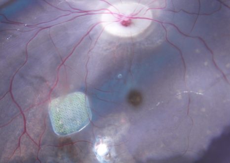
In a study published in Nature Biomedical Engineering, Picaud and his colleagues tested this device on non-human primates, demonstrating that it can restore significant visual acuity. In vitro tests showed first of all that each pixel activates different cells in the retina. This selectivity is expressed in the form of very high resolution, such that implanted animals can perceive the activation of just one of the implant’s pixels in a behavior test.
The high resolution of these implants led to the device being fitted in five French patients with AMD, for whom initial results show a gradual restoration of central vision. They are able to perceive light signals and some can even identify sequences of letters, with growing rapidity over time.
“The objective now is to conduct a Phase 3 trial in a larger group of patients with AMD. If the artificial retina works for them, there is no reason why it should not work in patients with retinitis pigmentosa – a disease also related to photoreceptor degeneration”, concludes Picaud.