Menu

Health Outcomes of the Nuclear Tests in French Polynesia

Polynésie française

Between 1966 and 1974, France performed 41 atmospheric nuclear tests in French Polynesia. © Unsplash

The health outcomes of the nuclear tests performed in French Polynesia in the 1970s have been the subject of epidemiological studies at Inserm for several years. Previous research published by a team from Inserm and Université Paris-Saclay with the Gustave-Roussy Institute had studied the relationship between the frequency of thyroid cancer in French Polynesia and the atmospheric nuclear tests performed by France. It had found a very probable slight increase in the thyroid cancer risk with the radiation dose received following the nuclear tests. In a new publication, the scientists confirm these findings and conduct a risk prediction analysis showing that these nuclear tests could be responsible for 2.3% of the thyroid cancer cases. The particularity of this study, whose results have been published in JAMA Network Open, lies in the access to the original internal reports by the radiation-protection services that relate to 41 atmospheric nuclear tests performed by France between 1966 and 1974 in French Polynesia, declassified by the French military in 2013.

Between 1966 and 1974, France conducted 41 atmospheric nuclear tests in Polynesia. For several years, research teams have attempted to evaluate the potential consequences of the radioactive fallout on population health. They have particularly focused on diseases that may be radiation-induced, such as cancer, cardiovascular diseases, and cataracts.

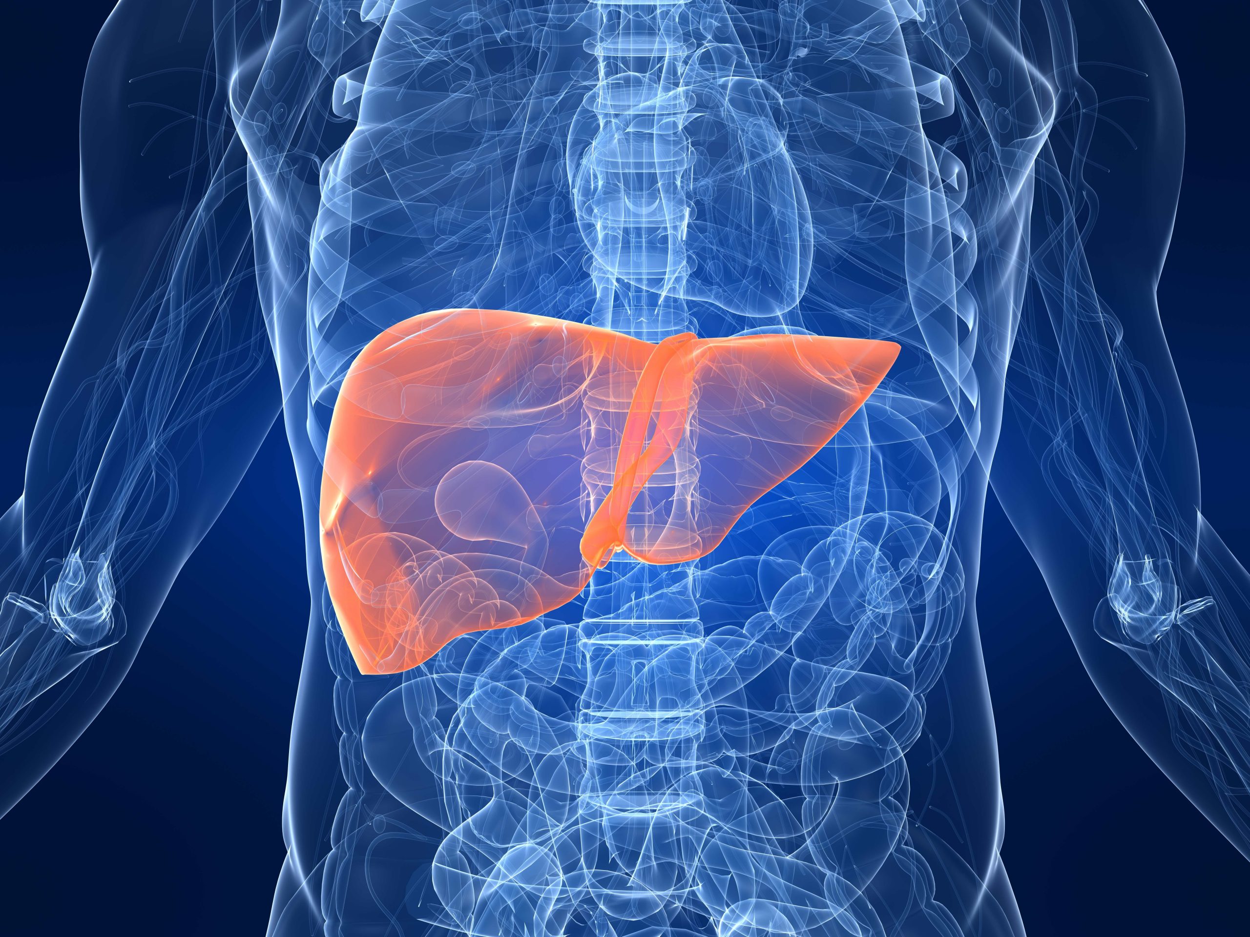
In an Inserm Collective Expert Review produced in 2021 (only available in French), researchers mentioned two epidemiological studies published on the subject, one of which concerns the risk of developing thyroid cancer. Due to the amount of radioactive iodine released in nuclear tests and its active absorption by the thyroid, this form of cancer is the most serious health risk for the population living near the nuclear test sites.

This is because when radioactive iodine binds to the thyroid instead of natural iodine, it exposes the gland to the radiation it emits. If high levels of this radioelement are found in the thyroid, this radiation may damage the DNA and lead to the development of cancerous tumors.

In a study published in 2010 in the British Journal of Cancer, researchers from Inserm and Université Paris-Saclay at the Center for Research in Epidemiology and Population Health (CESP) at the Gustave-Roussy Institute compared the radiation exposures of 229 Polynesians diagnosed with differentiated thyroid carcinoma between 1981 and 2003 with those of 373 “control” individuals who had not developed thyroid cancer in the same population. This case-control study conducted in the local population had shown a small increase in the risk of thyroid cancer in line with an increase in the dose received by the thyroid before the age of 15.

Although these findings are insufficient in providing a firm conclusion on the links between the fallout of the atmospheric nuclear tests and the occurrence of radiation-induced diseases in French Polynesia, they also did not rule out the existence of health impacts.

Continuing this research, the same team conducted a second epidemiological study, this time on 395 cases of thyroid cancer diagnosed between 1984 and 2016 in Polynesia and 555 controls from the general population. For the first time, they had access to the original radiation-protection service reports relating to the 41 atmospheric nuclear tests performed by France between 1966 and 1974 in French Polynesia, declassified by the French military in 2013.

Thanks to these declassified documents, meteorological data, and interviews with cases and controls (population movements, dietary habits, etc.), the researchers were able to simulate the radioactive cloud of each nuclear test and estimate the dose of radiation received to the thyroid of the study participants. This was on average 4.7 milligray for the cases and 4.6 milligray for the controls.

For all of the cancer cases (395 people), the scientists did not find a significant association between the dose of radiation received by the thyroid and the risk of thyroid cancer. However, the relationship was significant if the analysis only considered the invasive cancers requiring treatment, and excluded the noninvasive microcarcinomas that are common and usually do not need surgery.

Using these cross-referenced data (population movements, dietary habits and rates of thyroid cancer incidence), the researchers also made a risk prediction and showed that, based on current risk models, the nuclear tests performed by France could be responsible for a total of 2.3% of all thyroid cancer cases (confidence interval between 0.6 and 7.7%[1]).

“These findings are consistent with our previous conclusions and confirm that the nuclear tests are most likely responsible for a small increase in the incidence of thyroid cancers in French Polynesia. However, these findings should be considered with caution as the estimated radiation doses received by the thyroid over 50 years ago are bound to be inaccurate,” explains Florent de Vathaire, Inserm researcher at Gustave-Roussy, and first author of the study.

The scientists are continuing their research with the aim of adding to their analysis the evaluation of the genetic susceptibilities of the populations (hereditary factors and genetic combination specific to each individual) that could affect the development of thyroid cancers in the region.


[1] The confidence interval defines a minimum and maximum value between which, for a given risk of error, the exact value of the entire population is situated.

AI: A New GPT-Inspired French Algorithm Improves Trauma Surveillance

hôpital urgences

Scientists have developed an algorithm to better understand the traumas that represent one third of emergency department visits. © Unsplash

In France, one third of emergency department (ED) visits are the result of trauma. In order to better understand the mechanisms and improve the management of trauma, researchers from Inserm and Université de Bordeaux at the Bordeaux Population Health Research Center, with teams from Bordeaux University Hospital, have developed an algorithm capable of classifying ED visits for trauma through the artificial intelligence (GPT) analysis of clinical reports. This project, called TARPON[1], which has achieved performance accuracy levels of 97%, is described in the Journal of Medical Internet Research Artificial Intelligence. With these findings, the imminent establishment of a national trauma observatory can be envisaged.

Trauma accounts for 9% of mortality in France and often affects populations of young people. More than one third of the 21 million annual emergency department (ED) visits are for trauma. It is therefore a major public health problem that represents a significant health, social and economic burden for which the scientists are working to provide solutions.

The idea for the TARPON project conducted by researchers from Inserm and Université de Bordeaux with Bordeaux University Hospital is based on the observation that the medical staff write a report for each ED visit. These reports represent a wealth of information: symptoms, patient condition, as well as many details on the circumstances in which the trauma occurred.

However, these data remain untapped, with few statistics available on victims of daily life accidents, violence or suicide attempts. For road accidents, an observatory exists, but it is only complete for mortality and the majority of accidents related to cycle, pedestrian or scooter travel are not included. Therefore, the analysis of anonymized information from ED reports would make it possible to constitute the basis for a virtually-exhaustive trauma surveillance system.

These reports are unstructured texts written using a mixture of everyday, medical and technical terms, as well as abbreviations. In order to extract the relevant information without having to read them all, the research teams have developed an automatic language-processing technique based on artificial neural networks. They have adapted the GPT artificial intelligence model and trained it with a sample of over 500,000 reports from adult emergencies at Bordeaux University Hospital[2]. The result is a French-speaking clinical language-processing tool that is compliant with GDPR.

With the support of the Health Data Hub, Bpifrance, the Nouvelle Aquitaine region, the French medicines agency (ANSM) and the Road safety delegation (DSR), the researchers were able to fund the purchase of a powerful server dedicated to artificial intelligence and installed in the hospital itself. This server has been used to implement the algorithm developed by the scientists, in order to automatically classify trauma by type – with surprising precision.

The method developed by the researchers makes it possible to correctly classify 97% of the reports (compared with 86% using the former methods), as described by the researchers in their scientific article. Thanks to this first step, it will be possible to begin studying this data on the Health Data Hub’s technology platform by this summer.

These findings pave the way for the establishment of a national trauma surveillance system, as well as epidemiological analyses concerning, for example, the impact of medication use on the risk of accidents. Research which should therefore provide new insight into major public health issues. In the immediate future, TARPON will be extended to include fifteen EDs across the entire French territory.


[1] TARPON: Automatic processing of ED summaries to create a national observatory

[2] This research meets the obligations of the General Data Protection Regulation (GDPR).

Long COVID: The Persistence of SARS-CoV-2 in the Mucous Membranes May Be A Factor

SARS-CoV-2

This transmission electron microscope image shows SARS-CoV-2, isolated from a patient in the U.S., emerging from the surface of cells cultivated in a laboratory. © National Institute of Allergy and Infectious Diseases-Rocky Mountain Laboratories, NIH. Public domain.

Several months after infection with SARS-CoV-2, some patients still have symptoms: a phenomenon commonly referred to as “long COVID”. In new research, teams from Inserm and Université Paris Cité[1], in collaboration with the University of Minho in Braga (Portugal), have shown that this could be explained biologically by abnormalities of the immune system associated with the persistence of the virus in the mucous membranes. These findings, published in Nature Communication, could in the longer term pave the way for a diagnostic tool for long COVID.

Despite the fact that various studies consider long COVID to affect between 10 and 30% of people infected with SARS-CoV-2, its diagnosis and treatment remain difficult. The team of Inserm researcher Jérôme Estaquier, in collaboration with that of Ricardo Silvestre at University of Minho in Portugal, is conducting research to explain this phenomenon from the biological point of view.

At the present time, few biological criteria, apart from the persistence of symptoms beyond three months after the acute infection, enable its diagnosis. Once a patient is not fully recovered after this period, they are considered to have long COVID. Without a more reliable means of diagnosis, it is difficult to offer the appropriate care.

In order to better understand long COVID and find diagnostic markers, the researchers studied the immune systems of 164 people six months after they were infected.  They analysed the blood samples of 127 people, half of whom with long COVID (fatigue, shortness of breath, cough, muscle or chest pain, anxiety, etc.) and those of 37 controls who had not been infected.

The researchers focused on certain immune cells, namely the T cells (including CD8 cells) involved in eliminating the virus, and the SARS-CoV-2 antibodies. In addition, they had blood samples that were taken during the acute phase of the disease for 72 of these patients, enabling them to retrospectively compare the level of inflammation at the early stage in those who went on to develop long COVID or not.


Several immune markers identified

The researchers identified a number of blood markers present six months after infection in 70-80% of the subjects with long COVID, while those same markers were rare in the subjects who had not developed it.

In particular, the teams showed that a CD8 cell subtype expressing the inflammatory protein granzyme A is present in excess, whereas another CD8 subtype, this time expressing integrin b7, is present in small quantities. Yet it is the latter subpopulation that is essential for controlling viruses in the mucous membranes. In addition, virus-specific IgA antibodies are also present in excess whereas they should be rapidly eliminated if the virus is absent. These observations suggest the persistence of the virus in the body and especially in the mucous membranes.

The researchers hypothesise that SARS-CoV-2 could make itself at home in the intestinal mucosa as it is more “permissive” in immune terms than the rest of the body, insofar as the virus has to tolerate the bacterial flora. Other viruses, such as HIV, also use this escape strategy. Initially present in the lung mucosa, SARS-Cov-2 could therefore descend to the intestine and persist there without the immune system being able to eliminate it completely.

In the final stage of the study, when evaluating the initial level of inflammation during the acute phase, the scientists observed an association between an inflammatory response characterized particularly by very high levels of interferon IP-10 or interleukin IL-6 and the risk of going on to develop long COVID.

“This confirms clinical observations that the initial severity of COVID is associated with a higher risk of developing long COVID,” specify the researchers. “One hypothesis is that people with more exacerbated early immunodeficiency develop more severe initial forms of COVID-19 and fail to effectively eliminate the virus that passes into the intestinal mucosa, where it settles for a long time. The immune system kind of ends up tolerating it at the cost of persistent symptoms of varying intensity and nature,” explains Estaquier.

The objective is now to validate these findings in new cohorts to determine whether some of these markers could be used as a diagnostic tool.

“If measuring IgA some time after the acute phase and potentially CD8 b7 cells was able to diagnose long COVID, doctors could make an objective diagnosis. Then we could think about therapeutic targets based on this research,” concludes Estaquier.


[1]This research in France was supported by the Fondation pour la Recherche Médicale, the French National Research Agency (ANR), and ANRS | Emerging Infectious Diseases.

Inflammation and cancer: identifying the role of copper paves the way for new therapeutic applications

équipe CurieThe research team developed a “drug prototype” capable of mitigating both the mechanisms of inflammation and the processes potentially involved in metastatic spread. © Institut Curie / BELONCLE Frank

For the first time, researchers from Institut Curie, the CNRS and Inserm have uncovered a previously unknown chain of biochemical reactions. This chain involves copper and leads to metabolic and epigenetic alterations[1] that activate inflammation and tumorigenesis. But there is more; the research team developed a “drug prototype” capable of mitigating both the mechanisms of inflammation and the processes potentially involved in metastatic spread. Published in the journal Nature on April 26, 2023, these results provide hope for new therapeutic opportunities to control inflammation and cancer.

Inflammation is a complex biological process that can eradicate pathogens and promotes repair of damaged tissues. However, deregulation of the immune system can lead to uncontrolled inflammation and produce lesions instead. Inflammation is also involved in cancer. The molecular mechanisms underlying inflammation are not fully understood, and so developing new drugs represents a significant challenge.

As far back as 2020, Dr. Raphaël Rodriguez, CNRS research director and head of the Chemical Biology team at Institut Curie (Equipe Labellisé Ligue Contre le Cancer) at the Cellular and Chemical Biology laboratory (Institut Curie/CNRS/Inserm), had shed new light on a membrane receptor called CD44, which marks immune responses, inflammation and cancer progression. Dr. Rodriquez and his team showed that CD44 helped import iron into cell[2], triggering a series of reactions leading to activation of genes involved in the metastatic process.

“This is a cell plasticity phenomenon we continued to study, investigating other metals potentially internalized by CD44, notably copper,” he explains.


Copper causing epigenetic alterations

Along with his colleagues[3], Dr. Rodriguez has now reached a new milestone.

The research team managed to identify a signaling pathway involving copper and leading to the expression of pro-inflammatory genes in macrophages, the cells present in all tissues and playing an important role in innate immunity.

Once internalized in macrophages, copper enters into the mitochondria (the organelle responsible for cell respiration and energy production), where it catalyzes the oxidation of NADH into NAD+  (nicotinamide adenine dinucleotide, a molecule needed for the activity of certain enzymes). The increase of NAD+ in cells enables the activity of certain enzymes involved in the production of metabolites essential for epigenetic regulation. These metabolites thus, contribute to the activation of genes involved in inflammation.


Inflammation and cancer: shared molecular mechanisms

The scientists did not stop there, they also designed molecules able to bind to copper, inspired from the structure of metformin.[4] By testing these new molecules on models of acute inflammation, they found that a synthetic dimer of metformin, LCC-12 (also termed Supformin), reduced activation of macrophages and attenuated inflammation.

“Our work has enabled us to develop a drug prototype that inactivates copper chemistry in the cell’s metabolic machinery, thus blocking expression of the genes involved in inflammation”, explains Dr. Rodriguez.

To finish, they applied this therapeutic strategy to cancer cell models engaged in an epithelial-mesenchymal transition[5]. Here again, Supformin blocked the cellular mechanism and thus the cell transformation.

“The genes activated in cancer cells are not the same as those expressed in immune cells, but the chain reaction leading to epigenetic alterations is identical”, explains Dr. Rodriguez.

These results thus reveal the role of copper in cancer cells and their ability to adopt a metastatic nature.

Dr. Raphaël Rodriguez concludes: “Our study reveals that the inflammatory and cancer processes depend on similar molecular mechanisms and could therefore in the future benefit from similar innovative therapies, such as those tested with Supformin.”

The explanations of Dr. Raphaël Rodriguez in video :


[1]Epigenetics is the study of the mechanisms at play in gene regulation, which is essential to the action of cells and to maintaining their identity. Unlike genetic mutations, which are permanent, epigenetic modifications on DNA or histones are reversible.

[2] Read the press release “Cancer: a new mechanism that regulates cell activity involving iron”: https://curie.fr/sites/default/files/medias/documents/2020-08/CPCNRS-CD44ferCancer-FR-emb.pdf

[3] This study was conducted at Institut Curie, in the Cellular and Chemical Biology unit (Institut Curie, CNRS, Inserm), in collaboration with UVSQ, Raymond Poincaré hospital (AP-HP), Gustave Roussy hospital, the Institut de chimie moléculaire et des matériaux d’Orsay (CNRS/University Paris-Saclay), the Multimodal Imaging Center (CNRS/Institut Curie/Inserm/University Paris-Saclay), the Center for Infection and Immunity of Lille (CNRS/Inserm/Institut Pasteur de Lille/CHU of Lille/University of Lille), Institute of Pharmacology and Structural Biology (CNRS/University of Toulouse III) along with British and Australian researchers.

[4]Metformin is a treatment used for Type-2 diabetes, and is able to form a bimolecular complex with copper.

[5] Epithelial-mesenchymal transition is the first step in enabling cancer cells to metastasize.

Hypertension: A Mixture of Air Pollutants Could Cause Repeated High Blood Pressure Peaks

Pollution de l'airAir pollution is an acknowledged environmental factor in high blood pressure. © Adobe Stock

Air pollution is an acknowledged environmental factor in high blood pressure. It consists of a mixture of particles and gases whose combined effects on human health are not yet well known. A team from Inserm and Sorbonne Université, assisted by international collaborators, used continuous monitoring to study the daily life impact of a mixture of five air pollutants on the blood pressure of 221 MobiliSense[1] study participants in the Greater Paris area. With two models – one taking into account variations in ambient air pollutant levels, the other variations in the amounts inhaled – the researchers observed an association with acute repeated blood pressure increases. This study, published in Environmental Research, paves the way for a better understanding of the link between air pollution and hypertension.

Hypertension is a chronic disease that affects one in three adults. Linked to abnormally high pressure of the blood in the blood vessels, it can lead to cardiovascular, cerebrovascular, and even neurodegenerative complications.

Previous studies have shown that some air pollution molecules affect blood pressure and could therefore promote hypertension. However, in everyday life, the air pollution to which people are exposed generally consists of mixtures of air pollutants rather than a single component in isolation – mixtures that had been little researched until now.

An international team led by Basile Chaix, Inserm Research Director at the Pierre Louis Institute of Epidemiology and Public Health (Inserm/Sorbonne Université), wanted to characterize the effects on blood pressure of the daily-life exposure to a mixture of five air pollutants – black carbon, nitrogen dioxide (NO2), nitrogen oxide (NO), carbon monoxide (CO), and ozone (O3) – in 221 participants from the MobiliSense study.

In order to study the effects of this complex mixture, the research team developed new monitoring methods and used innovative measuring equipment. Each participant wore an ambulatory blood pressure measurement device[2], two portable sensors to continuously measure pollutants in the ambient air near the breathing zone, a GPS tracker to record mobility, and an accelerometer to measure physical activity and thus estimate the ventilation rate[3] (the volume of air inhaled or exhaled per unit of time). The measurements were taken over one day in the lives of the participants.

Their blood pressure was measured every 30 minutes in order to observe as closely as possible the time between variations in ambient air pollutant levels, the estimated amount of pollutants inhaled, and their potential impact on blood pressure.

Learn more about blood pressure and hypertension

Blood pressure results from the ejection of blood from the heart into the blood vessels and consists of the pressure it exerts on the vessel walls. It is characterized by two values:

  • the upper value or systole (systolic blood pressure), measured when the heart contracts. This pumps blood through the aorta to the peripheral arteries;
  • the lower value or diastole (diastolic blood pressure), measured when the heart relaxes. This enables the cardiac ventricles to receive the blood that enters the atria through the vena cava- and pulmonary veins.

Hypertension is when the resting systolic value is above 140 mmHg and/or the resting diastolic value is above 90 mmHg.

The researchers observed that when the levels of all the pollutants in the mixture increased within the 5 minutes prior to measuring blood pressure, there was an increase in systolic pressure (see box). A similar association was also found between an increase in the amount of pollutants inhaled within the 5 minutes prior to measuring blood pressure (related to an increase in the concentrations measured and/or physical activity and therefore in the ventilation rate) and an increase in systolic pressure.

“We chose to consider short windows of exposure (5 min, 15 min, 30 min, 1 hour) to study the time between exposure to pollution and blood pressure response, specifies Chaix. Here we see that the association is weaker when the exposure is observed over windows longer than 5 minutes, which indicates the immediacy of the blood pressure elevation in response to increased levels of air pollutants in the studied mixture,” adds the researcher. He continues: “These repeated increases in blood pressure linked to exposure to air pollutants in urban areas when out and about could contribute to a chronic rise in blood pressure, month after month, year after year. “

Another observation from these two models is that – when we consider the individual contribution of each pollutant to the effect of the mixture on blood pressure – ozone and black carbon present as being the greatest contributors to its increase.

With few existing studies having used these measurement and modeling methods to study mixtures rather than isolated pollutants, the research team clarifies that it does not currently have the possibility to compare its findings with other research, which means that they should be interpreted with caution.

However, should these findings be confirmed, it may be possible to extrapolate them to the populations of other major European cities whose pollution levels are similar to those of Greater Paris.

As for the implications of the study, Chaix concludes: “Our findings call for air pollution to be considered as a cause of hypertension and for the rollout of public policies aimed at reducing exposure to this pollution in everyday life – and particularly that of road traffic in the heart of our towns and cities. “

Next on the agenda is for the team to explore the physiological mechanisms and causes behind the associations observed in this study.


[1]The MobiliSense study is conducted on inhabitants of the Greater Paris area and aims to explore the effects of air and noise pollution exposure on cardiovascular and respiratory health.

[2]Unlike the blood pressure measurements taken when the individual is in a resting state, ambulatory blood pressure measurements are taken throughout the day and during the course of the individual’s activities using a wearable device.

[3]In previous studies, the team had shown that the amount of polluted air inhaled was not directly proportional to the levels of pollutants in the breathing zone but was also dependent on the ventilation rate, which varies with the intensity of physical activity. The ventilation rate was therefore estimated for each participant based on the accelerometer measurements.

A New Target to Regress Liver Fibrosis

foie

Cirrhosis is the final stage of fibrosis associated with chronic liver diseases. It affects 200,000 to 500,000 individuals in France and is responsible for 170,000 deaths per year in Europe. © Adobe Stock

Chronic liver diseases are characterized by persistent inflammation that contributes to their progression to more severe stages. They may progress to fibrosis and cirrhosis, and then require liver transplantation. Therefore, limiting the progression of fibrosis and bringing about its regression is a major therapeutic challenge. Several studies have recently suggested that one interesting approach could be to target the inflammatory response. In new research, scientists from Inserm and Université Paris-Cité at the Inflammation Research Center (CRI), in collaboration with teams from the Paris Public Hospitals Group (AP-HP)[1], have shown that blocking the activation of a particular population of T cells, the Mucosal-Associated Invariant T (MAIT) cells, could halt the progression of fibrosis and even bring about its regression. Targeting the MAIT cells involved in the inflammation seen in fibrosis and cirrhosis would therefore open up new avenues for better therapeutic care. This study has been published in Nature Communications.

Mainly alcoholic, viral, or metabolic in origin, cirrhosis is the last stage of fibrosis associated with chronic liver diseases. Cirrhosis affects between 200 000 and 500 000 people in France and is responsible for 170 000 deaths per year in Europe. Ultimately, it leads to liver failure for which the only cure is transplantation.

One characteristic of chronic liver diseases is persistent inflammation that contributes to their progression to more severe stages, particularly fibrosis and its final stage, cirrhosis. A better understanding of how to regulate this inflammatory response therefore constitutes a major challenge in developing new strategies to treat these diseases.

In 2018, the team of Inserm researcher Sophie Lotersztajn had shown that a population of T cells called MAIT promoted the progression of liver fibrosis. These immune cells are particularly abundant in the human liver and are involved in the inflammatory processes associated with fibrosis.

In their new study, the scientist and her colleagues worked on the basis of liver samples from cirrhotic patients as well as from mouse models of the disease.

They showed that the administration of a pharmacological agent to inhibit the activation of MAIT cells can limit liver inflammation and not only halt the progression of fibrosis, but also regress it.

It is now well known that other immune cells, such as the macrophages, play a central role in the progression and regression of fibrosis. Here, analysis of the mechanisms involved showed that blocking the activation of MAIT cells interrupts their dialog with profibrogenic macrophages, i.e. accelerators of fibrosis, and promotes the emergence of fibrosis-resolutive macrophages.

cellules MAIT

In the first image, the MAIT cells (in red, shown using arrows) are located near fibrogenic cells (in green) in the liver of cirrhotic patients. In the second image, the MAIT cells (in red) are activated (activation marker in green) in the liver of cirrhotic patients. This activation is blocked in the presence of a MAIT cell inhibitor. Credits: Sophie Lotersztajn


“Cirrhosis is a major public health problem. Even though the only treatment is liver transplantation, our research opens up other therapeutic avenues for targeting inflammation and halting or even regressing fibrosis. The research now needs to be continued, particularly in order to develop drug candidates that target and inhibit the MAIT cells,” concludes Lotersztajn.


[1] This research is the result of a collaboration between the team of Drs. Sophie Lotersztajn and Hélène Gilgenkrantz (Inflammation Research Center (CRI) Inserm-Université Paris Cité), the team of Dr. Valérie Paradis at the CRI (also Department of Pathology, Beaujon Hospital), Department of Anesthesiology and Critical Care (Prof. Emmanuel Weiss), the teams of Institut Curie (Dr. Olivier Lantz), Institut St Louis (Dr. Michèle Goodhardt) and Génosplice (Dr. Pierre de la Grange)

Restoring Vision Through a New Brain-Machine Interface: Sonogenetic Therapy

 thérapie sonogénétique

Sonogenetic therapy consists of genetically modifying certain neurons in order to activate them remotely by ultrasound. © Alexandre Dizeux/Physics for Medicine Paris

Restore vision using a combination of ultrasound and genetics? This is the goal of an international team led by Inserm research directors Mickael Tanter and Serge Picaud from Paris’ Physics for Medicine unit (ESPCI Paris/PSL Université/Inserm/CNRS) and Vision Institute (Sorbonne Université/Inserm/CNRS), respectively, in partnership with the Institute of Molecular and Clinical Ophthalmology in Basel. In a new study, they provide proof of concept of this so-called “sonogenetic” therapy in animals. This consists of genetically modifying certain neurons in order to activate them remotely by ultrasound. The results show that when used on rodent neurons sonogenetics can induce a behavioral response associated with light perception. This discovery makes it possible to envisage, in the longer term, an application in blind people with optic nerve atrophy. The study has been published in Nature Nanotechnology.

Sonogenetic therapy consists of genetically modifying certain neurons in order to activate them remotely by ultrasound. This technology had previously been tested in culture and the first in vivo tests did not enable the researchers to become aware of its therapeutic potential linked to its very high spatiotemporal resolution. The genetic modification in question consists of introducing the genetic code of a mechanosensitive ion channel into the cells. The neurons that express this channel can then be remotely activated by low-intensity ultrasound applied to the surface of the brain without the need for contact (see diagram below).

Ultrasound waves can access tissue deep down, such as in the visual cortex – even from the surface of the dura mater[1] that surrounds the brain – and target very specific areas. It is these waves that form the basis for high-resolution brain imaging or ultrasound technologies. In this case, they enable highly selective activation, because only those neurons carrying the mechanosensitive channel and targeted by the ultrasound beam are stimulated.

In a recent study, a team of researchers led by Inserm research directors Mickael Tanter and Serge Picaud tested the efficacy of this sonogenetic therapy in animals. The aim of this research is to provide a solution to restore vision to patients having lost the connection between their eyes and brain due to conditions such as glaucoma, diabetic retinopathy, or hereditary or dietary optic neuropathies.

Their findings show that sonogenetic stimulation of the visual cortex induces a behavioral response associated with light perception. The animal learns an associative behavior in which it seeks to drink as soon as it perceives light. Ultrasound stimulation of its visual cortex induces the same reflex, but only if the neurons in the cortex express the mechanosensitive channel. The animal’s behavior suggests that sonogenetic stimulation of its cortex induced the light perception at the origin of the behavioral reflex.

The study showed that therapy works on different types of neurons, whether in the retina or visual cortex of the rodents, thereby demonstrating the universal nature of this approach.

By converting the images of our environment into the form of a coded ultrasound wave to directly stimulate the visual cortex – at rates of several tens of images per second – sonogenetic therapy appears to offer genuine hope for restoring vision to patients who have lost optic nerve function.

More generally, this sonogenetic stimulation approach offers innovative technology for interrogating brain function. Unlike current neuron stimulators or prostheses, its “non-contact” and selective cell type functioning represents a major innovation in relation to electrode devices.

“This sonogenetic therapy to ultimately restore the vision of blind people illustrates the power of a multidisciplinary project and a beautiful human adventure between a retinal biologist like Serge Picaud, and myself, a wave physicist for medicine,” declares Tanter, Inserm research director at the Physics for Medicine unit in Paris (ESPCI Paris/PSL Université/Inserm/CNRS).

“The development of a clinical trial of sonogenetic therapy still has many steps to go through to validate its efficacy and safety. If the results are confirmed, this therapy could succeed in restoring patients’ vision in a stable and safe manner,” concludes Picaud, Inserm research director and director of the Vision Institute (Sorbonne Université/Inserm/CNRS).

[1] Outermost layer of the meninges that protect the brain

Towards a Better Understanding of the Role of Male Hormones in Women with Multiple Sclerosis

sclérose en plaque

The image shows the brain region where demyelination is typically induced. The red cells correspond to all of the microglial cells with inflammatory properties when demyelination has just occurred. If the spontaneous regeneration process of myelin is effective, their inflammatory nature then diminishes in favor of an anti-inflammatory and pro-regenerative nature. The green cells are a subpopulation of these microglial cells that become anti-inflammatory.© Zahaf et al.

Multiple sclerosis (MS), an autoimmune disease for which there is no cure as yet, affects three women for every one man. Faced with this observation, scientists are studying the role of the sex hormones in order to better understand the differences between men and women in relation to the disease and its progression. A team led by Inserm researcher Elisabeth Traiffort in Unit U1195 “Diseases and hormones of the nervous system” (Inserm/Université Paris-Saclay) has recently shown that although male hormones – androgens – are present at very low levels in women, their presence is necessary to regenerate the myelin sheath which is destroyed in MS. These findings have been published in Nature Communications.

Multiple sclerosis (MS) is an autoimmune disease. In its most common form, relapsing-remitting MS[1], which accounts for 85% of cases, it manifests as inflammatory flares during which the patient’s immune cells attack and destroy the myelin in the central nervous system (see box). This phenomenon causes lesions that lead to motor, sensory, and visual disorders.

These symptoms are reversible at the beginning of the disease, thanks to the spontaneous repair of the destroyed myelin. However, over time, symptoms gradually become irreversible, reflecting the failure of the repair process, marking entry to the progressive phase of the disease. While current treatments reduce the frequency and severity of the inflammatory flares, thereby improving patient quality of life, they remain ineffective against the progression of the disease.


What is Myelin?

An axon is the single extension through which a neuron communicates with its target cell. Myelin is a biological membrane that wraps around axons to form a sheath. The myelin sheath serves to isolate and protect the nerve fibers. It also acts as an accelerator to the propagation speed of the nerve messages that transport information along the axon. Demyelination is the destruction of the myelin sheath following a nervous system attack.


Current research aims to gain a better understanding of the mechanisms of the disease and develop new therapeutic avenues that would prevent patients from entering the progressive phase, particularly by promoting the regeneration of myelin. Inserm researcher Elisabeth Traiffort and her team at the “Diseases and hormones of the nervous system” unit (Inserm/Université Paris-Saclay) are working for example to better understand the differences between women and men in MS, in order to determine whether it could be beneficial or even necessary to adapt therapeutic management to the patient’s sex. Remember that the disease is predominantly female, since three out of every four patients are women.

Investigating the Role of Androgens in Women

While the hormonal environments of men and women are very different, they cannot be restricted to high androgen levels in men and fluctuating levels of estrogen and progesterone in women. We know that men also produce estrogens, particularly in the brain where an enzyme is found that converts androgens into estrogens, while women produce small amounts of androgens. It was on this last aspect that Inserm researcher Traiffort and her colleagues focused their latest study.

Research has already shown that androgens protect neurons in the central nervous system of men with relapsing-remitting forms of MS and induce the regeneration of myelin sheaths destroyed in males, in animal models of the disease. But what is the role of the small amounts of androgens that are also found in the central nervous system of women? Can these androgens present at much lower levels than in men also impact the progression of the disease in female patients?

The scientists worked with animal models of the disease and also on tissues from patients supplied by organ donation banks. They first showed that in regions where myelin is destroyed, the AR receptor that enables androgens to transmit their signal is strongly expressed in the nervous tissue of women with MS, as it is in the female mouse models of the disease. This observation would suggest the existence of an essential role of androgens in the demyelinated tissue of the affected women.

In accordance with this hypothesis, the scientists have shown that despite only being present in small quantities in female mice, androgens still have a beneficial effect on the optimal regeneration of the destroyed myelin. Indeed, when the signals transmitted by the androgens are completely absent, this regeneration is greatly reduced.

Finally, other findings in animals and in human tissues suggest that these same androgens also have major anti-inflammatory effects on demyelinated nerve tissue in females unlike what is observed in males. The beneficial effects of androgens in women with MS could therefore also be linked to a decrease in the level of local inflammation, in areas where myelin is destroyed. This finding is interesting if we consider the current hypothesis that disease progression could be closely associated with the inflammatory cells residing in nervous tissue.

“While the low levels of androgens detected in women could point to a minor role for these hormones in the disease, we show that this is not the case. Our data suggest the use of appropriate doses of androgens in women with MS and the need to consider the patient’s sex when treating this and in all likelihood other conditions involving the destruction of myelin in the central nervous system,” concludes Traiffort.


This research was carried out with the support of the ARSEP Foundation.

[1] There are two active forms of the disease. The most common is the relapsing-remitting form, which accounts for 85% of MS cases at diagnosis. It is characterized by flares, in which symptoms appear within a few hours or days, often associated with the extreme and unusual fatigue suggestive of the diagnosis. Then the symptoms disappear completely or partially within a few weeks. The “primary” progressive form accounts for only 15% of cases. It is characterized by the slow and continuous worsening of the neurological symptoms, with no flares or remission.

COVID-19: Infection-Vaccination is the Most Protective Combination Against Reinfection

SARS-CoV-2

Electron microscopy visualization of a cell infected with SARS-CoV-2. © Philippe Roingeard, Anne Bull-Maurer, Sonia Georgeault/Inserm.licence CC-BY-NC 4.0 international

A large part of the population has developed immunity against SARS-CoV-2 following infection, vaccination – or both. In addition, some infected patients enjoy “hybrid” immunity when they are vaccinated following their infectious episode. Scientists from Inserm, CNRS, Université Claude-Bernard Lyon 1 and ENS de Lyon at the International Center for Research on Infectious Diseases (CIRI) seek to characterize the imprint left by SARS-CoV-2 exposure through vaccination or the combination of the two events on immune memory. The objective? Deepen their understanding of the mechanisms of immune response to the virus in order to improve patient care and optimize vaccine strategies. In a new study, the scientists compared the immune memory of convalescent individuals, whether or not vaccinated against SARS-CoV-2, with that induced by vaccination in individuals having never been infected with the virus. Their findings show that those who are vaccinated following an infection are the best protected from SARS-CoV-2 reinfection. The full article has been published in Science Translational Medicine.

Our body keeps a memory of the infections it has already fought in order to protect us against possible reinfection. The efficacy of vaccination is based on a strategy of simulating an infection to induce protective immunity, i.e. the production of memory cells “trained” in recognizing the pathogen, which can protect the body in the event of infection.

In the case of COVID-19, immunity is conferred either by infection (natural immunity) or by vaccination (vaccine immunity). Some people also benefit from “hybrid” immunity since they have been vaccinated following an infectious episode.

In order to better understand the precise mechanisms of the immune response to SARS-CoV-2, researchers from Inserm, CNRS, Université Claude-Bernard Lyon 1 and ENS de Lyon compared different immune memory parameters from blood samples collected from individuals with natural immunity, vaccine immunity, or hybrid immunity to SARS-CoV-2.

They focused on the adaptive immune response and more specifically on the so-called “humoral” response (see box below).

More About Adaptive Immune Response

The adaptive immune response is established a few days after contact with the pathogen, unlike the innate immune response, which is immediate.

There are two main categories of adaptive immune response.

Cell responses, which are based on the recognition and destruction of the infected cells by the cytotoxic (killer) T cells.

Humoral responses, which are based on the production of antibodies by the B cells. These antibodies recognize the pathogen and neutralize it to prevent it from infecting the target cells.

Humoral immune memory has two compartments:

– serological memory, estimated by the levels of circulating antibodies produced by the memory plasma cells. These antibodies create a barrier that can prevent reinfection.

– cell memory, consisting of memory B cells that do not secrete antibodies but which can differentiate rapidly and massively into plasma cells to generate a new amplified antibody production. These memory B cells are called upon when the barrier of antibodies produced by the memory plasma cells is deficient or insufficient.

The findings show that six months after the last vaccine injection or after infection, people with hybrid immunity are those with the highest levels of neutralizing antibodies in the blood.

In addition to this quantitative variation in serological memory, the authors also show that hybrid immunity induces a qualitative change in the cell memory constituted by the B cells. This results in a multiplication of the number of certain memory B cells carrying receptors enabling their relocation in the respiratory and intestinal mucosa. This last point suggests that hybrid immunity could provide better protection to the SARS-CoV-2 penetration sites.

“As a whole, the findings of this study demonstrate the superiority of hybrid immunity over all other forms of immunity. They emphasize the importance of including previously infected individuals in vaccination campaigns,” explains Thierry Defrance, Inserm researcher and last author of the study.

“Finally, this study serves as a reminder that while serum antibody levels are certainly an important marker of immunity, they are not the sole determinant of protective immunity. Other components of immune memory, T cells and also memory B cells, may induce a rebound in antibody secretion when stimulated by the virus,” adds the scientist.

Using Modeling to Limit Infectious Disease Transmission at Airports and Train Stations

The model concerns London Heathrow airport. © Unsplash

In crowded places, such as airports and train stations, social distancing is difficult to maintain and the risk of infectious disease transmission is increased. In order to reduce this risk, it is essential that we improve our understanding of the dynamics of disease transmission within such places and the effective mitigation measures that can be implemented at low cost. This is the objective of a mathematical model developed by teams from Inserm and Sorbonne Université at the Pierre Louis Institute of Epidemiology and Public Health with the Spanish Institute CSIC-IFISC. Taking the example of London Heathrow airport and diseases such as H1N1 influenza and COVID-19, this model makes it possible to identify zones with the highest risk of transmission within crowded places. By targeting these hotspots with measures such as air filters or the use of Far-UVC lights[1], the scientists also show that it is possible to significantly reduce contamination. Their full findings have been published in Nature Communications.

Crowds and gatherings, with their prolonged contacts between individuals, are a crucial factor in the spread of infectious diseases. While it is possible to implement certain risk reduction measures such as the wearing of masks, the maintenance of social distancing cannot always be respected, especially in transportation hubs such as airports and train stations. After all, these locations are designed to optimize logistical efficiency rather than reduce crowding. They are characterized by a constant in and outflow of visitors, with a high risk of international disease transmission.

The study by the scientists from Inserm, Sorbonne Université and CSIC-IFISC describes a mathematical model that identifies, within these places, the hotspots for the transmission of infectious diseases. It is essential to know exactly where these hotspots are in order to implement appropriate “spatial immunization” strategies, i.e. specific prevention measures that target these zones and reduce contamination.

“In the hotspots that we have identified with our model, the development of dedicated approaches such as air filtration, systematic surface disinfection, and the use of Far-UVC lights can significantly reduce the risk of pathogen spread beyond the first cases arriving at an airport or train station without having been detected,” explains Mattia Mazzoli, Inserm researcher and first author of the study.


A Model Built From GPS Data

In this article, the scientists studied the example of Europe’s busiest airport: London Heathrow. Their model uses anonymized data concerning the movements of over 200 000 people within the airport, derived from the GPS tracking of cell phones between February and August 2017. Using this data, the researchers were able to visualize movements with a spatial resolution of 10 meters, reconstruct the contact networks between these different people, and thereby detect the zones where contacts were the most intense, with a higher risk of contamination.

In order to provide some practical examples, the scientists fed their mathematical model with data concerning the spread of diseases such as H1N1 influenza and COVID-19 in order to study their dissemination throughout the airport.


A Model That Can Be Applied to the Future

The results of this modeling show that the communal areas such as bars and restaurants are where the highest number of infections occur, as these are where travelers and airport staff are brought into contact for long periods of time.

“The danger of these contagion hotspots is driven by a balance between the number of people that use them and the time they spend there. While these are not always the busiest places in the airport, they do involve more sustained contacts for longer periods of time, enabling the spread of diseases,” emphasizes Mazzoli.

Although the model has only been tested with H1N1 influenza and COVID-19, it could still be used in the future to study any new and as yet uncharacterized pathogen. In addition, the method is immediately generalizable to other modes of transport such as trains, subways, bus stations or other crowded facilities where social distancing is impossible, such as malls and convention centers.

“Using spatial immunization measures reduces the number of infections among airport users and, to a lesser extent, among airport staff. When well-targeted and implemented in zones identified as presenting the highest risk, these measures are helpful in containing and/or delaying the spread of infectious agents to the rest of the world via airports or other crowded centers. Our model could be particularly useful in the early stages of a potential future epidemic, when the first cases imported into airports and train stations have not yet been detected,” concludes Mazzoli.

Extreme Temperatures During Pregnancy: A Possible Impact on the Lung Development of Newborn Girls

Grossesse

Exposure to extreme temperatures from the fetal stage could impact health. © Fotalia

Exposure to extreme temperatures from the fetal stage could impact health. This is what suggests a study by researchers from Inserm, Université Grenoble Alpes and CNRS, based on the SEPAGES cohort[1], intended to study the impact of various environmental factors on the health of pregnant women and their children. In this research, to be published in JAMA Network open, associations were found in newborn girls, between in utero exposure to very high or very low ambient temperatures from the second trimester of pregnancy and the alteration of several respiratory parameters.

The thermoregulation implemented by the body in response to variations of temperature requires the adaptation of maternal blood flow and cardiac function which, when this occurs during pregnancy, can be to the detriment of the fetus. Physiological alterations have also been observed in animals in response to heat stress exposures, such as impaired placental development with reduced blood flow, or oxidative stress which, outside of normal conditions, may affect the health of mother and child. External temperature could therefore have an impact on embryo-fetal development.

A team led by Inserm researchers Johanna Lepeule and Ariane Guilbert at the Institute for Advanced Biosciences (Inserm/Université Grenoble Alpes/CNRS), wished to verify this hypothesis using data from the SEPAGES cohort (Assessment of Air Pollution Exposure During Pregnancy and Effect on Health). Made up of pregnant women and children from their pregnancies, this cohort makes it possible to study the effect of various environmental factors on health.


Exposure Modeled Throughout Pregnancy

The researchers modeled the exposure to ambient temperatures of 343 women and their children, from conception to their first weeks of life. At the same time, they evaluated the respiratory function of the newborns at around 6 to 7 weeks after birth. Various measurements were used to calculate the tidal volume (volume of air that enters and leaves with each breath), respiratory rate (number of breaths per minute), and functional residual capacity (FRC) (volume of air remaining in the lungs after an expiration)[2].

Since fetal development and respiratory function differ slightly according to sex, the research team also compared outcomes between girls and boys.


Associations That Vary According to Sex

In boys, the scientists did not observe any significant alterations in lung function associated with external temperature during pregnancy. However, they found that girls exposed in utero from the second trimester of pregnancy to the highest or lowest temperatures had a lower FRC and a higher respiratory rate than those exposed to temperatures closer to the average.

In addition, girls exposed to very low temperatures in utero had decreased tidal volume.

Although the observed variations are not pathological in nature and do not make it possible to predict a future respiratory disorder, explains Lepeule, the various lung function measurements all converge towards an association between in utero exposure to high or low temperatures and poorer lung performance in newborn girls. “

New analyses of the respiratory data collected in children at 3 and 8 years of age will be needed in order to determine whether these associations persist over the long term or whether they are reversible over time.

In the meantime, “these findings underpin the importance of developing public policies to protect pregnant women and their children from extreme temperatures, particularly in the current context of climate change,” concludes Lepeule.


[1] The SEPAGES couple-child cohort (Assessment of Air Pollution Exposure During Pregnancy and Effect on Health), coordinated by Inserm and Université Grenoble Alpes, aims to characterize the exposure of pregnant women and their children to environmental contaminants and study their effect on the health of pregnant women, fetuses, and children.

[2] This residual volume plays an essential role in the maintenance of lung function: as the lungs are elastic, they retract during the muscle relaxation that enables expiration. At the end of expiration, the residual volume makes it possible to limit the retraction forces placed on the lungs so that the pulmonary territories remain open to gas exchange (O2 and CO2 essentially). Otherwise, the lungs would close on themselves, and the alveoli would collapse, meaning that gas exchange could no longer take place.

fermer